Brazilian Journal of Microbiology (2012): 418-428 ISSN 1517-8382
PROBIOTIC FEATURES OF TWO ORAL LACTOBACILLUS ISOLATES
Gordana Zavisic1; Sasa Petricevic1; Zeljka Radulovic1; Jelena Begovic2; Natasa Golic2; Ljubisa Topisirovic2; Ivana
Strahinic2*
1
Galenika a.d. Institute for R&D, Batajni ki drum bb, 11080 Belgrade, Serbia; 2 Institute of Molecular Genetics and Genetic
Engineering, University of Belgrade, Vojvode Stepe 444a, P. O. Box 23, 11010 Belgrade, Serbia.
Submitted: November 18, 2010; Returned to authors for corrections: May 24, 2011; Approved: August 15, 2011.
ABSTRACT
In this study, we checked lactobacilli strains of human origin for their potential as probiotic. Samples were
collected from oral mucosa of 16 healthy individuals, out of which twenty isolates were obtained and two of
them were selected and identified as Lactobacillus plantarum (G1) and L. casei (G3). Both isolates exhibited
antagonistic action towards pathogenic microorganisms such as Staphylococcus aureus, Pseudomonas
aeruginosa, Escherichia coli, Salmonella abony, and Clostridium sporogenes, but not on the growth of
Candida albicans. The bacteriocin activity against Staphylococcus aureus ATCC 6358-P was shown only by
L. plantarum G1. Moreover, the isolates G1 and G3 showed good viability in the acid gastric environment
and in the gut environment containing bovine bile salts. The viability of G1 and G3 isolates in the
gastrointestinal tract, and the adhesion to the intestinal mucosa were also confirmed in vivo. The biochemical
tests of blood samples revealed lower levels of serum triglycerides and cholesterol, as well as reduced
activity of alkaline phosphatase in all lactobacilli-treated Wistar rats, compared to control ones. No toxicity
for NMRI Ham mice was observed. According to our experimental results, these findings imply that L.
plantarum G1 and L. casei G3 could be characterized as potential probiotics.
Key word:Lactobacillus, probiotics, antimicrobial activity, hipolypemic effect
INTRODUCTION
According to definition of probiotic recommended by
FAO/WHO, probiotics are live microorganisms, which when
administered in adequate amounts confer a health benefit on
the host (13). Bacterial species that are currently of commercial
interest as probiotics mainly belong to the genus Lactobacillus
and Bifidobacterium (8, 20, 33). Their use in the recommended
doses is safe for humans. However, some health risks could
exist although they are very sparse and mostly detected in
immunocompromised persons (17, 23). It is known that 700 to
1000 different bacterial species reside in the human intestines
(18). Among them, Lactobacillus sp. pertains to the
sub-dominant gastrointestinal microbiota (27).
In particular, Lactobacillus species found in the
gastrointestinal tract have received tremendousattention due to
their health-promoting properties (39). Their useful action on
the intestinal microbiota results in the protection of the human
body from pathogens through different mechanisms, including
competitive binding to the intestinal mucosa and production of
antimicrobial substances, such as organic acid, primary lactic
acid, carbon dioxide, and bacteriocin (3, 4). In addition, they
are used for the prevention and treatment of gastrointestinal
disorders, overcoming intolerance to lactose, the host immune
responses modulation, and prevention of cancer (22). Also,
they showed a protective action against cardiovascular diseases
through the reduction of serum cholesterol and triglyceride
levels, the removal of cholesterol by the cholesterol micelles
and precipitation of the cholesterol with bile acids (1, 40).
In view of all these facts, the purpose of this study was to
isolate and characterize the lactobacilli originating from the
human oral mucosa, as well as to evaluate their probiotic and
some functional properties like their effects on serum lipids
content and the alkaline phosphatase (ALP) activity. The main
contribution of these investigations is discovering new
potential probiotic strains that could eventually be usefully
applied in practice.
MATERIALS AND METHODS
Bacterial isolation methods
MRS medium (DeMan-Rogosa-Sharpe; Merck GmbH,
Darmstadt, Germany) was used for isolation, multiplication,
and recovery of lactobacilli. The oral mucosa material was
obtained from 16 healthy subjects between 10 and 50 years of
age. Each swab was suspended in 10 ml of phosphate buffer
with 0.05% cystein and homogenised for 2 min. A set of
10-fold dilutions was made in the sterile phosphate buffer, pH 7.2.
Subsequently, 100 µl of each dilution was smeared on the
surface of MRS agar (Merck). The inoculated plates were
incubated at 37oC in the anaerobic environment (Gas pack
vessel, BioMerieux, France) for 72 h. Individual colonies were
stickled on fresh MRS plates and used as the starting material
for bacterial assessment.
Strains, media, and growth conditions
Staphylococcus aureus ATCC 6538-P, Escherichia coli
ATCC 8739, Pseudomonas aeruginosa ATCC 9027,
Salmonella abony NTCC 6017, Bacillus cereus ATCC 11778,
Clostridium sporogenes ATCC 19404, Candida albicans
ATCC 10231, and Enterococcus sp. were grown on selective
media, such as: Baird Parker medium for S. aureus,
MacConkeymedium for E. coli, Deoxycholate lactose medium
for S. abony , Cetrimide medium for P. aeruginosa, Sulphate
mediumfor C. sporogenes, Sabouraud dextrose mediumfor C.
albicans, andBile esculin medium for enterococci. Agar plates
were prepared by adding agar (1.5%, w/v) (Torlak, Belgrade,
Serbia) to each broth when used as a solid medium. C. albicans
was incubated at 25oC for 72 h. The other listed strains were
incubated at 37oC for 48 h. Triptone soy broth (TSB) was used
for cultivation of lactobacilli in mixed culture with pathogenic
bacteria. The above-mentioned media were obtained from
Torlak.
Identification of lactobacilli isolates
All isolates were preliminary identified as lactobacilli,
using the following physiological tests: the growth at different
temperatures (15°C, 30°C, 37°C and 45°C) in MRS broth for 5
days; the growth in MRS broth with 2%, 4% and 6.5% (w/v)
NaCl for 5 days; CO2 production from glucose in MRS broth
lacking beef extract and containing inverted Durham’s tubes;
L-arginine and esculin hydrolysis, and growth in 10% skimmed
milk medium (38). The biochemical identification of
lactobacilli isolates was carried out by standard API 50CH test
performed in accordance with the manufacturer's procedure
(Bio-Merieux, Montalien-Vecien, France). API ZYM was used
for testing the enzymatic activity of bacteria. All tests were
Zavisic, G. et al. Probiotic features of two oral Lactobacillus
DNA isolation and manipulations
Lactobacilli genomic DNA was isolated using the QIA
DNA Mini Kit (Qiagen GmbH, Hilden, Germany). PCR
amplicons were generated using Taq polymerase (Pharmacia,
Vienna, Austria), according to the supplier’s instructions. PCR
products were analyzed on 1% agarose gels and purified using
the QIAquick gel extraction kit (Qiagen GmbH, Hilden,
Germany). Species determination was done by PCR, using
primers complementary to 16S rDNA: UNI16SF (5'-GAG
AGT TTG ATC CTG GC-3) and UNI16SR (5'-AGG AGG
TGA TCC AGC CG-3'). PCR amplifications were performed
by using the GeneAmp PCR System 2700 thermal cycler
(Applied Biosystems, Foster City, CA, USA) and Taq
polymerase (Pharmacia, Vienna, Austria). PCR amplifications
were carried out in tubes containing 25 µl reaction mixture
composed of 1xTaq buffer, 1 U Taq polymerase, 1.5 mmol
MgCl2, 200 µ mol dNTPs each and 1.5 µ mol primer each. PCR
amplification conditions were as follows: 5 min at 96°C; 30
cycles of 96°C for 30 s, 55°C for 30 s, and 72°C for 30 s, and
an additional extension step of 5 min at 72°C. Resulting PCR
amplicons were purified with QIAGENE PCR Purification Kit
(QIAGEN GmbH Hilden, Germany), following the
manufacturer’s instruction. Sequencing was done in Central
Service of Macrogen (Macrogen, Seoul, South Korea) by using
the dideoxynucleotide DNA chain termination method. The
BLAST algorithm (http://www.ncbi.nlm.nih.gov/BLAST; RID:
1138633900-27581-131272740575. BLASTQ4) was used to
determine the most related sequence relatives in the NCBI
nucleotide sequence database. For the final L. plantarum
determination, a multiplex PCR assay with the recA gene based
specific primers plantF, paraF, pentF, and pREV was
performed as previously described (37).
Agar-well diffusion assay
The overnight cultures of the indicator strains were mixed
at 1% (106 CFU/ml) with melted nutrient agar poured in sterile
Petri dishes and allowed to solidify. A 6-mm wide well was cut
in the agar across the centre of the dish. Aliquots (100 µl) of
cell-free filtrate of the lactobacilli overnight cultures (18 h)
were poured in the wells. The plates were first incubated at 4oC
for 2 h to allow the test material to diffuse in the agar and then
incubated for 18 h at the specified temperature. After the
incubation, a clear zone of inhibition around the well was
measured. To detect the antimicrobial activity, the following
indicator strains were used: S. aureus ATCC 6538-P, E. coli
ATCC 8739, P. aeruginosa ATCC 9027, S. abony NTCC
6017, B. cereus ATCC 11778, C. sporogenes ATCC 19404,
and C. albicans ATCC 10231.
Bacteriocin assay
The overnight cultures of the G1 and G3 strains were
pelleted and the pH of supernatants was adjusted to 7.0. The
neutralized supernatants were passed through a sterile
microbiological filter (0.45 µm) and treated with 1 mg/ml
Proteinase-K (Merck, Darmstadt, Germany) at 37ºC for 60
min. The wells of the previously prepared Petri plates
containing inoculated indicator strain were filled with 100 µl of
the prepared supernatant. The plates were incubated as
described above. The proteinaceous nature of the antimicrobial
substances was assessed as the absence of clear inhibition
zones around the wells, which is the result of bacteriocin
degradation by the added proteinase-K (31). The MRS medium
buffered to 7.0 was used as the control.
Quantification of antimicrobial/bacteriocin production
Pelleted cells were resuspended in 1 ml of MRS, and were
used as inoculum for the new culture in MRS broth with
approximately 106 CFU/ml. To quantify the yield of
antimicrobial/bacteriocin production, unbuffered or neutralized
aliquots of cell-free filtrate were serially diluted in MRS broth
before loading 100 µl of each dilution onto indicator strains.
The activity of each dilution was determined by agar-well
diffusion assay. Antimicrobial/bacteriocin activity was
antimicrobial/bacteriocin was defined as the reciprocal of the
highest dilution showing a clear zone of growth inhibition on
the indicator lawn. Quantification of antimicrobial/bacteriocin
production was done in duplicate, variation of AU values was
less than 5%.
The effect of L. plantarum G1 and L. casei G3 on S. aureus ATCC 6538-P, E. coli ATCC 8739 and S. abony NTCC 6017 growth in mixed cultures
To test the effect of single or mixed cultures of L.
plantarum G1 and L. casei G3 on the growth of pathogenic
strains, the TSB medium was initially inoculated with
lactobacilli (107 CFU/ml) or single inoculations and 107
CFU/ml or 1010 CFU/ml when mixed cultures of G1 and G3
were used. The ratio of mixed lactobacilli G1 and G3 was 1:1.
Subsequently, S. aureus ATCC 6538-P (105 CFU/ml), E. coli
ATCC 8739 (107 CFU/ml), and S. abony NTCC 6017 (104
CFU/ml) were added and obtained cultures were incubated
under agitation (100 rpm) at 37oC during 24 h. Pure cultures of
pathogenic strains served as a control. To determine the
number of viable cells (CFU/ml) by the agar plate count
method, the following media were used: Baird Parker agar for
S. aureus, MacConkey agar (MCA) for E. coli, Deoxycholate
Lactose agar for S. abony, and MRS agar for lactobacilli. The
plates were incubated at 37oC for 48 h and each strain count
(CFU/ml) was determined. All the experiments were carried
out in triplicates.
Analytical method: HPLC assay of lactic acid in cells free
supernatant
The concentration of lactic acid (LA) was determined by
HPLC (HP1100, Hewlett Packard, Palo Alto, CA, USA) with
an ion exchange column (Supelcogel C-610H, Supelco, USA)
using 0.1% (w/v) H3PO4 as the mobile phase. The flow rate of
the mobile phase was 0.5 ml/minand absorbance at 210 nm
was measured by a Diode Array Detector (DAD 1100, Hewlett
Packard). LA verification was determined by HLPC (LC-6A,
Shimadzu, Kyoto, Japan), with the same column, mobile phase,
flow rate and Refraction Index Detector (RID, 9100 Varian,
Inc,Palo Alto, CA, USA). The reproducibility was checked by
triplicate tests. The system suitability and linearity for
concentration of LA was checked by standard organic acid kit
(cat. No. 47264, Supelco Inc, Bellefonte, PA, USA) (19).
In vitro testing – resistance to artificial gastric and intestinal fluids
The test of bacterial survival in artificial gastric juice
(AGJ) was performed as follows: 10 ml of MRS medium was
inoculated at 1% (v/v) with lactobacilli strains and incubated at
37oC for 18 h. After washing of the bacterial cells, 10 ml of
cell suspension (108 CFU/ml) was added to 90 ml of AGJ (0.03
M NaCl, 0.32% pepsin at pH 2.0 adjusted with 0.58 ml 10 M
HCl) and incubated with gently agitation (58 rpm) to simulate
peristalsis. Bacterial aliquots were taken for the enumeration of
viable cells at 0, 60, and 120 minutes. Bacterial survival was
expressed with reference to the initial bacteria count. Pepsin and
HCl were obtained from Sigma (Sigma-Aldrich, Sent Louis,
MO, USA). The effect of bile salts solution on bacterial
survival was studied by ressuspending the harvested cells
(grown in MRS medium at 37oC for 18 h) in PBS buffer (0.01
M K2HPO4, 0.01 M KH2PO4 and 0.15 M NaCl) containing
0.5% bovine bile salts and adjusting it to pH 8.0 with 1 M
NaOH. The suspensions were incubated at 37oC for up to 2 h
with gently agitation (58 rpm). The samples for total viable
counts were taken at 0, 30, 60, 90, and 120 min and expressed
with reference to the initial bacteria count.
In vivo testing – abnormal toxicity, bacterial viability, adhesiveness to the intestinal mucosa and serum lipid level
Animals: For these studies, Wistar rats (6-8 weeks old,
about 200 ± 10 g weight), and NMRI Ham mice (6 weeks old,
18-22 g weight) derived from the animal house of Galenika,
Serbia, were used. The animals were housed individually under
Zavisic, G. et al. Probiotic features of two oral Lactobacillus
humidity 40%-70%, 12 h light/12 h dark cycle), according to
the principles enunciated in the Guide for Care and Use of
Laboratory Animals, (NIH publication No. 85-23). They were
fed with commercial rat pellet and water ad libitum. Abnormal
toxicity was tested in NMRI Ham mice. The National Ethical
Committee (06/10, Faculty of Biology, University of Belgrade)
approved all experimental protocols.
Experimental procedure
The abnormal toxicity test was used to assess the effects
of the G1 and G3 isolates applied in a single daily oral dose of
107 CFU/0.5 ml, which, calculated with reference to human
body mass of 70 kg, would correspond to the daily probiotic
dose of 1011 CFU/kg. Clinically healthy NMRI Ham laboratory
mice (28) of both sexes and body mass of 18-22 g were used
for the test. The mice were kept in macrolon cages, with feed
and water given ad libidum. The lactobacilli were resuspended
in 0.5 ml of sterile saline and, using a gastric sonde,
administered into the stomach of each of experimental animals.
The test result was negative, i.e. the strain showed to be
non-toxic when the mice survived the 72-hour period following the
administration (12).
Studies of bacterial viability in the GIT and adhesiveness
to the intestinal mucosa were conducted on 15 Wistar rats. The
animals were randomly divided into three groups (G1-treated,
G3-treated and control). They were fed daily through a gastric
needle-tube with 1 ml of saline containing 106 CFU of
lactobacilli (treated rats) or saline (control rats) for 7 days. The
total number of lactobacilli was determined by plate count on
MRS agar, after anaerobic incubation at 37oC for 48 h.
Following lactobacilli administration, the animals were kept
under strict clinical supervision, particularly in the first 4
hours. The faecal samples were taken aseptically after emission
from each rat in the morning, before administration of the
lactobacilli, on days 0, 3, and 7 for microbiological analysis of
the microbiota. After 7 days, the animals were euthanized with
increased concentration of CO2 and their organs and tissues
subjected to observation. A portion of the middle part of ileum
(1 cm length fragment) was taken from each animal for
microbiological analysis, i.e. the assessment of bacterial
adhesiveness to the intestinal mucosa. A section of the ileum
was rinsed three times with saline to eliminate the faecal
content and, using a sterile tweezers, spread on previously
fresh prepared MRS plates and left at room temperature for 6 h.
Intestinal section was removed and the plates were incubated in
anaerobic conditions at 37oC for 48 h. The obtained material
was purified on MRS agar. DNA was isolated from 174
individual colonies both from faecal samples and from the
surface of the intestinal mucosa and PCR with UNI16SF and
UNI16SR was performed. Sequencing of obtained PCR
fragments was used to verify/identify re-isolated bacteria. The
PCR reactions and the sequencing were performed as described
above.
On day 7, blood samples were taken from the jugular vein
and biochemical serum tests were performed, i.e. serum
cholesterol and triglyceride levels were assessed, as well as
alkaline phosphatase (ALP) activity. Biochemistry of
triglycerides, cholesterol and ALP activity was tested
spectrophotometrically using the instrument Abbott Architect
C8000 (Abbott Laboratories. Abbott Park, IL, USA). Serum
triglycerides were assayed colorimetrically with an enzyme
that produces hydrogen peroxide (15, 29). Cholesterol was
assayed colorimetrically using cholesterol esterase and
cholesterol oxydase (5). ALP was assayed
spectrophotometrically, according to the modified method
using nitrophenyl phosphate as the substrate (35).
Statistical data analyses
Presented results are the mean values calculated based on
three independent measurement results: ( ±s), where: -
Mean value calculated on three independent measurement
results s – Standard deviation under the conditions of
RESULTS
Oral swabs (16 samples) from healthy subjects, between
10 and 50 years old, were used for the isolation of a new
potentially probiotic lactobacilli. Preliminary identification of
98 isolates (selected out of 167 colonies based on colony
morphology) was relied on the results obtained after
morphological, physiological, and biochemical testing.
According to the results obtained, 20 different groups of
lactobacilli were formed. One representive from each group
was chosen for antimicrobial testing and two out of 20 isolates
proved inhibitory effects against different indicator strains. The
isolates showing antimicrobial activity were assigned as G1
(40-year old female) and G3 (12-year old child) and selected
for the further investigations. Subsequently, the two selected
isolates were more accurately identified to the species level by
sequencing of the complete 16S rRNA genes. According to the
nucleotide sequence, strain G1 was identified as L.
plantarum/pentosus and strain G3 as L. casei. Finally, using
multiplex PCR assay, G1 isolate was recognized as L.
plantarum.
Both indigenous isolates L. plantarum G1 and L. casei G3
were able to inhibit the growth of S. aureus ATCC 6538-P, E.
coli ATCC 8739,P. aeruginosa ATCC 9027, S. abony NTCC
6017, and C. sporogenes ATCC 19404. Nevertheless, G1 and
G3 failed to inhibit C. albicans ATCC 10231. Unlike G1, no
antimicrobial activity of G3 was detected against the indicator
strains B. cereus ATCC 11778. No cross-inhibition among the
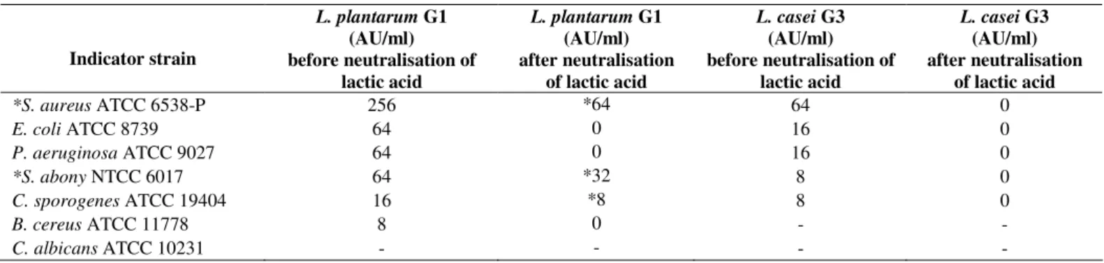
antimicrobial producers was observed. The cell free filtrate of
G1 showed strong antimicrobial activity against all used
pathogenic bacteria, particularly against S. aureus (Table 1). In
order to investigate the proteinaceous nature of the
antimicrobial substances (bacteriocin or bacteriocin-like
substance), the filtrate obtained from overnight culture was
neutralised and the enzyme Proteinase-K was added. No zone
of inhibition was detected only in the case when G1 cell free
filtrate was used against indicator S. aureus ATCC 6538-P.
Lactobacillus casei G3 did not exhibit bacteriocin activity
against the selected indicator strains (Table 1). According to
HPLC analysis LA concentration of cell free filtrates were
11.85 g/l and 11.41 g/l for L. plantarum G1 and L. casei G3,
respectively.
Table 1. Antimicrobial and bacteriocin (*) activity of cells free filtrate obtained from L. plantarum G1 and L. casei G3 overnight
cultures.
Indicator strain
L. plantarum G1 (AU/ml) before neutralisation of
lactic acid
L. plantarum G1 (AU/ml) after neutralisation
of lactic acid
L. casei G3 (AU/ml) before neutralisation of
lactic acid
L. casei G3 (AU/ml) after neutralisation
of lactic acid
*S. aureus ATCC 6538-P 256 *64 64 0
E. coli ATCC 8739 64 0 16 0
P. aeruginosa ATCC 9027 64 0 16 0
*S. abony NTCC 6017 64 *32 8 0
C. sporogenes ATCC 19404 16 *8 8 0
B. cereus ATCC 11778 8 0 - -
C. albicans ATCC 10231 - - - -
The effect of single L. plantarum G1 or L. casei G3 on S.
aureus ATCC 6538-P, E. coli ATCC 8739, and S. abony
NTCC 6017 growth was observed to be dependent of the
pathogen (Table 2). When G1 was mixed with these pathogens,
the growth of the pathogen was 6.5 log units lower for S.
aureus, 2.1 log units lower for E. coli and 0.8 log units for S.
abony, when compared to the growth of the pure culture of the
Zavisic, G. et al. Probiotic features of two oral Lactobacillus
reduction was in the range of 1-2 log units when G3 was used.
The cumulative effect of G1 and G3 (107 CFU/ml) resulted in
the reduction of S. aureus growth for 7.5 log units, of E. coli
for 7.2 log units, and of S. abony for 5.7 log units, when
compared to control cultures. The results for mixed cultures of
G1 and G3 (1010 CFU/ml) showed that the complete inhibition
of pathogenic bacteria growth was observed after 24 h
incubation.
Table 2. The effect of L. plantarum G1 and L. casei G3 on the growth of S. aureus ATCC 6538-P, E. coli ATCC 8739 and S.
abony NTCC 6017.
Indicator strain Indicator strain (control)
Indicator strain with 107 CFU/ml
G1
Indicator strain with 107 CFU/ml
G3
Indicator strain with 107 CFU/ml
G1 +G3
Indicator strain with 1010 CFU/ml
G1 +G3 S. aureus
ATCC 6538-P 6.5±0.10 x 10
8
2.0±0.08 x 10⁸ 7.1±0.13 x 10⁸ 2.1±0.09 x 101 0
E. coli
ATCC 8739 4.8±0.09 x 10
8
4.4±0.10 x 10⁶ 3.6±0.10 x 10⁷ 3.3±0.09 x 101 0
S. abony
NTCC 6017 1.2±0.05 x 10
8
2.2±0.06 x 10⁷ 9.9±0.12 x 10⁶ 3.7±0.08 x 101 0
Each value represents mean +/- SEM (n = 3)
Bacterial survival was tested in the conditions similar to
those in the proximal part of the gastrointestinal tract, at time
intervals corresponding to the actual presence of lactobacilli in
the intestines. Incubated at 37oC, both isolates showed a high
degree of the survival in AGJ and in the solution containing
0.5% bovine bile salts. After 120-min exposure of strains to
AGJ, a decreased count of viable G1 by 1.94 log CFU/ml, and
G3 by 1.74 log CFU/mlwas observed (Fig. 1A). In addition,
after 120-min exposure to the bovine bile salts solution, viable
G1 and G3 decreased, respectively, 0.45 log CFU/ml, and 0.22
log CFU/ml (Fig. 1B).
Figure 1. Survival of L. plantarum G1 and L. casei G3 in simulated artificial gastric juice at pH 2.0 (A) and in the solution containing
Studies of abnormal toxicity did not reveal any visible
changes in the behaviour of NMRI Ham mice treated with
lactobacilli. In addition, all mice survived the treatment with
lactobacilli.
The composition of microbial populations in the faecal
samples of Lactobacillus-treated and control animals was also
investigated and representatives of the genera Enterococcus
sp., Escherichia coli and Clostridium sp. were evaluated on
selective media. After 7-day treatment of Wistar rats with G1, a
significant decrease (in 3 log units) in E. coli populations was
estimated. No important changes in enterococci and clostridia
counts were detected.
Additionally, after 7 days of lactobacilli administration,
the strains G1 and G3 were re-isolated from the faecal samples,
as well as from the ileum surface. Among 174 colonies
recovered from both, faecal samples and ileum surface, 10
colonies were confirmed as G1 and 10 as G3 by 16S rDNA
sequencing.
Biochemical analysis of blood samples revealed the
influence of G1 and G3 on lipid metabolism in rats. Compared
to the control group, the 7-day treatment with lactobacilli
evidently decreased triglycerides and cholesterol levels, as well
as the ALP activity. In the case of G1-treated rats, a decrease in
triglycerides level of 28% (from 1.44 mM/l to 1.04 mM/l),
cholesterol level of 27% (from 1.86 mM/lto 1.35 mM/l), and
ALP activity of 17% (from 512.50 U/l to 420.00 U/l) was
demonstrated. However in G3-treated rats a decrease in
triglycerides level of 35% (from 1.44 mM/l to 0.95 mM/l),
cholesterol level of 19% (from 1.86 mM/l to 1.51 mM/l), and
ALP activity of 21% (from 512.50 U/lto 395.00 U/l), was
detected (Table 3).
Table 3. Lipid content and ALP activity in serum of Wistar rats treated with lactobacilli.
Cholesterol (mM/l)
Triglycerides (mM/l)
Alkaline phosphatase (ALP) (U/l)
Control 1.86 ± 0.09 1.44 ± 0.21 512.50 ± 33.77
L. plantarum G1 1.35 ± 0.06 1.04 ± 0.11 420.00 ± 29.44 L. casei G3 1.51 ± 0.03 0.95 ± 0.02 395.50 ± 6.06 Each value represents mean +/- SEM (n = 3)
DISCUSSION
Preservation of the microbiological balance in the human
gastrointestinal tract is of great importance, since disturbed
equilibrium, especially between Lactobacillus as one of the
most important Gram-positive and other mainly Gram-negative
bacteria might result in the occurrence of various diseases (8,
16). An estimated number of approximately 100 to 125 species
of the Lactobacillus genus exist and the number is constantly
increasing, due to the isolation and identification of new
lactobacilli species (10, 34). Since lactobacilli are mainly used
in pharmaceutical and food industry, precise identification to
the species level, as well as testing their probiotic features, are
needed. Species like L. acidophilus, L. plantarum, L.
bulgaricus, L. casei, L. rhamnosus, and L. fermentum are
among the most abundant lactobacilli used in probiotic
products. In this paper, two human isolates that showed
antimicrobial activity were analyzed. The isolate G1,
originated from an adult oral mucosa, was identified as L.
plantarum, and the isolate G3, originated from a child oral
mucosa, was identified as L. casei. It is well known that L.
plantarum and L. casei form the dominant oral microbiota of
healthy individuals, while other lactobacilli, like L. salivarius,
L. acidophilus, L. oris, and L. fermentum are also frequently
found (2, 7).
Our in vitro studies showed that G1 and G3 expressed
antimicrobial (especially G1) and bacteriocin activity (only
Zavisic, G. et al. Probiotic features of two oral Lactobacillus
Liasi and coauthors (24) demonstrated that L. plantarum LA22
inhibited the growth of bacteria S. aureus, B. cereus, E. coli,
Salmonella enterica, and Listeria monocytogenes. These results
were also corroborated with other literature data stating that
some L. plantarum strains, besides L. paracasei, L. rhamnosus,
and L. salivarius show the greatest antimicrobial activities (6,
11, 23). The strain G1 expressed its antimicrobial activity
against the pathogenic S. aureus, which is in correlation with
the results obtained for antimicrobial activity of L. plantarum
LA22 and L. paracasei subsp. paracasei BGBUK2-16 (26, 7).
Moreover, according to Todorov and Dicks (36) L. plantarum
isolated from molasses produced two thermostable bacteriocins
(ST28MS and ST26MS) that inhibited the growth of a broad
spectrum of pathogens, including, S. aureus, E. faecalis, E.
coli, P. aeruginosa, and Acinetobacter baumanii. The results
obtained from our study strongly indicate that the antimicrobial
activity of G1 is the result of a bacteriocin-like activity. The
nature of this bacteriocin-like substance(s) will be a focus of
our future studies. We can only speculate that the antimicrobial
activity of G1 may be the result of the cumulative effect of
lactic acid and bacteriocin, while the antimicrobial effect of G3
is the result of lactic acid activity alone. In addition, complete
growth reduction of S. aureus ATCC 6538-P, E. coli ATCC
8739, and S. abony NTCC 6017 pathogens was obtained when
a mixed culture of G1 and G3 at 1010 CFU/ml concentration
was used.
The resistance to specific conditions in stomach and
duodenum is another important factor that could explain the
efficiency of some probiotic preparations. When compared to
the literature data, both strains used in our study showed a high
degree of viability in the simulated conditions of the proximal
and the distal part of the gastrointestinal tract (24, 34). Gastric
digestion at pH 2 did not significantly affect the survival of G1
and G3 and even after 120 min the amount of viable bacteria
decreased by less than 2 log units. In similar experiment,
Pereira and Gibson (32) showed that the strain L. casei Shirota
exhibited a significant decrease of 3-4 log units after 60 min of
exposure to gastric digestion. The proposed bile concentration
to which a probiotic strain should be tolerant varies from 0.15
to 0.6% (14). Lactobacilli tested in this study survived in the
presence of 0.5% bile salts and appear to be resistant to the
intestinal conditions. Overall, obtained results indicate that the
viability of G1 and G3 strains in simulated GIT conditions was
sufficient for their successful and massive passage through this
system.
To assess the safety of G1 and G3 strains, NMRI Ham
mice were fed with bacteria in a dose 100 times greater than
the average most frequently administered probiotic dose for the
oral use. No feeding or behavioural changes were observed in
the treated mice, when compared to the control group. None of
the treated mice died during 72 h following lactobacilli
administration. Therefore, G1 and G3 most probably did not
induce toxic effects, i.e. they were considered safe after the
oral administration. In vivo studies on Wistar rats showed good
viability of lactobacilli in the GIT. Namely, G1 and G3 strains
were re-identified in faecal samples of the treated Wistar rats.
Moreover, a lower E. coli count in faecal samples was seenin
Wistar rats treated with G1. In addition, G1 and G3 strains
were also re-identified in the material taken from the surface of
ileum mucosa of Wistar rats. These results could be an
indicator of good colonization ability and bacterial
adhesiveness of G1 and G3 to the intestinal mucosa as
described previously (28).
The analysis of biochemical parameters of blood samples
revealed that the application of G1 and G3 improves lipid
metabolism and hepatic function. It was shown that both strains
from our study reduced the content of serum lipids, including
cholesterol and triglycerides, in Wistar rats. These results
comply with literature data indicating that specific strains of
lactobacilli, such as L. casei ASCC 292, present hypolipidemic
effect through different mechanisms, like cholesterol removal
through cholesterol micelles destabilization and, secondly,
through precipitation of the cholesterol with bile acids (9, 21,
atheromatous plaque, thus playing a key role in the prevention
of cardiovascular diseases development (30). The ALP is one
of four liver enzymes included in most routine laboratory tests,
because their raised levels may be an indication of a liver
disease. Also, G1 and G3 lowered the activity of liver
enzymes, such as alkaline phosphatase (ALP), indicating
possible general improvement of the liver function.
In conclusion, the results of our study suggest that the
indigenous oral strains L. plantarum G1 and L. casei G3
exhibit high resistance to GIT conditions, including low pH
and bile salts, antimicrobial activity against different human
pathogens, reduce serum cholesterol and triglyceride levels and
decrease the activity of ALP. Additionally, both strains showed
high viability in the GIT of Wistar rats and revealed to be safe
for the consumption when Ham mice were used as a model
organism. Finally, G1 and G3 strains have a promising
probiotic potential and after more detailed analyses, including
clinical trials, could be applied as nutraceuticals or
biotherapeuticals.
ACKNOWLEDGEMENT
These investigations were supported by the Ministry of
Education and Science, Republic of Serbia (grant No.
451-01-0065/2008-01/28 and grant No. 173019).
REFERENCES
1. Aboderin, F.I.; Oyetayo, V.O. (2006). Haematological studies of rats fed different doses of probiotic Lactobacillus plantarum, isolated from fermenting corn slurry. Pakistan. J. Nutr. 5, 102-105.
2. Ahrne, S.; Nobaek, S.; Jeppsson, B.; Adlerberth, I.; Wold, A.E.; Molin, G. (1998). The normal Lactobacillus flora of healthy human rectal and oral mucosa. J. Appl. Microbiol. 85, 88-94.
3. Adams, M.R.; Hall, C.J. (1988). Growth inhibition of food-borne pathogens by lactic and acetic acids and their mixtures. Int. J. Food Sci. Technol. 23, 287-292.
4. Al-Allaf, M.A.H.; Al-Rawi, A.M.M.; Al-Mola, A.T. (2009). Antimicrobial activity of lactic acid bacteria isolated from minced beef meat against some pathogenic bacteria. Iraqi. J. Vet. Sci. 23, 115-117. 5. Allain, C.C.; Poon, L.S.; Chan, C.S.H.; Richmond,W.; Fu, P.C. (1974).
Enzymatic determination of total serum cholesterol. Clin. Chem. 20, 470-475.
6. Amin, M.; Jorfi, M.; Khorsravi, A.D.; Samarbafzadeh, A.R.; Farajzadeh, S. (2010). Isolation and identification of Lactobacillus casei and Lactobacillus plantarum from plants by PCR and Detection of their
antibacterial activity. J. Biol. Sci. 9, 810-814.
7. Badet, C.; Thebaud, N.B. (2008). Ecology of Lactobacilli in the Oral Cavity: A Review of Literature. Microbiol. J. 2, 38–48.
8. Botina, S.G.; Koroban, N.V.;Klimina, K.M.; Glazova, A.A.; Zakharevich, N.V.; Zinchenko, V.V.; Danilenko, V.N. (2010). Genetic diversity of the genus Lactobacillus bacteria from the human gastrointestinal microbiome. Russian J. Gen. 46, 1399-1406.
9. Brashears, M.M.; Gilliland,S.E.; Buck, L.M. (1998). Bile salt deconjugation and cholesterol removal from media by Lactobacillus casei. J. Dairy Sci. 81, 2103-2110.
10. Canchaya, C.; Claesson, M.J.; Sinderen, van G.D.; Toole, W. O. ( 2006). Diversity of the genus Lactobacillus comparative genomics of five species. Microbiol. 152, 3185-3196.
11. Devine, D.A.; Marsch, P. (2009). Prospects for the development of probiotics and prebiotics for oral applications. J. Oral Microbiol.doi: 10.3402/jom.v1i0.1949.
12. European Pharmacopeia 6.0, p. 165. (2008). Abnormal toxicity.
13. FAO/WHO. (2002). Guidelines for the evaluation of probiotics in food drafting guidelines for the evaluation of probiotics in food. Report of a joint FAO/WHO working group on drifting guidelines for the evaluation of probiotics in food. London, Ont. Canada.
14. Fernandez, M.; Boris, S.; Barbes, C. (2003). Probiotic properties of human lactobacilli strains to be used in the gastrointestinal tract. J. Appl. Microbiol. 94, 449-455.
15. Fossati, P.A.; Prencipe, L. (1983). Serum triglycerides determined colorimetrically with an enzyme that produces hydrogen peroxide. Clin. Chem. 29, 538-542.
16. Goel, A.K.; Dilbaghi, N.; Kamboj, D.V.; Singh, L. (2006). Probiotics: Microbial therapy for health modulation. Defence Sci J. 56, 513-529. 17. Graf, C.; Sarasin, F.P.(2007). Efficacy and safety of probiotics. Rev.
Med. Suisse. 3, 2350-2354.
18. He, X.; Tian, Y.; Guo L.; Lux R.; Zusman D.R.; Shi W. (2010). Oral-Derived Bacterial Flora Defends Its Domain by Recognizing and Killing Intruders—A Molecular Analysis Using Escherichia coli as a Model Intestinal Bacterium. Microb. Ecol. 60, 655–664.
19. Huh, Y.S.; Jun, Y.S.; Hon, Y.K.; Song, H.; Lee,S.Y.; Hong, W.H. (2006). Effective purification of succinic acid from fermentation broth produced byMannheimia succinciproducens.Proc. Biochem. 41, 1461-1465.
20. Ishibashi, N.; Yamazaki, S. (2001). Probiotics and safety. Am J Clin Nutr. 73, 465S-470S.
Zavisic, G. et al. Probiotic features of two oral Lactobacillus
22. hypocholesterolemic effect of Lactobacillus casei ssp. casei (biodefensive properties of lactobacilli). Indian J. Med. Sci. 60, 361-370. 23. Kaushik, J.K.; Kumar, A.; Duary, R.K.; Mohanty, A.K.; Grover, S.; Batish, K. (2009). Functional and probiotic attributes of an indigenous isolate of Lactobacillus plantarum. PLoS One doi: 10.1371/journal.pone.0008099.
24. Koll-Klais, P.; Mandar, R.; Leibur, E.; Marcotte, H.; Hammarstrom, L.; Mikelsaar, M. (2005). Oral lactobacilli in chronic periodontitis and periodontal health species composition and antimicrobial activity. Oral. Microbial. Immunol. 6, 354-361.
25. Liasi, S.A.; Azmi, T.I.; Hassan, M.D.; Shuhaimi, M.; Rosfarizan, M.; Ariff, A.B. (2009). Antimicrobial activity and antibiotic sensitivity of three isolates of lactic acid bacteria from fermented fish product. Budi. Malaysian J. Microbiol. 51, 33-37.
26. Liong, M.T.; Shah, N.P. (2005). Acid and bile tolerance and cholesterol removal ability of lactobacilli strains. J. Dairy Sci. 88, 55-66.
27. Lozo, J.; Vukasovic, M.; Strahinic, I.; Topisirovic, L. (2004). Characterization and antimicrobial activity of bacteriocin 217 produced by natural isolate Lactobacillus paracasei subsp. paracasei BGBUK2-16. J. Food Protection. 67, 2727-27345.
28. Matsuda, K.; Tsuji, H.; Asahara, T.; Matsumoto, K.; Takada, T.; Nomoto. K. (2009). Establishment of an Analytical System for the Human Faecal Microbiota, Based on Reverse Transcription-Quantitative PCR Targeting of Multicopy rRNA Molecules. Appl. Environ. Microbiol. 75 (7), 1961–1969.
29. Minelli, E.B.; Benini, A.; Marzotto, M.; Sbarbati, A.; Ruzzenente, O.; Ferrario, R.; Hendriks, H.; Dellaglio, F. 2004. Assessment of novel probiotic Lactobacillus casei strains for the production of functional dairy foods. Int. Dairy J. 14, 723-736.
30. McGowan, M.W.; Artiss, J.D.; Strandbergh, D.R.; Zak, B. (1983). A peroxidase-coupled method for the colorimetric determination of serum triglycerides. Clin. Chem. 29, 2077-2080.
31. Naruszewicz, M.; Johansson, M.L.; Zapolska-Downar, D.; Bukowska, H. (2002). Effect of Lactobacillus plantarum 299v on cardiovascular
disease risk factors in smokers. Am. J. Clin. Nutr. 76, 1249-1255. 32. Oh, S.; Kim, S.H.; Worobo, R.W. 2000. Characterization and
purification of a bacteriocin produced by a potential probiotic culture, Lactobacillus acidophilus 30SC. J. Dairy Sci. 83, 2747-2752.
33. Pereira, D.I.; Gibson, G.R. (2002). Cholesterol assimilation by lactic acid bacteria and bifidobacteria isolated from the human gut. Appl. Environ. Microbiol. 68, 4689-4693.
34. Pham, M.; Lemberg, D.A.; Day, A.S. (2008). Probiotics: sorting the evidence from the myths. Med. J. Aust. 188, 304-308.
35. Pot, B.; Tsakalidou, E. (2009). Taxonomy and metabolism of
Lactobacillus. In Lactobacillus Molecular Biology:From genomics to
probiotics. Ljungh A., and Wadstrom, T. (eds) Norfolk, UK: Caister
Academic Press, p. 3-59.
36. Tietz, N.W. (1995). Quantitative determination of alkaline phosphatase. In Clinical Guide to Laboratory Tests. Wu, A., 3rd ed. Saunandders, Philadelphia, PA.
37. Todorov, S.D.; Dicks, L.M.T. (2005). Lactobacillus plantarum isolated from molasses produces bacteriocins active against Gram-negative bacteria. Enzyme Microb. Technol. 36, 318-326.
38. Torriani, S.; Felis, E.G.; Dellagio, F. (2001). Differentiation of
Lactobacillus plantarum, Lact. pentosus, and Lact. paraplantarum by
recA gene sequence analysis and multiplex PCR assay with recA
gene-derived primers. Appl. Environ. Microbiol. 67, 3450-3454.
39. Veljovic, K.; Terzic-Vidojevic, A.; Vukasinovic, M.; Strahinic, I.; Begovic, J.; Lozo, J.; Ostojic, M.; Topisirovic, L. (2007). Preliminary characterization of lactic acid bacteria isolated from Zlatar cheese. J. Appl. Microbiol. 103, 2142–2152.
40. Walter, J. (2008).Ecological Role of Lactobacilli in the Gastrointestinal Tract: Implications for Fundamental and Biomedical Research. Appl.
Environ. Microbiol.74, 4985-4966.
41. Wang, Y.; Xu, N.; Xi, A.; Ahmed, Z.; Zhang, B.; Bai, X. (2009). Effects
of Lactobacillus plantarum MA2 isolated from Tibet kefir on lipid
metabolism and intestinal microflora of rats fed on high-cholesterol diet. Appl. Microbiol. Biotechnol. 84, 341-347.