Occurrence of genes encoding enterotoxins in uropathogenic
Escherichia coli
isolates
Mahsa Mirzarazi
1, Seyedeh Elham Rezatofighi
1,
Mahdi Pourmahdi
2, Mohamad Reza Mohajeri
3 1Department of Biology, Faculty of Science, Shahid Chamran University of Ahvaz, Ahvaz, Iran. 2
Faculty of Veterinary Medicine, Shahid Chamran University of Ahvaz, Ahvaz, Iran. 3
Clinical and Pathological Laboratory of Dr. Mohajeri, Isfahan, Iran.
Submitted: August 08, 2013; Approved: June 6, 2014.
Abstract
To determine the presence of some toxins of diarrheagenicEscherichia coli(DEC) in uropathogenic E. coli(UPEC), 138 urinary tract infection (UTI)-causing UPECs were analyzed. TheastA,set,sen andcdtBgenes were detected in 13 (9.4%), 2 (1.3%), 13 (9.4%) and 0 (0%) of UPEC isolates respec-tively. The results show that some genes encoding toxins can be transferred from DEC pathotypes to UPECs therefore these isolates can transform into potential diarrhea-causing agents .
Key words: uropathogenicEscherichia coli, toxins, phylogenetic groups, set,sen, astAandcdt genes.
Escherichia coliis a commensal of the human intes-tine. However sometimes it causes extra-intestinal infec-tions such as urinary tract infecinfec-tions (UTIs), in this case they are named uropathogenicE. coli(UPEC) (Abeet al., 2008; Oliveiraet al., 2011). They differ from commensal and diarrheagenic strains with respect to phylogenetic groups and virulence factors (Sabateet al., 2006). Com-mensal strains mostly belong to phylogenetic group A and B1 while most extra intestinal pathogenicE. coli(ExPEC) strains fall into group B2 or D (Abdallah et al., 2011; Clermontet al., 2000; Johnson and Stell, 2000; Molina-Lópezet al., 2011).
Enteroaggregative heat stable toxin 1 (EAST-1), a 38 amino acid peptide, is encoded by theastAgene located on the 60-MDa pAA plasmid common to most enteroaggri-gativeE. coli (EAEC) strains (Mendez-Arancibia et al., 2008; Telliet al., 2010; Vilaet al., 2000). In addition to the astAgene, this plasmid contains genes encoding adherence fimbria (AAFI and AAFII) (Mendez-Arancibia et al., 2008). TheastAgene is present in commensal, aggregative, and nonaggregativeE. colistrains (Telliet al., 2010; Vilaet al., 2000). The toxin encoded by this gene stimulates the production of high levels of cyclic guanosine mono-phosphate (cGMP) in the cell such that sodium (Na)/chlo-ride (Cl) ions cotransport system is inhibited and
absorption of water and electrolytes from the intestine at villus tips is reduced, resulting in the elevation of secretion of Cl-and water in crypt cells (Telliet al., 2010).
Shigellaenterotoxin 1 (ShET1) , a virulence factor in EAEC, was detected for the first time inShigella flexneri 2a. This enterotoxin is encoded by chromosomalsetgenes located on the antisense strand of mucinase gene in S. flexneristrains and EAEC (Telliet al., 2010; Vilaet al., 2000). Thesetgenes encoding this toxin contain 2 contigu-ous open reading frames (ORFs) of 534 (setlA) and 186 (setlB) bp (Fasanoet al., 1997). These genes are located on the she pathogenicity island (PAI), a 46-kb chromosomal element that carries some genes having potential or estab-lished roles in bacterial virulence.The watery phase of diar-rhea in shigellosis is caused by this toxin (Thong et al., 2005).
Shigella enterotoxin 2 (ShET2), a 62-8 kDa single protein, is encoded by thesengene located on the 140-MDa invasion plasmid (Fasanoet al., 1997; Olesenet al., 2012; Telliet al., 2010). This toxin is found in most species of Shigellaas well as enteroinvasive E. coli (EIEC) strains (Farfán et al., 2011; Fasanoet al., 1997; Yavzori et al., 2002).Cytolethal distending toxin (CDT), a complex pro-tein, contains 3 polypeptides CdtA, CdtB, and CdtC. This toxin has DNase I activity and breaks double-strand DNA
Send correspondence to S.E. Rezatofighi. Department of Biology, Faculty of Science, Shahid Chamran University of Ahvaz, 6135743135 Ahvaz, Iran. E-mail: [email protected].
and therefore is called genotoxin or cyclomodulin. Five types of CDTs have been found inE. colistrains thus far. Some of these CDTs are encoded by genes located on plasmids; for example, gene encoding CDT-III is carried by pVir, a conjugative plasmid, while others are encoded by genes carried by a lambdoid or P2 phages (Vargaset al., 1999). Because some virulence factors (VFs) of diarrhea-genicE. coli(DEC) such as EAST, SHET1, ShET2, and CDT toxins are located on PAIs, plasmids and other mobile genetic elements, this study aimed to investigate the pres-ence of these toxins in UPEC isolates and their relationship with phylogenetic groups in order to understand the genetic diversity of UPEC strains .
One hundred and thirty-eight UPEC clinical isolates were investigated in this study. These bacteria were iso-lated from urine samples of patients with UTI referred to clinical laboratories of Isfahan, Iran. UPEC was confirmed by a positive urine culture with at least 105cfu ofE. coli /mL. These isolates were identified by standard laboratory protocols. In addition, 30E. coli isolates were collected from feces of healthy humans and were used as controls. The study protocol conformed to the ethical guidelines of the Declaration of Helsinki (No 63/21/8/90).E. coliisolates were inoculated in Luria Bertani broth and incubated over-night at 37 °C. Total DNA was obtained by using the boil-ing method. Bacteria were pelleted from broth, resuspend-ed in sterile distillresuspend-ed water, and boilresuspend-ed at 95 °C for 10 min. Next, the samples were centrifuged at 14,000 rpm for 5 min. The supernatants were collected used as DNA tem-plate and stored at -20 °C. For confirmingE. coliisolates,
PCR was performed to amplify a fragment of the gene en-coding for the highly specific E. coli universal stress protein A (uspAgene). PCR primers and conditions were described by Chen and Griffiths (1998).
For phylogenetic groups, two genes ofchuA,yjaA, and a DNA fragment TSPE4.C2 were investigated by a tri-plex PCR method designed by Clermontet al.(2000). This reaction was performed in a final volume of 20mL, contain-ing DNA (2 mL boiling lysate), 3 mM MgCl2, 0.4 mM dNTP, 2.5 U Taq DNA Polymerase (CinaGen, Iran), 1x Taq DNA Polymerase Buffer, and 0.4mM of each primer. The thermal cycler (Bio-Rad-icycler, America) conditions were as follows: 94 °C for 5 min followed by 30 cycles of 94 °C for 30 s, 60 °C for 30 s and 72 °C for 30 s and final ex-tension of 7 min at 72 °C. All primers used are listed in Ta-ble 1. Detection of ShET1, ShET2, and EAST-1 enterotoxins encoded byset,sen, andastAgenes, respec-tively, was done by amplifying these genes using primers reported previously (Abeet al. 2008). In addition, thecdtB gene encoding cytolethal distending toxin was also ampli-fied as suggested by Johnson and Stell (2000). TheastA gene PCR condition was set up as follows: 94 °C for 3 min followed by 30 cycles of 94 °C for 30 s, 55 °C for 1 min, 72 °C for 1 min and 5 min at 72 °C as final extension. PCR was carried out in a 20mL volume containing 2mL of 10x Taq Polymerase buffer, 3 mM MgCl2, 0.8 mM of each primer, 0.4 mM dNTP, 1 U of Taq polymerase (CinaGen, Iran), and 1mL of template DNA. Forsetandsengenes PCR conditions were similar and involved denaturation at 95 °C for 3 min, 30 cycles of denaturation at 95 °C for 50 s,
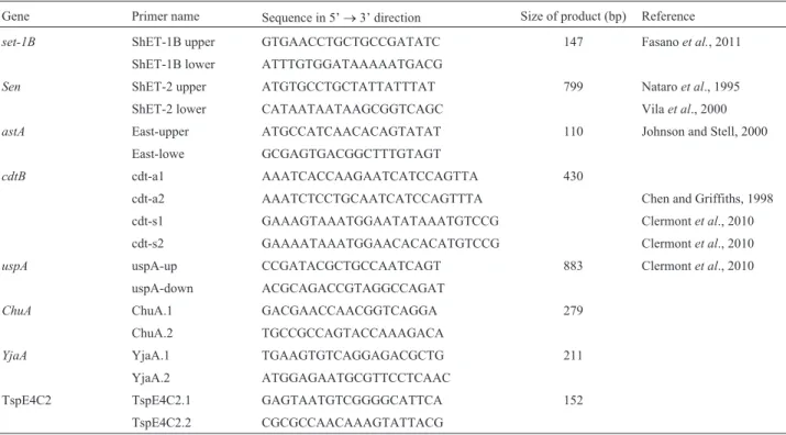
Table 1- Targets, names, sequences (5’-3’), and product sizes of DNA primers.
Gene Primer name Sequence in 5’®3’ direction Size of product (bp) Reference
set-1B ShET-1B upper GTGAACCTGCTGCCGATATC 147 Fasanoet al., 2011 ShET-1B lower ATTTGTGGATAAAAATGACG
Sen ShET-2 upper ATGTGCCTGCTATTATTTAT 799 Nataroet al., 1995
ShET-2 lower CATAATAATAAGCGGTCAGC Vilaet al., 2000
astA East-upper ATGCCATCAACACAGTATAT 110 Johnson and Stell, 2000
East-lowe GCGAGTGACGGCTTTGTAGT
cdtB cdt-a1 AAATCACCAAGAATCATCCAGTTA 430
cdt-a2 AAATCTCCTGCAATCATCCAGTTTA Chen and Griffiths, 1998
cdt-s1 GAAAGTAAATGGAATATAAATGTCCG Clermontet al., 2010
cdt-s2 GAAAATAAATGGAACACACATGTCCG Clermontet al., 2010
uspA uspA-up CCGATACGCTGCCAATCAGT 883 Clermontet al., 2010
uspA-down ACGCAGACCGTAGGCCAGAT
ChuA ChuA.1 GACGAACCAACGGTCAGGA 279
ChuA.2 TGCCGCCAGTACCAAAGACA
YjaA YjaA.1 TGAAGTGTCAGGAGACGCTG 211
YjaA.2 ATGGAGAATGCGTTCCTCAAC
TspE4C2 TspE4C2.1 GAGTAATGTCGGGGCATTCA 152
annealing at 55 °C for 90 s, extension at 72 °C for 2 min and one final extension cycle at 72 °C for 7 min. The PCR as-says were performed in a final volume of 25mL, containing DNA (1mL boiling lysate), 3 mM MgCl2, 0.4 mM dNTP, 1 U Taq Polymerase (CinnaGene, Iran), 1x PCR Buffer, and 0.4mM primers. ThecdtBgene was also amplified as de-scribed previously by Johnson and Stell (2000). The associ-ation between different groups and presence of investigated genes was assessed using Pearson Chi-square test or Fisher’s exact test with the SPSS 16.0 software. Results were considered as statistically significant at p < 0.05.
All the 168 UPEC and commensal isolates were con-firmed asE. coliby standard laboratory protocols.In addi-tion theuspAgene was detected in all the UPEC isolates. Of the 138 UPEC isolates, 16 (12%), 76 (55%), 29 (21%), and 17 (12%) strains belonged to phylogenetic groups B1, B2, D, and A respectively. Concerning to the 30 commensalE. coliisolates, 9 (30%), 12 (40%), and 9 (30%) bacteria were allocated into B2, D, and A groups, respectively but none of the isolates tested clustered in B1 group. A comparison of UPEC strains in different phylogenetic groups showed that B2 group isolates were significantly higher than those be-longing to other phylogenetic groups (p£0.001). For the commensal isolates, D group isolates were statistically more significant than those belonging to other groups (p£0.001). These details are shown in Table 2. Presence of 4 genes ofset,sen,astA,cdtBwas investigated in UPEC and commensalE. coliisolates. TheastAgene was detected in 13 (9.4%) UPEC and 5(16.6%)E. coliisolates collected from feces. Theset gene was detected in only 2 (1.3%) UPEC isolate and was not amplified for any of the com-mensalE. coliisolates tested. Thesengene was detected in 13 (9.4%) UPEC isolates and in 2 (6.6%) commensal iso-lates. ThecdtBgene was not found among the UPEC andE. colicommensal strains analyzed. None of these differences were statistically significant (p > 0.05). Subsequently, the association between phylogenetic groups and these toxins was investigated. No statistically significant correlation was seen between phylogenetic groups and these factors (p > 0.05). Of the 13 astA-positive UPEC isolates, 9
(69.2%), 2 (15.4%), 1 (7.7%), and 1 (7.7%) belonged to B2, B1, D, and A phylogenetic groups, respectively. Both the set-positive isolates belonged to the B2 phylogenetic group. Six (46%) UPEC isolates carrying thesengene be-longed to the D group, 4 (31%) to the B2 group, 2 (15%) to the B1 group and 1 (8%) to the A group. Although more enterotoxins were observed in the B2 phylogenetic group than in other groups, the different was not statistically sig-nificant.Three UPEC isolates had bothsetandsengenes and belonged to the B2 phylogenetic group.
In the present study, we investigated the prevalence of different toxins and phylogenetic groups in UPEC iso-lates. Our data showed few differences and similarities with other studies performed on UPECs. The high prevalence of the B2 phylogenetic group observed in our study is in agreement with that observed in previous studies by other researchers who found a high prevalence of the B2 phylo-genetic group in ExPEC pathogenic strains (Abdallahet al., 2006; Clermontet al., 2000; Molina-Lópezet al., 2011). The structuralastAgene, identified first in EAEC, encodes a low-molecular weight enterotoxin EAST-1 (Yatsuyanagi et al., 2003). In addition to EAEC, this gene has been found in enterohemorragic E. coli (EHEC), enteropathogenicE. coli(EPEC), atypical enteropathogenicE. coli(A-EPEC), Enterotoxigenic E. coli (ETEC), and Shiga Toxin-ProducingE. coli(STEC), (Contreraset al., 2011; Paiva de Sousa et al., 2001; Yatsuyanagi et al., 2003) and other members ofEnterobacteriaceaesuch asSalmonella(Paiva de Sousaet al., 2001). Abeet al.(2008) found that some UPEC isolates carried the gene sequences aggR, aggC, aap,andastA, which are located in the conserved and large plasmid pAA. In their study, 7.1% out of 225 UPEC iso-lates were found to beastApositive. Sotoet al.(2009) de-tected theastAgene in 8% of 170 UPEC clinical isolates; in the present study, this gene was detected in 9.4% UPEC isolates . Furthermore, theastAgene was found in com-mensal isolates. This result is in accordance with that re-ported by Vilaet al.(2000), who found theastAgene in a significant proportion ofE. coliintestinal isolates that did not cause diarrhea, suggesting that this toxin is insufficient to cause diarrhea without presence of other virulence
fac-Table 2- Distribution of different genes and relationship with phylogenetic groups in clinical and commensal isolates ofE. coli.
Gene Number of cases
UPEC isolates
Phylogenetic groups of UPEC isolates Commensal
E. coli
isolates
Phylogenetic groups of commensalE. coliisolates
A B1 B2 D A B1 B2 D
138 (%) 17 (%) 16 (%) 76 (%) 29 (%) 30 (%) 9 (%) 0 (%) 9 (%) 12 (%)
set 2 (1.3) 0 (0) 0 (0) 2 (2.6) 0 (0) 0(0) 0 (0) 0 (0) 0 (0) 0 (0)
sen 13 (9.4) 1 (5.8) 2 (12.5) 4 (5.2) 6 (20.7) 2(6.6) 0 (0) 0 (0) 2 (22) 0 (0)
astA 13 (9.4) 1 (5.8) 2 (12.5) 9 (11.8) 1 (3.4) 5 (16.6) 0 (0) 0 (0) 4 (44) 1 (8)
cdtB 0 (0) 0 (0) 0 (0) 0 (0) 0 (0) 0 (0) 0 (0) 0 (0) 0 (0) 0 (0)
astA+sen 3 (2.2) 0 (0) 0 (0) 3 (3.9) 0 (0) 0 (0) 0 (0) 0 (0) 0 (0) 0 (0)
tors. ShET1 is a 55-kDa protein encoded by thesetgene lo-cated on the antisense strand of a mucinase gene in S. flexneriand EAEC (Royet al., 2006; Telliet al., 2010). This PAI has been found in other bacteria such asYersinia enterocolitica,Salmonella typhimurium, pathogenicE. coli isolates but not in any diarrhea-causing bacteria (Telliet al., 2010, Vila et al., 2000). ShET2, encoded by thesen gene located in the large invasion plasmid, has been re-ported in different Shigella species as well as in EIEC, EAEC, ETEC-ST, and amongE. coliisolates not associ-ated to diarrhea (Farfánet al., 2011; Nataroet al., 1995; Royet al., 2006; Vilaet al., 2000). In a study onE. coli iso-lates causing bacteremia, 21 and 8 out of 100 UTI cases were positive forset andsengenes, respectively. In this study UTI- causing agents were not distinguished, and bac-teria were isolated from blood not urine (Telliet al., 2010). Another study performed by Sotoet al.(2009) analyzing the presence ofset andsen genes in 170 UPEC isolates showed that 16% of the isolates had thesetgene; however, thesenwas not detected in any of the isolates. In contrast, in our study, thesetgene was detected in 1.3% isolates, which is lesser than that detected by Sotoet al.(2009) while the sengene was detected in 13 (9.4%) UPEC isolates . We could not find any report on the presence of thesengene in UPEC isolates. Thecdtgene was detected for the first time in DEC and subsequently among other gram-negative bac-teria such as Campylobacter spp., Shigella spp.,
Helicobacter spp. Aggregatibacter
actinomycetemcomitans, Escherichia albertii,
Haemophilus ducreyi, and Providenica alcalifaciens (Asakuraet al., 2007; Okudaet al., 1995; Scottet al., 1994; Vargaset al., 1999). InC. coliandC. jejuni,cdtgenes are not associated with any mobile genetic element, whereas in E. coliCDT encoding-genes are present on a plasmid or bacteriophage (Vargaset al., 1999). In the study by John-son and Stell (2000) on urosepsis isolates ofE. coli, 8% of the isolates werecdtB positive, suggesting that cdt gene should also be investigated as possible extraintestinal VF even though they have been primarily regarded as an en-teric VF. In contrast to the study by Johnson and Stell (2000),cdtgenes were not found in any case or control iso-lates. Although the presence of enterotoxins in the B2 phylogenetic group was more frequently detected than that of other phylogenetic groups, there seems to be no relation-ship between the presence of enterotoxins and the B2 phylogenetic group. Our results, along with those by Soto et al.(2009) and Abeet al.(2008), raise the probability that E. colistrains acquire these toxins to become potential diar-rhea-causing agents. However it should be remarked that not all isolates carrying these genes express these toxins (Vilaet al., 2000). The presence of EAST, ShET-1, and ShET-2 in UPEC strains shows that horizontal transfer of virulence factors present on plasmids, PAIs, and other mo-bile genetic elements in bacteria belonging to different or similar species can take place. A study on DEC pathotypes
in Brazil showed that 45% and 22% EAEC and EPEC strains, respectively, carried at least one of the urovirulence sequences (Regua-Mangiaet al., 2010). Although this and other studies report the presence of some enterotoxin genes inShigellaand DEC pathotypes of UPEC strains, we do not know whether these genes are expressed in vivo and play any role in bacterial pathogenesis. These questions remain to be answered (Abeet al., 2008).
Acknowledgments
This study was supported by research grant 91/4/06/636410 from the Shahid Chamran University of Ahvaz.
References
Abdallah KS, Cao Y, Wei DJ (2011) Epidemiologic Investigation of Extra-intestinal pathogenic E. coli (ExPEC) based on PCR phylogenetic group and fimH single nucleotide poly-morphisms (SNPs) in China. Int J Mol Epidemiol Genet 2:339-353.
Abe CM, Salvador FA, Falsetti INet al.(2008) Uropathogenic
Escherichia coli(UPEC) strains may carry virulence proper-ties of diarrhoeagenicE. coli. FEMS Immunol Med Micro-biol 52:397-406.
Asakura M, Samosornsuk W, Taguchi Met al.(2007) Compara-tive analysis of cytolethal distending toxin (cdt) genes amongCampylobacter jejuni,C. coliandC. fetusstrains. Microb Pathog 42:174-183.
Chen J, Griffiths MW (1998) PCR differentiation ofEscherichia colifrom other Gramnegative bacteria using primers derived from the nucleotide sequences flanking the gene encoding the universal stress protein. Lett Appl Microbiol 27:369-371.
Clermont O, Bonacorsi S, Bingen E (2000) Rapid and Simple De-termination of the Escherichia coli Phylogenetic Group. Appl Environ Microbiol 66:4555-4558.
Contreras CA, Ochoa TJ, Ruiz Jet al.(2011) Phylogenetic rela-tionships of Shiga toxin-producingEscherichia coliisolated from Peruvian children. J Med Microbiol 60:639-646. Farfán MJ, Toro CS, Barry EMet al. (2011) Shigella
entero-toxin-2 is a type III effector that participates in Shigella-induced interleukin 8 secretion by epithelial cells. FEMS Immunol Med Microbiol 61:332-339.
Fasano A, Noriega FR, Liao FMet al.(1997) Effect of shigella enterotoxin 1 (ShET1) on rabbit intestine in vitro and in vivo. Gut 40:505-511.
Johnson JR, Stell AL (2000) Extended virulence genotypes of
Escherichia colistrains from patients with urosepsis in rela-tion to phylogeny and host compromise. J Infect Dis 181:261-272.
Mendez-Arancibia E, Vargas M, Soto Set al.(2008) Prevalence of different virulence factors and biofilm production in enteroaggregativeEscherichia coliisolates causing diarrhea in children in Ifakara (Tanzania). Am J Trop Med Hyg 78:985-989.
Molina-López J, Aparicio-Ozores G, Ribas-Aparicio RMet al.
Nataro JP, Seriwatana J, Fasano Aet al.(1995) Identification and cloning of a novel plasmid-encoded enterotoxin of entero-invasiveEscherichia coliandShigellastrains. Infect Immun 63:4721-4728.
Okuda J, Kurazono H, Takeda Y (1995) Distribution of the cyto-lethal distending toxin A gene (cdtA) among species of
ShigellaandVibrio, and cloning and sequencing of thecdt
gene fromShigella dysenteriae. Microb Pathog 18:167-172.
Olesen B, Scheutz F, Andersen RLet al. (2012) Enteroaggre-gativeEscherichia coliO78:H10, the cause of an outbreak of urinary tract infection. J Clin Microbiol 50:3703-3711.
Oliveira FA, Paludo KS, Arend LNet al.(2011) Virulence char-acteristics and antimicrobial susceptibility of uropathogenic
Escherichia colistrains. Genet Mol Res 10:4114-4125.
Paiva de Sousa C, Dubreuil JD (2001) Distribution and expression of the astA gene (EAST1 toxin) inEscherichia coliand Sal-monella. Int J Med Microbiol 291:15-20.
Regua-Mangia AH, Irino K, da Silva Pacheco Ret al.(2010) Mo-lecular characterization of uropathogenic and diarrheagenic
Escherichia coli pathotypes. J Basic Microbiol 50:S107-S115.
Roy S, Thanasekaran K, Dutta Roy ARet al.(2006) Distribution of Shigella enterotoxin genes and secreted autotransporter toxin gene among diverse species and serotypes of shigella isolated from Andaman Islands, India. Trop Med Int Health 11:1694-1698.
Sabate M, Moreno E, Perez Tet al.(2006) Pathogenicity island markers in commensal and uropathogenicEscherichia coli
isolates. Clin Microbiol Infect 12:880-886.
Scott DA, Kaper JB (1994): Cloning and sequencing of the genes encodingEscherichia colicytolethal distending toxin. Infect Immun 62:244-251.
Soto SM, Guiral E, Bosch Jet al.(2009) Prevalence of theset-1B
and astA genes encoding enterotoxins in uropathogenic
Escherichia coli clinical isolates. Microb Pathog 47:305-307.
Telli M, Guiral E, Martínez JAet al.(2010) Prevalence of entero-toxins amongEscherichia coliisolates causing bacteraemia. FEMS Microbiol Lett 306:117-121.
Thong KL, Hoe SL, Puthucheary SDet al.(2005): Detection of virulence genes in Malaysian Shigella species by multiplex PCR assay. BMC Infect Dis14:5-8.
Vargas M, Gascon J, Jimenez De Anta MTet al.(1999) Preva-lence of Shigella enterotoxins 1 and 2 among Shigella strains isolated from patients with traveler’s diarrhea. J Clin Microbiol 37:3608-3611.
Vila J, Vargas M, Henderson IRet al.(2000) Enteroaggregative
Escherichia coli virulence factors in traveler’s diarrhea strains. J Infect Dis 182:1780-1783.
Yatsuyanagi J, Saito S, Miyajima Yet al.(2003) Characterization of atypical enteropathogenicEscherichia colistrains harbor-ing the astA gene that were associated with a waterborne outbreak of diarrhea in Japan. J Clin Microbiol 41:2033-2039.
Yavzori M, Cohen D, Orr N (2002) Prevalence of the genes for shigella enterotoxins 1 and 2 among clinical isolates of shigella in Israel. Epidemiol Infect 128:533-535.
Associate Editor: Agnes Marie Sá Figueiredo