M alnutrition and H epatic Fibrosis in M urine Schistosomiasis
Eridan M Coutinho
Laboratório de Imunopatologia, Departamento de Imunologia, Centro de Pesquisas Aggeu Magalhães-Fiocruz, Caixa Postal 7472, 50670-420 Recife, PE, Brasil
In this paper, four different approaches attempting to reproduce the schistosomal liver fibrosis in undernour-ished mice are reported: shifting from a deficient to a balanced diet and vice-versa, repeated infections, influence of the genetic background, and immunological response. Infections were performed with 30 cercariae of Schisto-soma mansoni and lasted at least four months. Undernourished mice were unable to reproduce the picture of “pipestem” fibrosis, except the C57 BL/10 inbred strain, four out of 21 mice developing the liver lesion. A link of this histological finding to the type of parasite strain can not be discarded at the moment. Repeated infections increased collagen deposition mainly in well nourished animals (seven out of 16 Swiss mice developed “pipestem”-like fibrosis). In undernourished infected Swiss mice the serum levels of soluble egg antigen specific antibodies IgG1, IgG2a, IgG2b, and IgG3 were two to four times lower than those detected for well nourished controls. The decreased humoral immune response coupled to the morphological, morphometric, and biochemical results reinforce the influence of the host nutritional status on the connective tissue changes of hepatic schistosomiasis.
Key words: Schistosoma mansoni - undernutrition - hepatic fibrosis - mouse
Schistosomal hepatic fibrosis (Symmers’ fibrosis) is a typical primary fibrosis involving essentially portal spaces, whose aetiopathogenesis is still incompletely understood. In humans, both periovular granulomatous lesions, and the diffuse progressive periportal fibrosis participate in the formation of fibrotic tissue (Andrade 1965, 1987b, Andrade & Bina 1983, Grimaud & Borojevic 1986).
The elucidation of the pathophysiology and patho-genesis of a disease almost invariably requires the devel-opment of an animal model. Concerning Symmers’ fibro-sis (clay pipestem fibrofibro-sis), different experimental hosts have been tried (Cheever et al. 2003), such as monkeys (Lichtemberg & Sadun 1968, Sadun et al. 1970, Lich-temberg et al. 1971, Damian et al. 1976, Farah et al. 2000), rabbits (Cheever et al. 1980), pigs (Hurst et al. 2000), and mice (Warren & DeWitt 1958, Andrade & Warren 1964, Cheever 1965, Warren 1968, Andrade 1987a, Andrade & Cheever 1993, Andrade et al. 1997). In this last animal model, during mild (one to two pairs of worms) and pro-longed (16 weeks or more) infections with Schistosoma mansoni, a picture mimicking “clay pipestem” fibrosis seen in humans with advanced schistosomiasis has been re-produced in outbred and in some strains of well-nour-ished inbred mice (Andrade & Cheever 1993), but not in undernourished outbred specimens (Coutinho et al. 1997). The importance of an adequate nutritional status for a healthy condition of the host is well recognized. Accord-ing to 1990 figures from the Disease Control Priorities Project in Developing Countries (Mason et al. 2003), it is estimated that 32% of the global burden of disease (mor-tality and morbidity) in these countries would be removed
by eliminating malnutrition. In endemic zones for S. mansoni infection, an overlapping of undernutrition and parasite infection is frequently observed. However, in-complete and sometimes conflicting results have been reported in the literature on the role of the host nutritional status as a probable co-factor in the pathogenesis of ad-vanced clinical forms of schistosomiasis.
Since time and parasite load are not sufficient to ex-plain all the cases, it was decided to study the liver pa-thology in the undernourished murine model under dif-ferent experimental situations. It is known, based upon parasitologic, histopathologic, biochemical, and mor-phometric data, that undernourished mice develop smaller circumoval granulomas, less intense portal inflammation, and minimal liver fibrosis, when compared with control animals. On the other side, undernourished mice are un-able to develop the “pipestem” – like portal lesion (Coutinho et al. 1997) as approximately 30-50% of well-nourished controls usually do (Andrade & Cheever 1993). The purpose of this paper is to report on four different approaches that are being tried in our laboratory, in suc-cessive attempts to reproduce and explain the pathogen-esis of the “pipestem” – like schistosomal hepatic fibro-sis in undernourished mice.
MATERIALS AND METHODS
Animals - Male albino Swiss mice (21 day-old), weigh-ing 11 to 15 g (Experiments I, II, IV) and inbred female mice (BALB/c and C57BL/10 strains), from the Animals Breeding Center-Oswaldo Cruz Foundation (Fiocruz), Rio de Janeiro, Brazil (Experiment III) were used. Animals were kept in individual wire bottom cages. Water and food were provided ad libitum. All animals received humane care in compliance with the guidelines of the Animal Care and Use Committee of the Fiocruz.
Infection - Each mouse was exposed to 30 cercariae of S. mansoni (percutaneous route) shed from laboratory raised and infected B. glabrata. The BH strain (Belo Horizonte, MG, Brazil) was used in Experiments I, II, IV; Fax: +55-81-3453.2449. E-mail: ecoutinho@cpqam.fiocruz.br
8 6 8 6 8 6 8 6
8 6 M alnutrition and Schistosomal Fibrosis in M ice • Eridan M Coutinho
and the SLM strain (São Lourenço da Mata, PE, Brazil) in Experiment III (see Experimental groups). Infections lasted 24 weeks (Experiment I), 20 weeks (Experiment III), and 16 weeks (Experiment IV). In Experiment II, reinfections were performed starting 45 days after the primary infection (5 reinfections with 15 cercariae each per mouse, at 15 day intervals). In all the experiments, mice were first infected after a period of four weeks ingesting their respective diet.
Diets - Undernutrition was induced by feeding mice with a multideficient and essentially low-protein diet (7-8% protein), planned to simulate that usually ingested by low-income individuals living in endemic areas of Manson’s schistosomiasis in Northeast Brazil (Coutinho et al. 1997) and is thus referred to as regional basic diet (RBD). Control diet (NUVILAB) was a pelleted commer-cial balanced chow for mice produced by Nuvital Nutri-ents Ltda. (Colombo, PR, Brazil), with 22% protein con-tent.
Evaluation of nutritional status - Body weight weekly recorded and food consumption measured every day were the parameters used to evaluate the nutritional status of the mice. The experimental model used has extensively been studied in previous investigations (Coutinho 1980, Teodósio et al. 1990, Coutinho et al. 1992, 1997).
Experimental groups
Experiment I - Shifting from a deficient to a balanced diet and viceversa: Group G1, “early undernutrition” -RBD was offered during 16 weeks and then replaced by NUVILAB for a further 8 weeks period; Group G2, “late undernutrition” - Mice were maintained on NUVILAB for 16 weeks, and then shifted to RBD for 8 weeks; Group CG3, “long-lasting undernutrition” (Controls for group G1) - The RBD-diet was offered throughout the whole experiment; Group CG4, “long-lasting” normal feeding (Controls for group G2) - Mice were maintained on NUVILAB throughout the experiment.
Results concerning Experiment I were fully reported in Mem Inst Oswaldo Cruz 98(7): 919-925, 2003 and are only summarized in this paper.
Experiment II - Repeated infections: Group A1, un-dernutrition, single infection; Group A2, normal feeding, single infection; Group B1, undernutrition, repeated in-fections; Group B2, normal feeding, repeated infections. Experiment III - Host genetic background: Group A1, undernourished BALB/c mice; Group B1, well-nourished BALB/c mice; Group A2, undernourished C57BL/10 mice; Group B2, well-nourished C57BL/10 mice; Group C1, un-dernourished outbred Swiss mice (controls); Group C2, well-nourished outbred Swiss mice (controls).
Experiment IV - Immunological response: Group A, undernourished, non-infected; Group B, undernourished, infected; Group C, well-nourished, non-infected; Group D, well-nourished, infected.
Morphological studies - Mice were sacrificed by
cer-vical dislocation. The livers were removed, rinsed with PBS (phosphate buffered saline), weighed, and divided into several portions. One section was placed in Bouin’s fixative and/or in buffered (pH 7.4) 10% formaldehyde for histologic examination. Tissue was embedded in paraffin
and the 5 µm thick sections obtained were stained with haematoxylin-eosin and picrosirius red method for col-lagen (Junqueira et al. 1979). A portion of the liver was placed in 4% potassium hydroxide for egg counting (Cheever 1970). Another sample was frozen at –70oC for
further biochemical quantification of hydroxyproline. Morphometry - Randomly sampled 5 µm-thick liver histological sections stained with picrosirius-red for col-lagen, were examined by semiautomatic morphometry us-ing the LEICA Qwin 2.6 Image Processus-ing and Analysis System (Leica Cambridge, Cambridge, England) coupled to a LEICA DC 300F digital camera. For morphometric measurements a total sectional area of 6.6 mm2 per animal
was evaluated. All periovular granulomas were included. A spherical shape and normal size distribution were as-sumed. The following parameters were calculated for granulomas: size, volume density, and numerical density. The sectional area of the fibrous tissue red stained was directly measured and calculated as a percentage of the total area examined, as previously described (Barbosa Jr. 2001, Coutinho et al 2003).
Biochemical study - A sample of the liver was frozen at –70oC for determination of collagen, measured as
hy-droxyproline by the spectrophotometric method B of Bergman and Loxley (1963). Hydroxyproline levels were corrected for intensity of infection with help of a simpli-fied electronic spread sheet elaborated by Cheever (1986) and used in subsequent papers.
Parasitologic studies - Worms recovered after perfu-sion of the portal system (Duvall & DeWitt 1967) were counted and separated by sex. Quantification of the num-ber of eggs in liver tissue was performed after digestion (Cheever 1970).
Immunological studies - Preliminary results are based on lymphoproliferative assays using cultures of spleen cells stimulated with mitogen concanavaline A (1 µg/ml) or soluble egg antigen – SEA (10 µg/ml); production of the cytokines IFN-γ, Il-4, Il-5 by cultured spleen cells af-ter stimulation with SEA and con A (ELISA immuno-enzimatic assay); and titration of SEA serum anti-bodies IgG1, IgG2a, IgG2b, IgG3 (ELISA).
Statistical analysis- Data were analyzed by the fol-lowing methods: Student’s “t-test (when appropriate), one-way ANOVA with Tukey’s and Tamhane’s post tests, Mann-Whitney’s and Kruskal-Wallis’non parametric tests (SPSS v.8 – Statistical Package for Social Sciences Incor-poration, US) and EXCEL (Microsoft, US). Values of p < 0.05 were taken to be significant in the four experiments.
RESULTS
Experiment I - Shifting from a deficient to a balanced diet and vice-versa - The influence of the type of diet on host nutritional status was quite evident, mice ingesting the balanced diet permanently (group CG4) or temporarily (groups G1, G2) showing the best performance regarding weight gain (Fig. 1). Food intake curves showed good correlation with body weight gain.
The histologic picture presented by mice in groups CG3 and G1 did not susbstantially differ. It included scat-tered small periovular granulomas, with variable amount of peri-sinusoidal and septal fibrosis and mild signs of non-specific reactional hepatitis (mononuclear leukocytes admixed with few polymorphonuclear eosinophils in por-tal spaces and around central veins). A few isolated foci of ischemic necrosis or moderate fatty changes were some-times observed. No instance of portal concentration of granulomas, associated with portal expansion and fibrotic connections between portal spaces (“pipestem”-like fi-brosis) was recorded. Well-nourished animais (group CG-4) also presented scattered, small periovular granulomas
of variable sizes and cellular composition. In some of them, there was a large amount of lymphocytes and eosino-phils, while in others fibroblasts and macrophages pre-dominated. Granuloma clusters in portal spaces were fre-quently observed, but the clear-cut picture of “pipestem”-like fibrosis did not develop. Besides focal cellular infil-trations in portal, parenchymal, and centrolobular areas, there were also foci of coagulative necrosis and peripor-tal or diffuse fatty changes. After shifting to a deficient diet (group G-2), the histologic picture underwent mild to moderate changes: several granulomas appeared large, round, with fusiform cells within a loose fibrilar matrix (Fig. 3), subsiding the signs of non-reactive hepatitis. The
Fig. 1
Fig. 2
Fig. 3 Fig. 4 Fig. 5
8 8 8 8 8 8 8 8
8 8 M alnutrition and Schistosomal Fibrosis in M ice • Eridan M Coutinho
“pipestem”-like fibrosis (Figs 4, 5) was detected in two mice belonging to the “late undernourished” group (G2). Morphometric analysis showed that in the well-nour-ished controls (Group CG4) and “late undernourwell-nour-ished” mice (group G2), the volume density and the mean size of periovular granulomas were significantly higher in com-parison with the other groups.
Total liver fibrosis measured morphometrically as per-centage of hepatic tissue (Fig. 2) and biochemically as hydroxyproline in liver tissue homogenates showed good correlation by both methods of evaluation, malnourished groups having the worst performance.
Experiment II - Repeated infections - The liver and spleen/body weight ratios from undernourished mice were always lower than those from their counterparts (reinfected well-nourished mice), as seen in Fig. 6.
Histologic examination detected in undernourished single infected mice (A1 group) only scattered small pe-riovular granulomas and mild non-specific reactional hepa-titis (mononuclear and polymorphonuclear eosinophils in some portal spaces and around central veins). Among well-nourished single infected mice only one animal de-veloped the “pipestem” lesion, but in reinfected mice im-ages of portal concentration of circumoval granulomas causing fibrotic expansion and development of thin fi-brous tracts connecting portal spaces (murine “pipestem”-like fibrosis) were seen in four mice (Fig. 7) and in three additional animals a mixed histologic picture was detected (scattered circumoval granulomas and “pipestem”-like fi-brosis). Reinfected undernourished mice, however, were not able to develop “pipestem”-like fibrosis, although thin fibrous strands could eventually be seen from one portal-space to the other.
Morphometric and biochemical evaluations regarding the amount of collagen deposited in the liver showed excellent correlation between the two methods, higher concentrations of fibrous tissue having been detected in the reinfected groups. (Fig. 9A, B).
Experiment III - Genetic background (inbred mice from BALB/c and C57BL/10 strains) - In all mice strains (inbred and outbred), the liver and spleen/body weight ratios were higher in the well-nourished groups.
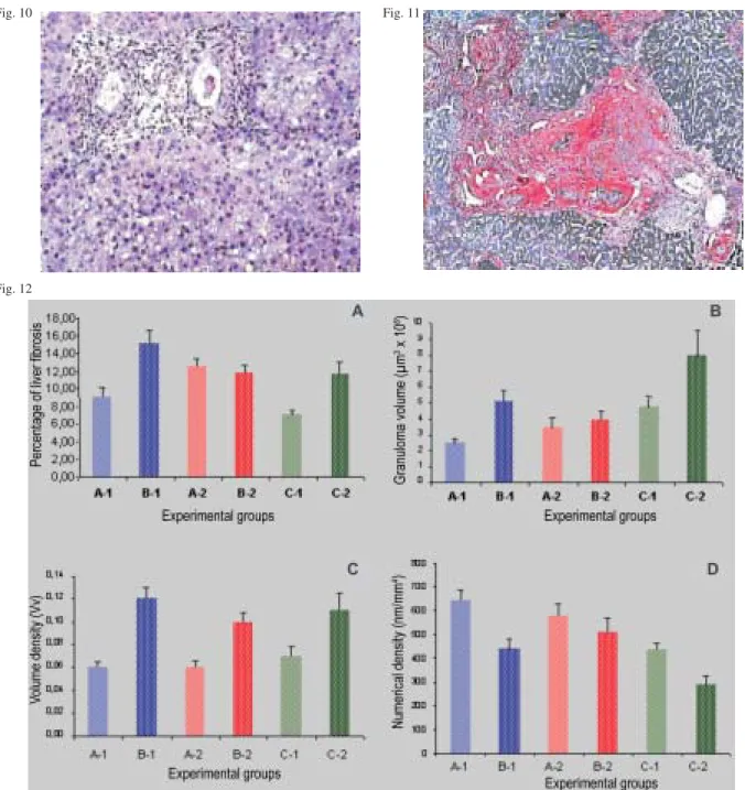
In undernourished BALB/c mice, scattered small fi-brotic granulomas around disintegrating miracidia or only fragments of S. mansoni eggshells were seen on histo-logic examination. Some polymorphonuclear cells and macrophages could sometimes be found at the periphery of the granulomatous reaction. Thin fibrous tracts were eventually found connecting granulomas to each other or linking them to portal spaces. No reactional hepatitis was present, but small foci of ischemic necrosis and some degree of periportal fatty metamorphosis were detected in a few instances. (Fig. 10).
In undernourished mice of the C 57BL/10 strain the granulomas seemed to be more cellular and collagen depo-sition less abundant than in the BALB/c mouse strain. Reactive hepatitis was intense, the acute inflammatory cells invading not only the portal spaces but also the sinusoids (polymorphonuclear cells including eosino-phils). Collagen deposition had a fibrilar aspect and low density. “Pipestem”-like murine fibrosis was detected in two animals (Fig. 11) and in two other mice a mixed lesion
(scattered granulomas and fibrous enlargement of portal spaces with fibrous conections with some granulomas) suggested the onset of “pipestem”-like lesions.
The livers from well-nourished BALB/c inbred mice and Swiss outbred controls did not significantly differ in their general histologic appearance. Foci of ischemic ne-crosis, acute reactive hepatitis of variable intensity and collagenized circumoval granulomas were the most out-standing features of the histopathologic picture. In well-nourished C57BL/10 mice, fibrous tracts between granu-lomas and portal spaces and reactive hepatitis seemed to occur in a higher frequency.
Morphometric evaluation (Fig. 12A, B, C, D) detected that undernourished inbred C57BL/10 mice had higher concentrations of total liver collagen than undernour-ished outbred Swiss mice. Significant results were also found between well-nourished inbred mice of both strains and undernourished Swiss outbred controls. Morphomet-ric studies showed that granuloma size and volume den-sity were greater in well-nourished mice, but grabuloma numerical density was higher in undernourished animals in both inbred and outbred mouse strains.
Experiment IV - Immunological response - Prelimi-nary results based upon immunological studies in out-bred Swiss mice of both sexes, showed that the lym-phoproliferative response (spleen cultured cells stimu-lated with Con-A and SEA) and the production of the cytokines IFN-γ and Il-5 in undernourished infected mice did not differ from those of well-nourished controls. How-ever, the serum levels of SEA-specific antibodies IgG1, IgG2a, IgG2b, and IgG3 in undernourished mice were two to four-fold lower than those detected for well-nourished control animals (Fig. 13).
DISCUSSION
Although it is assumed that the chimpanzee and the baboon (Farah et al. 2000) models most closely mimics the pathogenesis of Symmers’ fibrosis observed in man than does the mouse model, the use of the latest is relevant and widespread for several reasons, offering a convenient tool for examination of genetic regulation of schistosomal hepatic fibrosis and for elucidation of mechanisms of he-patic fibrosis in human and experimental hosts.
Undernourished mice have proved to be unable to develop the morphologic picture of “pipestem” peripor-tal-like fibrosis (Coutinho et al. 1997) described in several investigations on the murine model (DeWitt & Warren 1959, Andrade & Warren 1964, Cheever 1965, Warren 1966, Andrade & Cheever 1993, Henderson et al 1993, Andrade et al. 1997). In this paper experimental attempts designed to explain the non-occurrence of the lesion in undernour-ished mice are reported.
The way undernutrition can interfere with the process of fibrous resorption deserves further investigation. It is known that the rate of collagen degradation depends on several known and unknown factors, among them the interplay of collagenase and tissue collagenase in-hibitors (Truden & Boros 1988) and the degree of col-lagen maturation or “cross-linkings” (Ricard-Blum et al. 1992). In this way, nutrition may interfere in all these steps, once protein synthesis occur in all these pro-cesses. For this reason, the host nutritional status may be considered as a contributory factor to the remodeling of periovular granulomas of hepatic schistosomiasis and shistosomal granuloma in undernourished mice can be taken as an adequate model for studies on the dynamics
of hepatic fibrosis (Coutinho et al. 2003).
Collagen deposition seemed to be increased in well-nourished mice and significantly reduced in undernour-ished animals where it appeared looser and more fibrilar than in controls. Repeated infections lead to a higher col-lagen concentration in the liver of undernourished and well nourished mice, higher values being ascribed to well-nourished controls. These results were detected by both morphometric and biochemical evaluations and confirmed by histological examination. In spite of this, no case of “pipestem”-like periportal fibrosis was seen in undernour-ished outbred Swiss mice, even after repeated infections. However, seven out of 16 well-nourished Swiss mice de-veloped the lesion.
Experiment II - Fig. 6:organomegaly (liver, spleen) in undernourished and well-nourished mice submitted to single and/or repeated infections with Schistosoma mansoni. A1: undernourished (single infection); A2: well nourished (single infection); B1: undernourished (reinfected); B2: well nourished (reinfected). Fig. 7: “pipestem”-like liver fibrosis with vascular neoformation, in well-nourished reinfected Swiss mouse. Picrosirius x 100. Fig. 8: scattered small periovular granulomas connected by thin fibrous bridges, in undernourished reinfected Swiss mouse. Pricrosirius red x 50. Fig. 9: morphometric and biochemical evaluations on the amount of hydroxyproline (collagen) in the liver of undernourished and well-nourished mice submitted to single and/or repeated infections with S. mansoni. A1: undernourished (single infection); A2: well-nourished (single infection); B1: undernourished (reinfected); B2: well-nourished (reinfected)
Fig. 6
Fig. 7 Fig. 8
9 0 9 0 9 0 9 0
9 0 M alnutrition and Schistosomal Fibrosis in M ice • Eridan M Coutinho
Liver and spleen/body weight ratios, in the four re-ported trials, showed higher values for the well-nourished groups, being closely related to the type of diet ingested by the animal (i.e., deficient or balanced diet). By morpho-metric evaluation, considered to be a most sensitive method (Barbosa Jr. 2001), undernourished inbred mice of the C57BL/10 strain had higher concentrations of total liver collagen than undernourished Swiss (outbred)
con-trols. In those mice, the signs of reactive hepatitis were more intense, numerous granulomas were still in the exsudative acute phase, the deposited collagen was loose and fibrilar in appearance. The unexpected finding of “pipestem”-like fibrosis occurring in four out of 21 un-dernourished animals still rests unexplained. The influ-ence of another parasite strain (SLM strain-PE) different from the one used in the other three experiments (BH
strain-Experiment III - Fig. 10: scattered small sized cellular granulomas and periportal fatty chnges in the liver of undernourished BALB/c mouse. H & E x 100. Fig. 11: “pipestem”-like lesion in the liver of a C 57BL/10 mouse, showing fibrous enlargement, telangiectasia and periovular granulomas in a portal space. Picrosirius red x 100. Fig. 12: morphometric parameters (percentage of hepatic fibrous tissue, granulomas size, volume, and numerical densities of circumoval granulomas) in the liver of undernourished and well-nourished BALB/c and C57 BL/10 inbred mice infected with Schistosoma mansoni, in compararison with outbred controls. A1: undernourished BALB/c mice; B1: well-nourished C57 BL/10 mice; A2:undernourished BALB/c mice; B2: well-nourished C57BL/10 mice; C1: undernourished Swiss mice (outbred); C2: well-nourished Swiss mice (outbred)
Fig. 10 Fig. 11
MG), due to a laboratory accident occurring during the trial, cannot be discarded. Studies are now being carried out focusing on the role of different parasite strains in the pathogenesis of the lesion in undernourished mice.
In Experiment III, inbred BALB/c mice had the worst performance regarding growth curves and none of the mice, in either diet, developed the murine “pipestem”-like periportal fibrosis. This is in disagreement with Flannery’s observations (2003), who mentions that 59% of well nour-ished BALB/c mice developed “pipestem” fibrosis upon S. mansoni infection. However, in the reported studies the duration of infection was longer (20-24 weeks) and this may explain the lack of success in reproducing the lesion in our own trial.
The influence of genetic factors on the manifestations of disease associated with infection with S. mansoni (portal hypertension, liver granulomas, hepato-splenomegaly) and their modulation were studied in inbred strains of mice by Fanning et al. (1981). Granuloma size, organo-megaly, and portal venous pressure have proven to be strain dependent and determined by more than one gene. According to these authors, disease associated with in-fection with S. mansoni and its modulation in mice are influenced by the genetic (non- H-2) background of the host and dependent in part on cell-mediated immunity.
Body, liver, and spleen weights seem to vary among different strains. C3H/HeJ mice were found to develop the most pronounced degree of hepatosplenomegaly and granuloma formation. (Fanning et al. 1981). According to these investigators, more than one gene appears to be involved in the inheritance of the propensity to develop severe disease.
Hepatic fibrosis in S. mansoni infected mice is mainly associated with the circumoval granulomas (Dunn et al 1977), although some portal fibrosis not associated with granulomas is also present (Cheever 1965).
Marked differences in granuloma volume and in he-patic fibrosis were found between strains of well-nour-ished mice (Dunn 1980, Cheever et al. 1987). Besides, those strains with the largest granulomas also showed the most hepatic fibrosis. According to the last author, the regula-tion of granuloma size and of hepatic fibrosis is clearly complex and involves genes both outside of and within
the major histocompatibility complex. Studies on differ-ences in hepatic fibrosis in outbred ICR mice and several inbred mice strains by Cheever et al. (1983) showed that hepatic fibrosis was much more marked in S. mansoni infected mice of an outbred ICR strain.
In this investigation, decreased total liver fibrosis observed in undernourished infected mice may have re-sulted from an impaired mechanism of repair, a lower anti-gen load or a low antibody production due to low protein synthesis in a low-protein fed host (Oliveira et al. 2004). Although the lymphoproliferative response and the cytokine production (IFN-γ and IL-5) from undernour-ished outbred infected mice had been similar to those of well-nourished controls, the humoral immune response based on the serum levels of SEA-specific antibodies obtained in Experiment IV, coupled to the gathered mor-phological, morphometric, and biochemical data, suggest that the host nutritional status may interfere in the con-nective tissue changes occurring in hepatic schistoso-miasis.
ACKNOWLEDGMENTS
To Zilton A Andrade, Aryon A Barbosa Jr., Ricardo Ribeiro dos Santos, Milena BP Soares, Sheilla A Oliveira, Luciana S Flannery, Andréia F Barros, Fabiana I Silva, Sandra L Carvalho, Claudia L Cavalcanti, and Carlos F Luna for their valuable par-ticipation in different steps of the experiments here reported, and to Mr Roni Evencio de Araújo, for skilled technical assis-tance.
REFERENCES
Andrade ZA 1965. Hepatic schistosomiasis. Morphological aspects. In H Papper, F Schaffner (eds), Progress in Liver Disease 2, Grune & Stratton, New York, p. 228-242. Andrade ZA 1987a. Pathogenesis of “pipestem” fibrosis of the
liver (Experimental observation on murine schistosomia-sis). Mem Inst Oswaldo Cruz 82: 325-334.
Andrade ZA 1987b. Pathology of human schistosomiasis. Mem Inst Oswaldo Cruz 82 (Suppl. IV): 17-23.
Andrade ZA, Bina JC 1983. Patologia da forma hepato-esplênica da esquistossomose mansoni em sua forma avançada (Estudo de 232 necropsias completas). Mem Inst Oswaldo Cruz 78: 285-305.
Andrade ZA, Cheever AW 1993. Characterization of the mu-rine model of schistosomal hepatic periportal fibrosis (“pipestem” fibrosis). Int J Exp Pathol 74: 195-202. Andrade ZA, Warren KS 1964. Mild prolonged
schistosomia-sis in mice: alterations in host response with time and the development of portal fibrosis. Trans R Soc Trop Med Hyg 58: 53-57.
Andrade ZA, Silva LM, Souza MM 1997. An experimental approach to the pathogenesis of “pipestem” fibrosis (Sym-mers’fibrosis of the liver). Mem Inst Oswaldo Cruz 92: 699-706.
Barbosa Jr. AA 2001. Morphological computer-assisted quan-titative estimation of stained fibrous tissue in liver sec-tions: applications in diagnosis and experimental research. J Bras Patol 37: 197-200.
Bergman I, Loxley R 1963. Two improved and simplified meth-ods for the spectrophometric determination of hydroxypro-line. Ann Chem 35: 1961-1965.
Cheever AW 1965. A comparative study of Schistosoma mansoni infections in mice, gerbils, multimmate rats, and hamsters II. Qualitative pathological differences. Am J Trop Med Hyg 14: 227-238.
9 2 9 2 9 2 9 2
9 2 M alnutrition and Schistosomal Fibrosis in M ice • Eridan M Coutinho
Cheever AW 1970. Relative resistance of the eggs of human schistosomes to digestion in potassium hydroxide. Bull WHO 43: 601-603.
Cheever AW 1986. The intensity of experimental schistosome infections modulates hepatic pathology. Am J Trop Med Hyg 35: 124-133.
Cheever AW, Dunn MA, Dean DA, Duvall RH 1983. Differ-ences in hepatic fibrosis in ICR, C3H and C57BL/6 mice infected with Schistosoma mansoni. Am J Trop Med Hyg 32: 1364-1369.
Cheever AW, Duvall RH, Hallack Jr TA, Minker RG, Malley JD, Malley KG 1987. Variation of hepatic fibrosis and granuloma size among mouse strains infected with Schisto-soma mansoni. Am J Trop Med Hyg 37: 85-97.
Cheever AW, Duvall RH, Minker RG, Nash TE 1980. Hepatic fibrosis in rabbits infected with Japanese and Philippine strains of Schistosoma japonicum. Am J Trop Med Hyg 29: 1327-1339.
Cheever AW, Lenzi JÁ, Lenzi HL, Andrade ZA 2003. Experi-mental models of Schistosoma mansoni infection. Mem Inst Oswaldo Cruz 97: 917-940.
Coutinho EM 1980. Patobiologia da desnutrição nas doenças parasitárias. Mem Inst Oswaldo Cruz 75: 63-76.
Coutinho EM, Barros AF, Barbosa Jr. A, Oliveira SA, Silva LM, Araújo, RE, Andrade ZA 2003. Host nutritional status as a contributory factor to the remodeling of schistosomal hepatic fibrosis. Mem Inst Oswaldo Cruz98: 919-925. Coutinho EM, Freitas LPCG, Abath FGC 1992. The influence
of the regional basic diet from Northeast Brazil on health and nutritional conditions of mice infected with Schisto-soma mansoni. Rev Soc Bras Med Trop 25: 13-20. Coutinho EM, Souza MM, Silva LM, Cavalcanti CL, Araújo
RE, Barbosa Jr. AA, Cheever AW, Andrade ZA 1997. Patho-genesis of schistosomal “pipestem” fibrosis: a low-protein diet inhibits the development of “pipestem” fibrosis in mice. Int J Exp Pathol 78: 337-341.
Damian RT, Greene ND, Meyer KF, Cheever AW, Hubbard WJ, Hawes ME, Clark JD 1976. Schistosoma mansoni in baboons III. The course and characteristics of infection, with additional observations on immunity. Am J Trop Med Hyg 25: 299-305
DeWitt WB, Warren KS 1959. Hepato-splenic schistosomiasis in mice. Am J Trop Med Hyg 8: 440-446.
Dunn MA, Cheever AW, Dean DA, Duvall RH, Kelley EP 1980. Diminished liver fibrosis in murine schistosomiasis in an inbred mouse strain. Clin Res28: 274A (Abstract). Dunn MA, Kojhind M, Warren KS, Hait PK, Rifas L, Seifter S
1977. Liver collagen synthesis in murine schistosomiasis. J Clin Invest 59: 666-674.
Duvall RH, DeWitt WB 1967. An improved perfusion tech-nique for recovering adult schistosomes from laboratory animals. Am J Trop Med Hyg 16: 483-486.
Fanning MM, Peters PA, Davis RS, Kazura JW, Mahmoud AAF 1981. Immunopathology of murine infection with Schistosoma mansoni: relationship of genetic background to hepatosplenic disease and modulation. J Infect Dis 144: 148-153.
Farah IO, Nyindo M, King CL, Hau J 2000. Hepatic granulo-matous response to Schistosoma mansoni eggs in BALB/c mice and olive baboons (Papio cynocephalus anubis). J Comp Pathol 123: 7-14.
Flannery LMS 2003. Estudo sobre a Patogenia da Fibrose
He-pática Periportal na Esquistossomose do Camundongo, PhD Thesis, Universidade Federal da Bahia-Fundação Oswaldo Cruz, Salvador, BA, 96 pp.
Grimaud J-A, Borojevic R 1986. Portal fibrosis: intrahepatic portal vein pathology in chronic human schistosomiasis mansoni. J Submicrosc Cytol 18: 783-793.
Henderson GS, Nix NA, Montesano MA, Gold D, Freeman Jr GL, McCurley TL, Colley DG 1993. Two distinct patho-logical syndromes in male CBA/J inbred mice with chronic Schistosoma mansoni infections. Am J Pathol 142: 703-713.
Hurst MH, Willingham III AL, Lindberg L 2000. Tissue re-sponses in experimental schistosomiasis japonica in the pig: a histopathologic study of different stages of single low-or high-dose infections. Am J Trop Med Hyg 62: 45-56. Junqueira LCV, Bignolas G, Brentani R 1979. Picrosirius stain-ing plus polarization microscopy, a specific method for collagen detection in tissue section. Histochemistry J 11: 447-455.
Lichtemberg L, Sadun EH 1968. Experimental production of bilharzial pipe stem fibrosis in the chimpanzee. Exp Parasitol 22: 264-278.
Lichtemberg FV, Sadun EH, Cheever AW, Erickson DG, Johnson AJ, Boyce HW 1971. Experimental infection with Schisto-soma japonicum in chimpanzees. Parasitologic, clinical, se-rologic and pathological observations. Am J Trop Med Hyg 20: 850-893.
Mason JB, Musgrove P, Habicht J-P 2003. At least one-third of poor countries disease burden is due to malnutrition. Working Paper no. 1, Disease Control Priorities Project, Bethesda, Maryland; Fogarty International Center, National Institutes of Health, US.
Oliveira SA, Silva LM, Barbosa Jr. AA, Ribeiro-dos-Santos R, Coutinho EM, Andrade ZA, Soares MBP 2004. Decreased humoral and pathologic responses in undernourished mice infected with Schistosoma mansoni. Parasitol Res 93: 30-35.
Ricard-Blum S, Ville G, Grimaud JA 1992. Pyridinoline, a ma-ture collagen cross link, in fibrotic liver from Schistosoma mansoni infected mice. Am J Trop Med Hyg 47: 816-820. Sadun EH, Lichtemberg FV, Cheever AW, Erickson DG 1970.
Schistosomiasis mansoni in the chimpanzee. The natural history of chronic infection after single and multiple expo-sures. Am J Trop Med Hyg 19: 258-277.
Teodósio NR, Lago ES, Romani SAM, Guedes RCA 1990. A regional basic diet from Northeast Brazil as a dietary model of experimental malnutrition. Arch Lat Amer Nutr 40: 533-547.
Truden JL, Boros DL 1988. Detection of ∝2-macroglobulin, ∝-protease inhibitor, and neutral protease anti-protease complexes within liver granulomas of Schistosoma mansoni infected mice. Am J Pathol 130: 281-288.
Warren KS 1966. The pathogenesis of clay pipestem cirrhosis in mice with chronic schistosomiasis mansoni, with a note on the longevity of the schistosomes. Am J Pathol 49: 479-489.
Warren KS 1968. Pathophysiology and pathogenesis of hepatosplenic schistosomiasis. Bull Ny Acad Med 44: 280-294.