online | memorias.ioc.fiocruz.br
Co-administration of plasmid-encoded granulocyte-macrophage
colony-stimulating factor increases human immunodeficiency virus-1
DNA vaccine-induced polyfunctional CD4
+T-cell responses
Vinicius Canato Santana1,2, Rafael Ribeiro Almeida1,2, Susan Pereira Ribeiro1,2,
Luís Carlos de Souza Ferreira3, Jorge Kalil2,4, Daniela Santoro Rosa2,5, Edecio Cunha-Neto1,2,4/+
1Universidade de São Paulo, Faculdade de Medicina, Divisão de Imunologia Clínica e Alergia, São Paulo, SP, Brasil
2Instituto Nacional de Ciência e Tecnologia, Instituto de Investigação em Imunologia 3Universidade de São Paulo, Instituto de Ciências Biomédicas, Departamento de Microbiologia, São Paulo, SP, Brasil 4Universidade de São Paulo, Faculdade de Medicina, Instituto do Coração,
São Paulo, SP, Brasil 5Universidade Federal de São Paulo, Faculdade de Medicina, Divisão de Imunologia, São Paulo, SP, Brasil
T-cell based vaccines against human immunodeficiency virus (HIV) generate specific responses that may limit both transmission and disease progression by controlling viral load. Broad, polyfunctional, and cytotoxic CD4+ T-cell responses
have been associated with control of simian immunodeficiency virus/HIV-1 replication, supporting the inclusion of CD4+ T-cell
epitopes in vaccine formulations. Plasmid-encoded granulocyte-macrophage colony-stimulating factor (pGM-CSF) co-admin
-istration has been shown to induce potent CD4+ T-cell responses and to promote accelerated priming and increased migration
of antigen-specific CD4+ T-cells. However, no study has shown whether co-immunisation with pGM-CSF enhances the number
of vaccine-induced polyfunctional CD4+ T-cells. Our group has previously developed a DNA vaccineencoding conserved, mul
-tiple human leukocyte antigen (HLA)-DR binding HIV-1 subtype B peptides, which elicited broad, polyfunctional and long-lived CD4+ T-cell responses. Here, we show that pGM-CSF co-immunisation improved both magnitude and quality of vaccine-in
-duced T-cell responses, particularly by increasing proliferating CD4+ T-cells that produce simultaneously interferon-γ, tumour
necrosis factor-α and interleukin-2. Thus, we believe that the use of pGM-CSF may be helpful for vaccine strategies focused on the activation of anti-HIV CD4+ T-cell immunity.
Key words: granulocyte-macrophage colony-stimulating factor - DNA vaccine - polyfunctional CD4+ T-cells - HIV
doi: 10.1590/0074-02760150283
Financial support: CNPq (420166/2005-0), FAPESP (2004/15856-9, 2006/50096-0, 2008/57881-0)
VCS and RRA contributed equally to this work. + Corresponding author: [email protected] Received 29 July 2015
Accepted 20 October 2015
A safe and effective human immunodeficiency vi-rus (HIV) vaccine is still the most promising strategy for controlling the acquired immune deficiency syndrome pandemic. T-cell based vaccines generate HIV-specific re-sponses that may limit both transmission and disease pro-gression by controlling viral loads (Watkins et al. 2008). A recent Phase III clinical trial in Thailand (RV144) enrolling more than 16,000 individuals showed 31% efficacy in pre-venting viral acquisition (Rerks-Ngarm et al. 2009), which indicates that an HIV-1 vaccine is a feasible aim.
Mounting evidence suggests that
polyfunction-al CD4+ T-cell responses may be important to confer
protection against Mycobacterium tuberculosis and
Leishmania major challenges (Darrah et al. 2007, Lin-denstrøm et al. 2009) and also for controlling HIV-1 replication (Porichis & Kaufmann 2011). Although
HIV-specific CD4+ T-cells are preferentially targeted
by the virus, the vast majority of these cells remains
vi-rus-free at any time in vivo (Douek et al. 2002), which
may allow for their antiviral function. In fact, strong
vi-rus-specific CD4+ T-cell responses have been associated
with natural control of HIV-1 infection and prediction of disease outcome (Rosenberg et al. 1997, Gloster et
al. 2004, Soghoian et al. 2012). Cytotoxic CD4+ T-cells
were shown to suppress viral replication in both simian immunodeficiency virus (SIV) and HIV-1-infected cells (Sacha et al. 2009, Zheng et al. 2009) and the frequency
of polyfunctional mucosal CD4+ T-cells was shown to
be augmented in elite viral controllers when compared to noncontrollers or individuals on highly active antiret-roviral therapy (Ferre et al. 2010).
While the clinical associations of CD4+ T-cell
re-sponses and HIV-1 control face a cause-effect issue, the
finding that CD4+ T-cell depletion contributes to
reduc-ing vaccine-mediated protection against SIV (Vaccari et al. 2008) supports a direct role of such cells in
an-tiviral immunity. Also, increased polyfunctional CD4+
responses induced in recombinant simian varicella
vi-rus-SIVEnv/Gag vaccinated rhesus macaques were shown
to be key correlates of vaccine-mediated protection against SIV infection (Pahar et al. 2012). Thus, inclusion
of CD4+ T-cell targets deserves particular attention in
the design of potential anti-HIV vaccine constructs.
In order to induce HIV-specific CD4+ T-cell
respons-es, our group developed a DNA vaccine encoding mul-tiple human leukocyte antigen (HLA)-DR binding HIV-1 subtype B conserved peptides (HIVBrHIV-18). We have
responses in mice transgenic to common HLA class II alleles (HLA-DR2, -DR4, -DQ6, -DQ8) (Ribeiro et al. 2010). In addition, HIVBr18 immunisation activated polyfunctional and long-lived central and effector
mem-ory CD4+ T-cells in BALB/c mice (Rosa et al. 2011).
However, further improvements in HIVBr18 immuno-genicity will be required before the vaccine formulation could be submitted to clinical trials.
DNA vaccine immunisation leads to direct transfec-tion of antigen presenting cells (APCs) and tissue-res-ident-cells, providing local and systemic expression of target antigens and subsequent induction of cellular and humoral immunity. Professional APCs are not typically found in muscle tissue and they need to migrate to the inoculation site in response to inflammatory or chem-otactic signals before an efficient immune response is mounted (Kutzler & Weiner 2008).
Plasmid-encoded granulocyte-macrophage colony- stimulating factor (pGM-CSF) has been shown to mediate the recruitment of neutrophils, macrophages and imma-ture dendritic cells (DCs) to the immunisation site (Had-dad et al. 2000) and to enhance immune responses induced by DNA vaccines in different animal models (Weiss et al. 1998, Ahlers et al. 2002, Song et al. 2006). Remark-ably, it has been shown that a bicistronic DNA vaccine encoding HIV-1 gp120 and GM-CSF evoked an
exten-sive inflammatory infiltrate and induced potent CD4+
T-cell responses (Barouch et al. 2002). Also, accelerated
antigen-specific CD4+ T-cell priming and increased
mi-gration of activated CD4+ T-cells were observed in mice
immunised with a recombinant GM-CSF-encoding BCG vaccine (Nambiar et al. 2010). However, to our knowl-edge no study has shown that pGM-CSF
co-administra-tion improves the quality of vaccine-induced CD4+ T-cell
responses, which is fundamental for the development of new anti-HIV vaccine approaches.
MATERIALS AND METHODS
Plasmids - The plasmids used in this study were the HIVBr18 vaccine, previously developed by our group (Ribeiro et al. 2010, Rosa et al. 2011), a pGM-CSF kind-ly provided by Dr Sergio Costa Oliveira, from the Fed-eral University of Minas Gerais, Brazil, as previously reported (Diniz et al. 2010), and the pVAX1 vector. The plasmids were purified using the Endofree Plasmid Giga Kit (Qiagen) according to manufacturer’s instructions.
Peptides - The HIVBr18-encoded peptides (Fonseca et al. 2006) were synthesized by solid phase technology using 9-fluorenylmethoxycarbonyl strategy, with the C’ terminal carboxyl group in amide form (GL Biochem, China). Peptide purity and quality were assessed by re-verse-phase high performance liquid chromatography and mass spectrometry, and were routinely above 90%.
Ethics - Inbred BALB/c mice were obtained from the
Animal Facility at the School of Medicine of the Univer-sity of São Paulo (USP) and housed and manipulated un-der specific-pathogen-free conditions in the animal care facility at the Institute of Tropical Medicine, USP. Ex-periments were performed in accordance to the guide-lines of the Ethical Commission for Animals Use of the USP and approved under protocol 197-12.
Mice and immunisations – Six-eight week-old female BALB/c mice were used in this study. HIVBr18 combined with pVAX1 or pGM-CSF was administered intramuscu-larly at days 0, 14, and 28. Each animal received 200 µg of plasmid combination (100 µg of each) per dose, diluted in sterile saline. The doses were divided in two injections of 50 µL, one in each quadriceps. Two weeks after the last
dose, mice were euthanized in a CO2 chamber.
Spleen cell isolation for immune assays - Two weeks after the last immunisation, mice were euthanized and spleens were aseptically removed. After obtaining single cell suspensions, cells were washed in 10 mL of RPMI-1640 (Gibco, USA). Cells were then suspended in RPMI supplemented with 10% of foetal bovine serum (FBS) (Gibco), 2 mM L-glutamine (Sigma-Aldrich, USA), 10 mM Hepes (Sigma-Aldrich), 1 mM sodium piruvate, 1% vol/vol nonessential amino acid solution (Gibco), 40 mg/
mL of gentamicin, 20 mg/mL of peflacin and 5 x 10⁻⁵ M
2-mercaptoetanol (Sigma-Aldrich). The viability of cells was evaluated using 0.2% Trypan Blue exclusion dye to discriminate between live and dead cells. Cell concen-tration was estimated with the aid of a Neubauer cham-ber and adjusted in cell culture medium.
Detection of interferon (IFN)-γ-secreting T-cells by ELISPOT assay - The frequency of IFN-γ-secreting T-cells
was determined by incubating splenocytes (3 × 10⁵ cells/
well) from immunised mice with 5 µM of individual or pooled HIVBr18-encoded peptides for 18 h at 37ºC and
5% CO2. The ELISPOT assay was performed using
mu-rine IFN-γ BD kit according to manufacturer’s instruc -tions (Becton, Dickinson and Company, USA). Spots were counted using an AID ELISPOT reader (Autoimmun Diag-nostika GmbH, Germany). The number of antigen-specific
T-cells, expressed as spot-forming units (SFU)/106
sple-nocytes, was calculated after subtracting negative control values (medium only). Responses were considered positive when above cut-off, which was calculated as the mean re-sponse plus 3 standard deviations (SD) of splenocytes from mock-immunised mice (pVAX or pGM-CSF only), stimu-lated with each peptide. A positive control was performed in all experiments by incubation of splenocytes from each immunised group with concanavalin A (ConA) (final con-centration 2 µg/mL) (Sigma-Aldrich).
Analysis of polyfunctional CD4+ T-cell responses
- Briefly, freshly isolated splenocytes were suspended
(5 x 10⁶/mL) in phosphate-buffered saline (PBS) and
la-belled with 1.25 µM of carboxyfluorescein succinimidyl ester (CFSE) (Molecular Probes, USA) at 37ºC for 10 min. The reaction was quenched with RPMI-1640 sup-plemented with 10% FBS. CFSE-labelled cells were
in-cubated at a density of 2.5 x 10⁶ cells/mL and cultured
in 96 well round-bottomed plates (5 x 10⁵/well) for four
days at 37ºC and 5% CO2 with medium only or pooled
fluorescence-activated cell sorting (FACS) buffer (PBS + 0.5% bovine serum albumin + 2mM ethylenediamine tetraacetic acid) and surface stained using CD4-PercP Cy5.5 and CD8-Alexa Fluor 700 monoclonal antibodies for 30 min at 4ºC. Cells were fixed and permeabilised using the Cytofix/CytopermTM kit (BD Bioscienc-es). Permeabilised cells were washed with Perm/Wash buffer (BD Biosciences) and stained with CD3-APC-Cy7, interleukin (IL)-2-PE, tumour necrosis factor
(TN-F)-α-PECY7 and IFN-γ-APC for 30 min at 4ºC. Follow -ing stain-ing, cells were washed twice and suspended in FACS buffer. All antibodies were from BD Biosciences. Samples were acquired on a FACSCanto flow cytome-ter (BD Biosciences) and then analysed using the Flow-Jo software (FlowFlow-Jo, LLC, USA). Cells were gated on
forward scatter/side scatter, CD3+, and CD4+/CD8+. The
frequency of proliferating T-cells producing cytokines
was determined by gating cells on CFSElow population
that were positive for IFN-γ, TNF-α, or IL-2 expression.
A Boolean analysis was performed to obtain the
fre-quency of proliferating CD4+ or CD8+ T-cells producing
any combination of cytokines. As a positive control in all experiments, pooled splenocytes from each immunised group were incubated with ConA (final concentration 2 µg/mL). Background response from mock-immunised mice splenocytes (pVAX or pGM-CSF only) submit-ted to the same experimental conditions was subtracsubmit-ted from respective experimental group.
Data analysis and statistics - Statistical significance
(p-values) was calculated by using t test for all
experi-ments. The significance of single cytokine or combined
cytokines produced by CFSElow T-cells was calculated
us-ing Student’s t test (since we had 2 experimental groups
for each condition) and all bars were put in the same graph to facilitate visualisation. Statistical analysis and graphi-cal representation of data were performed using Graph-Pad Prism 5.0 software, and p-values < 0.05 were consid-ered significant (*: p < 0.05; **: p < 0.01; ***: p < 0.001).
RESULTS
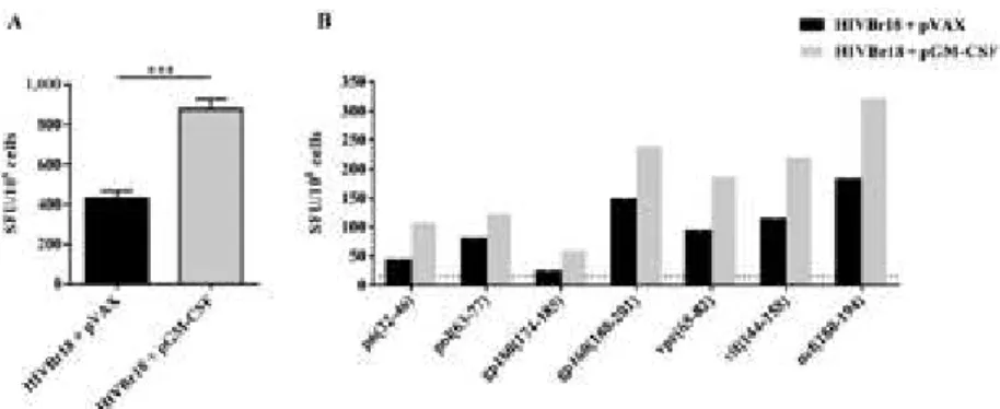
Co-administration of pGM-CSF enhances HIVBr18-induced T-cell responses - In order to evaluate whether GM-CSF would enhance HIVBr18 DNA vaccine im-munogenicity, we co-immunised BALB/c mice with a pGM-CSF. As a control, we co-immunised mice with HIVBr18 DNA vaccine and an empty pVAX1 vector. We observed that co-immunisation with pGM-CSF
en-hanced the frequency of IFN-γ-secreting T-cells against
pooled HIVBr18-encoded peptides, from 432-876
SFU/106 cells (Fig. 1A). To evaluate the impact of
GM-CSF on the breadth of T-cell responses we measured the
frequency of IFN-γ-secreting T-cells against individual
HIVBr18-encoded peptides and observed that although co-administration of pGM-CSF enhanced the magni-tude of T-cell responses, no difference was observed in the number of recognised peptides (Fig. 1B). There-fore, our data suggest that GM-CSF had an impact on HIVBr18 immunogenicity which was not sufficient to broaden T-cell immune responses.
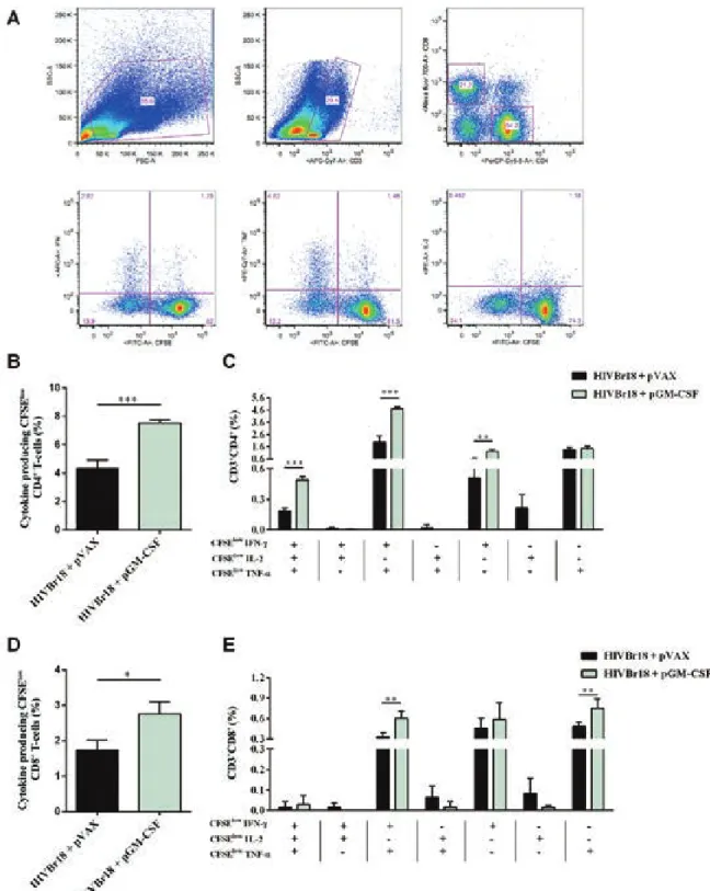
Co-administration of pGM-CSF increases the fre
-quency of HIVBr18-induced polyfunctional CD4+
T-cells - We also addressed the question whether GM-CSF
would improve the quality of HIVBr18-induced im-mune responses by measuring the frequency of
antigen-specific polyfunctional CD4+ T-cells. We immunised
BALB/c mice as previously mentioned and evaluated the
frequency of CD4+ or CD8+ T-cells that were both
pro-liferating and producing the cytokines IFN-γ, TNF-α,
and IL-2 (Fig. 2A). We observed that co-administration of pGM-CSF increased the frequency of proliferating
CD4+ T-cells producing any cytokine against pooled
HIVBr18-encoded peptides, from 4.33-7.52% (Fig. 2B).
In order to determine whether the proliferating CD4+
T-cells were single cytokine producers or polyfunctional cells, we analysed the ability of those cells to proliferate
and produce all possible combinations of IFN-γ, TNF-α, Fig. 1: co-administration of plasmid-encoded granulocyte-macrophage colony-stimulating factor (pGM-CSF) enhances human immunodefi-ciency virus-1 subtype B conserved peptides (HIVBr18)-induced T-cell responses. Two weeks after the last immunisation with HIVBr18 co-administrated with pVAX1 or pGM-CSF, pooled spleen cells from six BALB/c mice were cultured in the presence of both pooled and individual
HIVBr18-encoded peptides (5 µM), or medium only. Frequencies of interferon (IFN)-γ-secreting T-cells against pooled (A) and individual (B)
HIVBr18-encoded peptides were measured by ELISPOT and shown as spot forming units (SFU) per 106 cells. Mean plus standard deviation of
Fig. 2: co-administration of pasmid-encoded granulocyte-macrophage colony-stimulating factor (pGM-CSF) increases the frequency of hu-man immunodeficiency virus-1 subtype B conserved peptides (HIVBr18)-induced polyfunctional CD4+ T-cells. Two weeks after the last
im-munisation with HIVBr18 co-administrated with pVAX1 or pGM-CSF, pooled spleen cells from six BALB/c mice were collected, labelled with carboxyfluorescein succinimidyl ester (CFSE) (1.25 mM) and cultured for four days in the presence of pooled HIVBr18-encoded peptides or medium only. On day 4, cells were pulsed for 12 h with pooled peptides in the presence of Brefeldin A and co-stimulatory antibody (anti-CD28),
and then stained for CD3, CD4, CD8, interferon (IFN)-γ, tumour necrosis factor (TNF)-α, and interleukin (IL)-2. Multiparameter flow cytom -etry strategy used to determine the frequency of polyfunctional T-cells (A). Frequency of cytokine producing CFSElow CD4+ T-cells (B) and
CD8+ T-cells (D). Frequency of CFSElow CD4+ (C) and CD8+ (E) T-cells producing all the combinations of IFN-γ, TNF-α, and IL-2. Background
and IL-2. We observed that co-administration of pGM-CSF significantly improved the frequency of
prolifer-ating CD4+ T-cells producing IFN-γ (from 0.5-1.2%),
IFN-γ, and TNF-α (from 1.98-4.70%), or all three cy
-tokines (from 0.18-0.49%) (Fig. 2C). Similarly to CD4+
T-cells, co-administration of pGM-CSF increased the
frequency of proliferating CD8+ T-cells producing any
cytokine, from 1.73-2.75% (Fig. 2D). On the other hand, we observed that co-administration of pGM-CSF
signif-icantly improved the frequency of proliferating CD8+
T-cells producing, exclusively, TNF-α (from 0.48-0.75%), IFN-γ and TNF-α (from 0.33-0.60%), and no difference
was observed among CD8+ T-cells producing all three
cytokines (Fig. 2E). Thus, our data indicate that GM-CSF enhanced the frequency of HIVBr18-induced
poly-functional CD4+ T-cell responses.
DISCUSSION
In this study, we have shown that co-administration of pGM-CSF and the DNA vaccine HIVBr18 improved both the magnitude and the quality of antiHIV-1 T-cell immune responses, particularly by increasing the
fre-quency of polyfunctional CD4+ T-cells displaying
pro-liferation and capable to secrete different cytokines
(IFN-γ, TNF-α, and IL-2). Natural or vaccine induced
CD4+ T-cells populations can exert wide effects, for
ex-ample, direct cytotoxic effect on infected cells (Sacha et al. 2009, Zheng et al. 2009). Also, it can provide co-stim-ulatory signals to APCs for the activation and induction of effector and memory CD8 T-cell populations (Arens & Schoenberger 2010). Those functions are dependent of cytokine profile production, and in particular for HIV-1 infection, a better quality and protective immune re-sponse can be sustained if those polyfunctional T-helper 1 CD4+ T-cell are present (Ferre et al. 2010).
The finding that co-administration of a plasmid encoding GM-CSF enhanced DNA vaccine immuno-genicity, measured by the frequency of antigen-specific
IFN-γ-secreting T-cells, is in accordance with previous
evidence (Barouch et al. 2002, Nambiar et al. 2010). In fact, the role of GM-CSF as a vaccine adjuvant has been extensively studied, showing that both T and B-cell im-mune responses may be improved. Locally recruited antigen-presenting cells were shown to be critical for the magnitude and nature of such responses (McKay et al. 2004, Robinson et al. 2006, Qiu et al. 2007). Fur-thermore, intramuscular GM-CSF co-administration has been shown to increase the number of infiltrating
CD11c+ DC) and splenic DCs, expression of major
his-tocompatibility complex class II on splenic DC, and en-hance the antigenic capture, processing and presentation functions of splenic DCs (Zhai et al. 2015).
Here, we have shown that although the magnitude of HIVBr18-induced T-cell responses against both pooled and individual peptides was augmented by the co-ad-ministration of pGM-CSF, no difference in the breadth of epitopes recognised by T-cells was observed. As far as we know only one study has explored the impact of GM-CSF on the breadth of T-cell responses induced by vacci-nation (Rodríguez et al. 2012). In this case, it was shown that pGM-CSF co-administered to DNA-prime followed
by modified Vaccinia Ankara-boost broadened T-cell responses to pooled peptides representing five different HIV-1 Nef domains. Such results could not be reproduced in mice immunised with HIVBr18, which encodes isolat-ed peptides previously selectisolat-ed by the potential
immu-nogenic profile to CD4+ T-cells (Ribeiro et al. 2010). In
fact, the multiepitope vaccine approach was originally de-signed to overcome immunodominance effects leading to
broad epitope recognition per se (Livingston et al. 2002).
The fact that our DNA vaccine was rationally designed
for inducing anti-HIV-1 CD4+ T-cell immune responses
(despite having internal CD8 recognition epitopes) led to the question whether co-administration of pGM-CSF would change the immune responses mediated by those
cells. We found that the frequency of polyfunctionalCD4+
T-cells was significantly enhanced in mice co-immunised with HIVBr18 and pGM-CSF, particularly those
produc-ing IFN-γ and TNF-α, or IFN-γ, TNF-α, and IL-2.
Our results demonstrated that, besides enhancing the
magnitude of CD4+ and CD8+ T-cell responses to a
co-ad-ministered DNA vaccine-encoded antigen, expression of GM-CSF had a significant impact on the activation of
CD4+ T-cells capable to secrete different cytokines after
antigen-specific stimulation. It has been described that im-munisation with a bicistronic plasmid co-expressing HIV-1 gpHIV-120 and GM-CSF under control of a single promoter resulted in a dramatic augmentation of both
vaccine-in-duced proliferating and IFN-γ-secreting CD4+ T-cells
(Barouch et al. 2002). Also, a vaccine based on a BCG
en-coding GM-CSF led to accelerated antigen-specific CD4+
T-cell priming and increased migration of activated CD4+
T-cells into the lung, resulting in significantly increased
protection against M. tuberculosis (Nambiar et al. 2010).
The ability of GM-CSF to improve the frequency of
polyfunctional T-cells was previously described for CD8+
T-cells. It was shown that co-expression of GM-CSF and ovalbumin (OVA) in a DNA-prime adenoviral-boost immunisation resulted in a striking expansion of
poly-functional OVA-specific CD8+ T-cells (Tenbusch et al.
2008). Moreover, the use of pGM-CSF as an adjuvant for DNA vaccines expressing HIV-1 Gag and Nef-Tat-Vif in-creased the frequency of antigen-specific polyfunctional
memory CD8+ T-cells (Xu et al. 2008). Co-expression
of GM-CSF has also been shown to increase the titres and antigen avidity of antibodies, and antibody-depend-ent-cellular cytotoxicity induced by a DNA vaccine en-coding six SIV proteins (Lai et al. 2011). However, that study failed to show any improvement in the frequency of
DNA vaccine-induced polyfunctional CD4+ T-cells.
In our study, co-administration of pGM-CSF and HIVBr18, which encodes multiple HLA-DR binding HIVBr18 peptides, increased the frequency of
polyfunc-tional CD4+ T-cells as measured by antigen-induced
proliferation and concomitant expression of IFN-γ, IL-2, and TNF-α. Although the specific mechanisms driving
the GM-CSF-mediated enhancement of
antigen-specif-ic polyfunctional CD4+ T-cells activation have been not
elucidated, we believe that this new finding may con-tribute to the development of DNA vaccines focused on
ACKNOWLEDGEMENTS
To Luis Roberto Mundel and Edilberto Postól, for assis-tance at the animal facility.
REFERENCES
Ahlers JD, Belyakov IM, Terabe M, Koka R, Donaldson DD, Thomas EK, Berzofsky JA 2002. A push-pull approach to maximize vac-cine efficacy: abrogating suppression with an IL-13 inhibitor while augmenting help with granulocyte/macrophage colony-stimulat-ing factor and CD40L. Proc Natl Acad Sci USA99: 13020-13025.
Arens R, Schoenberger SP 2010. Plasticity in programming of effec-tor and memory CD8 T-cell formation. Immunol Rev235: 190-205.
Barouch DH, Santra S, Tenner-Racz K, Racz P, Kuroda MJ, Schmitz JE, Jackson SS, Lifton MA, Freed DC, Perry HC, Davies ME, Shiver JW, Letvin NL 2002. Potent CD4(+) T-cell responses
elic-ited by a bicistronic HIV-1 DNA vaccine expressing gp120 and GM-CSF. J Immunol168: 562-568.
Darrah PA, Patel DT, De Luca PM, Lindsay RW, Davey DF, Flynn BJ, Hoff ST, Andersen P, Reed SG, Morris SL, Roederer M, Seder RA 2007. Multifunctional Th1 cells define a correlate of vaccine-me-diated protection against Leishmania major. Nat Med13: 843-850.
Diniz MO, Lasaro MO, Ertl HC, Ferreira LCS 2010. Immune respons-es and therapeutic antitumor effects of an experimental DNA vaccine encoding human papillomavirus type 16 oncoproteins genetically fused to herpesvirus glycoprotein D. Clin Vaccine
Immunol17: 1576-1583.
Douek DC, Brenchley JM, Betts MR, Ambrozak DR, Hill BJ,
Oka-moto Y, Casazza JP, Kuruppu J, Kuntsman K, Wolinsky S,
Grossman Z, Dybul M, Oxenius A, Price DA, Connors M, Koup RA 2002. HIV preferentially infects HIV-specific CD4(+)
T-cells. Nature417: 95-98.
Ferre AL, Hunt PW, McConnell DH, Morris MM, Garcia JC,
Pol-lard RB, Yee HF, Martin JN, Deeks SG, Shacklett BL 2010. HIV
Controllers with HLA-DRB1*13 and HLA-DQB1*06 alleles have strong, polyfunctional mucosal CD4(+) T-cell responses. J Virol 84: 11020-11029.
Fonseca SG, Coutinho-Silva A, Fonseca LAM, Segurado AC, Moraes SL, Rodrigues H, Hammer J, Kallas EG, Sidney J, Sette A, Kalil J, Cunha-Neto E 2006. Identification of novel consensus CD4 T-cell epitopes from clade B HIV-1 whole genome that are frequent-ly recognized by HIV-1 infected patients. AIDS20: 2263-2273.
Gloster SE, Newton P, Cornforth D, Lifson JD, Williams I, Shaw GM, Borrow P 2004. Association of strong virus-specific CD4 T-cell responses with efficient natural control of primary HIV-1 infec-tion. AIDS18: 749-755.
Haddad D, Ramprakash J, Sedegah M, Charoenvit Y, Baumgartner
R, Kumar S, Hoffman SL, Weiss WR 2000. Plasmid vaccine ex-pressing granulocyte-macrophage colony-stimulating factor at-tracts infiltrates including immature dendritic cells into injected muscles. J Immunol165: 3772-3781.
Kutzler MA, Weiner DB 2008. DNA vaccines: ready for prime time?
Nat Rev Genet9: 776-788.
Lai L, Kwa S, Kozlowski PA, Montefiori DC, Ferrari G, Johnson WE, Hirsch V, Villinger F, Chennareddi L, Earl PL, Moss B, Amara RR, Robinson HL 2011. Prevention of infection by a granulocyte-macrophage colony-stimulating factor co-expressing DNA/mod-ified Vaccinia Ankara simian immunodeficiency virus vaccine. J Infect Dis204: 164-173.
Lindenstrøm T, Agger EM, Korsholm KS, Darrah PA, Aagaard C, Sed-er RA, Rosenkrands I, AndSed-ersen P 2009. TubSed-erculosis subunit vac-cination provides long-term protective immunity characterized by multifunctional CD4 memory T-cells. J Immunol182: 8047-8055.
Livingston B, Crimi C, Newman M, Higashimoto Y, Appella E, Sid -ney J, Sette A 2002. A rational strategy to design multiepitope immunogens based on multiple Th lymphocyte epitopes. J Im -munol168: 5499-5506.
McKay PF, Barouch DH, Santra S, Sumida SM, Jackson SS, Gor-gone DA, Lifton MA, Letvin NL 2004. Recruitment of different subsets of antigen-presenting cells selectively modulates DNA vaccine-elicited CD4(+) and CD8(+) T lymphocyte responses. Eur J Immunol34: 1011-1020.
Nambiar JK, Ryan AA, Kong CU, Britton WJ, Triccas JA 2010. Mod-ulation of pulmonary DC function by vaccine-encoded GM-CSF enhances protective immunity against Mycobacterium tubercu -losis infection. Eur J Immunol40: 153-161.
Pahar B, Gray WL, Phelps K, Didier ES, Deharo E, Marx PA, Traina-Dorge VL 2012. Increased cellular immune responses and CD4+T-cell proliferation correlate with reduced plasma
vi-ral load in SIV challenged recombinant simian varicella virus-simian immunodeficiency virus (rSVV-SIV) vaccinated rhesus macaques. Virol J9: 160.
Porichis F, Kaufmann DE 2011. HIV-specific CD4 T-cells and immune control of viral replication. Curr Opin HIV AIDS6: 174-180.
Qiu JT, Chang TC, Lin CT, Chen YM, Li FQ, Soong YK, Lai CH
2007. Novel codon-optimized GM-CSF gene as an adjuvant to enhance the immunity of a DNA vaccine against HIV-1 Gag.
Vaccine25: 253-263.
Rerks-Ngarm S, Pitisuttithum P, Nitayaphan S, Kaewkungwal J, Chiu J, Paris R, Premsri N, Namwat C, de Souza M, Adams E, Benenson M, Gurunathan S, Tartaglia J, McNeil JG, Francis DP, Stablein D, Birx DL, Chunsuttiwat S, Khamboonruang C, Thongcharoen P, Robb ML, Michael NL, Kunasol P, Kim JH 2009. Vaccination with ALVAC and AIDSVAX to prevent HIV-1 infection in Thailand. N Engl J Med361: 2209-2220.
Ribeiro SP, Rosa DS, Fonseca SG, Mairena EC, Postol E, Oliveira SC, Guilherme L, Kalil J, Cunha-Neto E 2010. A vaccine encoding conserved promiscuous HIV CD4 epitopes induces broad T-cell responses in mice transgenic to multiple common HLA class II molecules. PLoS ONE5: e11072.
Robinson HL, Montefiori DC, Villinger F, Robinson JE, Sharma S, Wyatt LS, Earl PL, McClure HM, Moss B, Amara RR 2006. Studies on GM-CSF DNA as an adjuvant for neutralizing Ab elicited by a DNA/MVA immunodeficiency virus vaccine. Virol -ogy352: 285-294.
Rodríguez AM, Pascutti MF, Maeto C, Falivene J, Holgado MP, Turk G, Gherardi MM 2012. IL-12 and GM-CSF in DNA/MVA immuniza-tions against HIV-1 CRF12_BF Nef induced T-cell responses with an enhanced magnitude, breadth and quality. PLoS ONE7: e37801.
Rosa DS, Ribeiro SP, Almeida RR, Mairena EC, Postol E, Kalil J, Cunha-Neto E 2011. A DNA vaccine encoding multiple HIV CD4 epitopes elicits vigorous polyfunctional, long-lived CD4(+) and
CD8(+) T cell responses. PLoS ONE6: e16921.
Rosenberg ES, Billingsley JM, Caliendo AM, Boswell SL, Sax PE, Kalams SA, Walker BD 1997. Vigorous HIV-1-specific CD4(+)
T-cell responses associated with control of viremia. Science
278: 1447-1450.
Sacha JB, Giraldo-Vela JP, Buechler MB, Martins MA, Maness NJ, Chung C, Wallace LT, Leon EJ, Friedrich TC, Wilson NA, Hi-raoka A, Watkins DI 2009. Gag- and Nef-specific CD4(+) T-cells
recognize and inhibit SIV replication in infected macrophages early after infection. Proc Natl Acad Sci USA106: 9791-9796.
responses during acute HIV infection predict disease outcome.
Sci Transl Med4: 123ra25.
Song RJ, Liu SQ, Adams RJ, Leong KW 2006. Enhancing efficacy of HIV Gag DNA vaccine by local delivery of GM-CSF in murine and macaque models. J Interferon Cytokine Res26: 380-389.
Tenbusch M, Kuate S, Tippler B, Gerlach N, Schimmer S, Dittmer U, Ueberla K 2008. Co-expression of GM-CSF and antigen in DNA prime-adenoviral vector boost immunization enhances polyfunc-tional CD8+ T-cell responses, whereas expression of GM-CSF
an-tigen fusion protein induces autoimmunity. BMC Immunol9: 13.
Vaccari M, Mattapallil J, Song K, Tsai WP, Hryniewicz A, Venzon D, Zanetti M, Reimann KA, Roederer M, Franchini G 2008. Reduced protection from simian immunodeficiency virus SIVmac251 in-fection afforded by memory CD8(+) T-cells induced by vaccination
during CD4(+) T-cell deficiency. J Virol82: 9629-9638.
Watkins DI, Burton DR, Kallas EG, Moore JP, Koff WC 2008. Non-humam primate models and the failure of the Merck HIV-1 vac-cine in humans. Nat Med14: 617-621.
Weiss WR, Ishii KJ, Hedstrom RC, Sedegah M, Ichino M, Barnhart K, Klinman DM, Hoffman SL 1998. A plasmid encoding murine gran-ulocyte-macrophage colony-stimulating factor increases protection conferred by a malaria DNA vaccine. J Immunol161: 2325-2332.
Xu R, Megati S, Roopchand V, Luckay A, Masood A, Garcia-Hand D, Rosati M, Weiner DB, Felber BK, Pavlakis GN, Sidhu MK, Eldridge JH, Egan MA 2008. Comparative ability of various plasmid-based cytokines and chemokines to adjuvant the activity of HIV plasmid DNA vaccines. Vaccine26: 4819-4829.
Zhai Y, Zhou Y, Li X, Feng G 2015. Immune-enhancing effect of
nano-DNA vaccine encoding a gene of the prME protein of Japa-nese encephalitis virus and BALB/c mouse granulocyte-macro-phage colony-stimulating factor. Mol Med Rep12: 199-209.
Zheng N, Fujiwara M, Ueno T, Oka S, Takiguchi M 2009. Strong abil-ity of Nef-specific CD4(+) cytotoxic T-cells to suppress human