Bull Pan Am Health Organ 18(l), 1984.
MYCOBACTERIA
ISOLATED
FROM APPARENTLY
NORMAL
SWINE LYMPH
NODES IN URUGUAY1
A. Saenzz and F. Errico3
Nontuberculow mycobact& such as Mycobacterium kansasii and M. avium intracellular-e currently pose signzj?cant problems for human hea&th. The epidemiologic mechanisms involved in their transmission are uncertain, but recent evidence suggests swine could be involved. The work presented here sought to isolate nontuberculous mycobacteria j?om apparently normal swine bmph no&s in Uruguay, to iofentz3 the isolates, and to assess the isolated organisms’ pathogenici& in laboratory animals.
Introduction
At present, increasing importance is being
ascribed to the study of nontuberculous myco-
bacteria in both humans and animals (1-3).
The principal reason is that as more is learned
about these organisms’ culture requirements,
morphology, pigment formation, and re-
sponses to biochemical tests, they are being
encountered with increasing frequency. It is
true that the tuberculous mycobacteria Myco-
bacterium tuberculosis and Mycobacterium bovis
continue to pose relatively major human and
animal health problems in Latin America (4),
and that the less-known nontubercnlous myco-
bacteria have thus far been of relatively slight
epidemiologic importance. Nevertheless, the
problems posed by the latter types may be-
come more important in the future-as is sug-
gested by increasingly frequent isolation of
strains from human patients that pose difficult clinical diagnostic problems and that are hard to treat because of resistance to most currently
employed antituberculosis drugs.
The epidemiologic mechanisms producing
sources of infection are still not clear in many
cases. It is evident, however, that the inci-
‘Also appearing in Spanish in the B&tin de la Oficina Sanitaria Panamericanq 94(4), 1984.
ZDoctor of medicine; Laboratory Chief and Bacteriolo- gist, Zoonoses Section, Ministry of Public Health of Uruguay.
SVeterinary doctor; Technician, Department of Bacte- riology, Miguel C. Rubino Veterinary Research Center, Casilla Correo 6577, Montevideo, Uruguay.
dence of human and animal infections caused
by nontuberculous mycobacteria that prolifer-
ate in the environment (water, soil, and food)
is growing (I, 2, 5). It is also evident that
swine, because oftheir susceptibility to various
mycobacterial species (particularly M. bovis,
M. avium, and M. tuberculosis), are also capable
of constituting an important reservoir and
source of mycobacterial transmission (4, 6, 7).
As early as 1963, Scannon et al. reported
the presence of nonchromogenic bacteria at a
hog-breeding facility (6). Later, in 1969,
Kleeberg highlighted the significance of find-
ing nontuberculous mycobacteria in swine,
considered as a reservoir, and the value of this
finding for subsequent epidemiologic work,
especially work on cattle (1). Then, a few
years thereafter, Gotijo and colleagues isolated
nontuberculous mycobacteria from 14.5 per
cent of 200 lymph nodes taken from apparent-
ly healthy hogs in Brazil (5). However, a bac-
teriologic study performed by Kantor and
Leslie in Argentina, using 715 hog lymph
nodes with granulomatous lesions, found that
nontuberculous mycobacteria accounted for
only 3.7 per cent of the lesions, the remainder
being infected with M. bovis (88.6 per cent)
and M. avium (7.5 per cent) (4); while another
survey, based on bacteriologic study of granu-
lomatous lesions in Uruguayan swine, re-
ported that 63.9 per cent of the 187 specimens
examined yielded M. bovis, 31.7 per cent
yielded M. a&m, and only 4.4 per cent
yielded other mycobacteria (8).
64 PAHO BULLETIN l vol. 18, no. 1, 1984
The purpose of the present work was to
determine whether or not nontuberculous
mycobacteria were present in the lymph nodes
of a sample of apparently healthy swine, to
identify each type of mycobacteria isolated,
and to study the isolates’ pathogenicity in
laboratory animals.
Materials and Methods
Over a period of 18 months, 250 specimens
of apparently normal hog lymph nodes were
obtained at two slaughterhouses-one in the
capital city of Montevideo and the other in
Canelones, a department capital some 45 kilo-
meters from Montevideo. All the specimens
collected from each hog (including the sub-
maxillary, retropharyngeal, bronchial, media-
stinal, and mesenteric nodes) were placed in
aseptic plastic bags. They were then trans-
ported under refrigeration (4-7OC) to the
laboratory, where they were dissected and
classified as being normal in appearance, ede-
matous, hemorrhagic, or anthracic. Each
specimen was then treated according to the
method described by Thorel and Boisvert (9).
After treatment and neutralization, 0.3 ml of
the specimen was placed in each of six tubes
containing Lowenstein-Jensen media and six
containing Stonebrink media; and two tubes
of each type were then incubated at 22OC,
37”C, and 45’C. The cultures were read after
4,8, 15,30, and 60 days of incubation. In ad-
dition, smears were prepared from each speci-
men and stained by the Ziehl-Neelsen
method.
TO classify the types of mycobacteria iso-
lated, the macroscopic appearance of the pre-
pared smears was assessed; the appearance of
the cultures was noted; and determinations
were made regarding the cultures’ chromo-
genicity, photochromogenicity, development
time, optimal growth temperature, and
growth in the Lowenstein-Jensen tubes as
compared to growth in the Stonebrink tubes.
In addition, a number of cytochemical tests
were performed to determine the specimens’
levels of beta glucosidase, catalase (at room
temperature and at SS’C), niacin, urease,
iron uptake, nitrate reduction, potassium tel-
lurite reduction, tween 80 hydrolysis, and
growth in 5 per cent sodium chloride.
Experiments to assess certain isolates’
pathogenicity were performed using the triple
inoculation method developed by Saenz and
colleagues (10) with merions,4 hamsters, and
mice. The merions and hamsters were inocu-
lated intraperitoneally with bacillary masses
ranging from 0.5 to 1.0 mg. The mice were
given 0.03 mgintravenously and 0.5 mg intra-
peritoneally. All the inoculated animals were
then kept under observation for a period of
one year following their inoculation.
Results
In all, 30 sets of specimens (12 per cent of the 250 tested) yielded isolates of mycobacte- ria. These were classified as indicated in Table 1. A more detailed listing of the test criteria
used to arrive at these classifications is pro-
vided in Table 2.
Pathogenicity experiments were performed
on the 14 isolates listed below.
Strains of isolated mycobacteria No. of isolates tested for pathogenicity tested
M. avium intracelluture 4
M. terrae-triviale 4
M. scrafulaceum 1
M. kunsasii 1
M. chelonei 1
M. vaccae 1
M. aurum 1
M. gastri 1
The detailed findings of these pathogenicity tests on the 14 isolates are to be described in a
subsequent communication. The basic
results, however, can be summarized as
follows:
1) The five isolates in the MAIS group (M.
avium intracellulare, M. scrofulaceum) were found
Saenz and En-iio l NONTUBERCULOUS MYCOBACTERIA IN URUGUAYAN SWINE 65
Table 1. Strains of mycobacteria isolated from 250 sets of apparently normal swine lymph nodes.
Strains of mycobacteria No. of % of isolated isolates isolates
M. nuium iniracdlulare M. smfilaccum M. term+triviale M. kanmsii M. g&i M. uaccae M. chelonei M. aurum
Total
14 2 5 3 2 2 1 1 30
46 7 17 10 7 7 3 3 100
to produce similar lesions in gerbils, hamsters, and mice, the gerbil being the most sensitive.
During the first three months a pathologic pic-
ture of a Yersin type developed; but continued
observation showed that the animals could
present pulmonary pictures of a bronchopneu-
monial type at the time of death.
2) The tested M. kansasii isolated produced a picture similar to the classical one described
by Buhler and Pollak (II). The animal of
choice for pathology testing of M. kansasii is
the hamster.
3) Certain of the strains classically regarded
as saprophytic (M. tewae-triviale, M. chelonei,
M. vaccae, and M. aurum) were found to
possess important pathogenic abilities, pro-
ducing specific and significant lesions.
4) The M. gashi strain tested showed no evidence of pathogenicity.
Discussion
The proportion of swine (30 of 250) yielding
mycobacterial isolates should be considered
significant, especially since the samples were
obtained from animals destined for human
consumption.
Swine, which are usually infected through the digestive tract (via food or water) or some- times via fomites in the air, can serve as im-
portant mycobacterial reservoirs and trans-
mission vehicles (2, 4, 6). There is thus a need to pursue further studies aimed at determining
the importance of these animals in mycobacte-
rial transmission and the type of lesions that
transmitted mycobacteria can cause in both
humans and animals. Until now, most studies
on the isolation and classification of mycobac-
teria in swine have been done on specimens
exhibiting granulomatous lesions (I, 3, 4, 6,
8, IZ), with the result that these studies ob-
tained a high frequency of M. bovis isolates.
However, previous studies of swine lymph
nodes without macroscopic lesions have not
yielded isolates of M. bovis, and the overall
percentages of mycobacterial species isolated
were similar to those obtained by ourselves (5, 7, 13).
Fourteen of the isolates obtained in the
present study (46 per cent of the total) con- sisted of M, avium intracehlare. This finding is
consistent with the fact that this mycobacte-
rium has accounted for a high percentage of
the isolates obtained by other studies of lymph
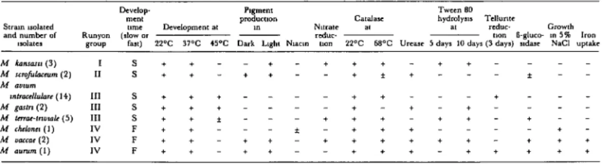
Table 2. Test criteria uacd to identify the 30 strains of mycobacteria k&ted from the lymph nodes of 250 apparently normal swine.
lhclop PlgllW Twwccn 80
men, Cat&x hydmlyr,~ Tcllurnc
SIral” rrolarcd ume Lk”clopmenr at pCdUCU0” L” N,,ratc a, at 4°C Growth
and null&r of Runyon (rlow or mduc- tnn I-gluco- 3” 5% Iron
,lolater WuP fax) 22% VT *5oc Dark l&t N,ac,n uon 22T 68% “rcasc 5 day. 1” dayr (3 day.) ndax N&l up*&
M knnr~rr (3) I S++--+-+++-++-
M rrmfulnccvm (2) II s++-++--+*+---*--
M D”fYrn
*“lrue/h&rr (14) III s+++----++---+---
M gartn (2) III S+++----+-+-+- ---
M Lmoc-m”mlr (5) III s++*---+++-++-+--
M rhdona (1) IV F++---*-+++--- + -
M u(lfcoe (2) IV F++-++-++++++-+++
66 PAHO BULLETIN l vol. 18, no. 1, 1984
nodes, both with and without lesions (4, 5, 8, 12, 13).
It appears that humans contract M. avium
intracellulare infections from some natural re-
servoir, since no transmission between
humans has been found (4). The results of the present study suggest swine as a likely source
of M. avium intracellulare infection in man.
There is also evidence that exposure to this
mycobacterium may be fairly common. Spe-
cifically, partial sample surveys of the Uru-
guayan population’s reaction to avian tu-
berculin have found positive response rates
ranging from 5.3 to 36 per cent (2). Despite
these high percentages, however, only four
clinical cases produced by M. avium intracellu- lare have been reported (14).
In regard to the M. scrofulaceum and M. kan- sasii isolated in our study, these have been iso-
lated previously from swine lymph nodes with
and without lesions (4, 5, 7, 13). Bacilli of the
M. scrofulaceum type have been implicated by
authors in several countries as a cause of
adenitis in children (10, 15, 1 S), and M. kansa- sii is known to cause severe illness in man.
The natural habitat of M. kansasii is not
known, but it has been isolated from cattle,
swine, water, and milk (16).
The rest of the mycobacteria isolated in our
study (M. tewae-triviale, M. gastri, M. chelonei,
M. vaccae, and M. aurum) are considered
saprophytic in both humans and animals;
nevertheless, as noted above, we found some
of these to demonstrate pathogenicity in labo-
ratory animals. It is also worth noting that in two instances M. vaccae has been isolated from
retropharyngeal nodes of dairy cattle with
macroscopic lesions, and isolates from two
human hospital patients have also been identi-
lied by one of us (Errico) as consisting of M.
vaccae.
Also, the finding of a relatively high per-
centage of atypical mycobacteria, regarded as
nonpathogenic but capable of sensitizing
humans and animals, indicates that a share of
the positive human reactions to tuberculin
may be due to paraspecific sensitization by
these mycobacteria (2). This suggests that
their possible role as sensitizing agents could
prove a worthwhile subject of future study.
Conclusions
In our opinion, several findings emerging
from the present study have implications of
epidemiologic importance. These findings are
(1) that swine appear to serve as a natural re-
servoir for nontuberculous mycobacteria; and
(2) the relatively high rate of mycobacterial
isolations from our specimens (12 per cent)
supports comparable values reported by Goti-
jo et al. in Brazil (14.5 per cent) and Garcia
Rodriguez et al. in Spain (14 per cent) (5, 13).
It also seems noteworthy that 16 of the 30
isolates obtained from our swine specimens in
Uruguay belonged to the MAIS complex, and
that three others were identified as M. kansasii.
This means that a total of 63.3 per cent of the
mycobacteria isolated were identified as
pathogens capable of infecting people. It also
appears that these pathogenic species, like
those that are saprophytic, may interfere with
the diagnosis of tuberculosis by producing
paraspecific sensitization in both humans and
animals.
ACKNOWLEDGMENTS
We wish to express our appreciation to Center, for their assistance in preparing ma-
Mrs. Maria Alba Boga de Frost of the Minis- terial for isolation and classification. We are
try of Public Health Zoonoses Laboratory and also very grateful to Dr. Isabel M. de Kantor
to Mr. Miguel Castro, Laboratory Assistant of the Pan American Zoonosis Center’s Tu-
Saenz and Ewico l NONTUBERCULOUS MYCOBACTERIA IN URUGUAYAN SWINE 67
of these isolates. In addition, we would like to belonging to the old Runyon Group IV, one thank Ms. F. Thorel of the Alfort School who of these being the isolate of M. aumn and the provided confirmatory typing of two isolates other an isolate of M. vaccae.
SUMMARY
While the importance of nontuberculous myco- bacteria such as Mycobacterium avium intracellukzre and M. kansasii as a source of human and animal disease is far less than that of M. tuberculosis and M. bovis in the Americas, the former are being isolated with in- creasing frequency from human patients and are posing significant clinical diagnosis and drug treat- ment problems. Furthermore, though it appears that swine can serve as reservoirs and transmission vehicles for such bacteria, the epidemiologic mech- anisms involved are still unclear. The purpose of the work reported here was to determine whether the lymph nodes of apparently healthy swine were harboring nontuberculous mycobacteria, to identi- fy the types of mycobacteria isolated, and to assess the isolates’ pathogenicity in laboratory animals.
Accordingly, sets of lymph nodes from 250 swine were obtained at Uruguayan slaughterhouses. Smears were prepared from these specimens, samples were cultured on Lowenstein-Jensen and Stonebrink media, and a number of cytochemical and other tests were performed.
Thirty of these swine (12 per cent) yielded iso- lates of mycobacteria, including 14 of M. avium in-
tracellulare; five of M. ternze-triviale; three of M. kun- sasii; two each of M. scrojhxum, M. gas&i; and M. vaccae; and one each of M. chelonei and M. aurum. Subsequent testing demonstrated that selected iso- lates of M. avium intracehlare, M. scrofukzceum, and M. hnsasii exhibited pathogenicity in gerbils, hamsters, and mice, and also that isolates of certain types generally regarded as saprophytic (M. tewae- triviale, M. chetonk, M. vaccae, and M. aurum) pro- duced significant lesions in laboratory animals.
The presence of these atypical mycobacteria in a relatively high percentage of the test animals sug- gests that such mycobacteria-both saprophytic and pathogenic varieties-might be sensitizing humans and animals to tuberculin, thereby inter- fering with the diagnosis of tuberculosis by means of tuberculin testing. In addition, the findings sup- port the hypothesis that swine serve as a natural re- servoir for nontuberculous mycobacteria. In this regard it appears significant that the swine tested were destined for human consumption, and that 19 of the 30 isolates obtained (63.3 per cent) consisted of pathogenic mycobacterial types capable of infect- ing people.
REFERENCES
(1) Kleeberg, H. H., and E. Nel Ellen. Porcine mycobacterial lymphadenitis. J S Aj? Vet Assoc 40 (3):233-250, 1969.
(2) Saenz, A., W. Pedreira, et al. Sensibilidad cu- tdnea a 10s ant&nos de micobacterias at&has en el Ulu- guay. Montevideo, 1979.
(3) Thoen, C. O., J. L. Jarnagin, and M. L. Champion. Isolation and identification of mycobac- teria from porcine tissues: A three year summary. AmJ Vet Res 36(9):1383-1386, 1975.
(4) de Kantor, I. N., and I. W. Lesslie. Aisla- miento y clasificacion de micobacterias de ganglios de cerdos en la Argentina. Bol Of Sanit Panam 77(6): 495-499, 1971.
(5) Gotijo, P. P. (fdho), D. Do Nascimento, and L. Souza Fonseca. Isolamento de micobacterias atipicas a partir de ganglios linfaticos de suinos. Rev&a Brasileira de Microbiologia 5(3):59-62, 1974.
(6) Scammon, L. A., M. J. Pickett, S. Froman, and D. W. Will. Nonchromogenic acid-fast bacilli
isolated from tuberculous swine: their relation to M. avium and the “Battey” type of unclassified mycobacteria. Am Rev Resp Dis 87:97-102, 1963. (7) Viallier, J., J. Dabrigeon, and G. Viallier. Isolement de mycobactiries atypiques 5 partir de ganglions de ports presumis sains. Bulletin de la Societe’ de Medicine Veterinaire Compar&e (Lyon) 78: 137-
140, 1976.
(8) Errico, F., and J. Bermudez. Identification de micobacterias en suinos. Rev&a de la Sociedad de Medicina Veterinarza de1 Uruguay 16( 74): 117- 119, 1980.
(9) Thorel, M. F., and H. Boisvert. Action de l’acide sulfurique a 4 per cent sur les diverses especes de mycobacteries. Am Biol Clin 34:431-435, 1976.
68 PAHO BULLETIN l vol. 18, no. 1, 1984
(11) Buhler, V. B., and A. Pollak. Human in- fection with atypical acid-fast organisms: Report of two cases with pathologic findings. Am J Clin Path01 23:363, 1953.
(12) Songer, J. G., E. J. Bicknell, and C. 0. Thoen. Epidemiological investigation of swine tu- berculosis in Arizona. Can J Comp MedN 115-120, 1980.
(13) Garcia-Rodriguez, J. A., F. Martin-Luego, M. C. Saenz-Gonzalez, and M. Marreno SuLez. Isolement de mycobactCries atypiques a partir de ganglions bovins et porcins prCsum& sains. Revue
d’Epidkmiologie, Medecine Sociale et Sante Publique 23:269-276, 1975.
(14) Tellerin, M., et al. Micobacteriosis. Un- published document, 1982.
(25) Galiana, J., and W. Pedreira. Adenitis cer- vical por micobacterias atfpicas. Archives de Pediattia de1 Urugzw,y 42(2):65-86, 1971.
(16) Meyer, L., and H. David. Mycobacteriologie en Sante Publique: Centre National de Reference pour la Tuberculose et des Mycobacteries. Institut Pasteur, Paris, 1980.
RUBELLA SURVEILLANCE IN THE UNITED STATES
Although the incidence of rubella reported in the United States has fluctuated slightly over the past several years, a downward trend has been observed for most of the country. A review of data for the period 1 January 1980 through 24 September
1983 indicates that if no sudden change in reporting patterns occurs, the annual in- cidence of rubella in 1983 should be the lowest ever.
In 1980, a total of 3,904 cases of rubella were reported to the U.S. Centers for Disease Control; 2,077 cases were reported in 1981; and 2,325 cases were reported in 1982. During the first 38 weeks of 1983 (ending 24 September 1983), 791 cases were reported, a 61% decrease from the number reported during the same period in 1982.
Regarding congenital rubella syndrome (CRS), detailed reports of CRS cases are collected at the U.S. National Congenital Rubella Syndrome Registry. The cases reported are classified as confirmed or as compatible with CRS according to specific criteria and are reported by year of birth.
According to the registry, the incidence of confirmed and compatible cases has declined substantially since 1979. Fifty-five cases were reported in 1979, 14 in 1980, nine in 1981, and nine in 1982. California reported seven of the nine cases in 1982 and is the only state that has reported cases in 1983 (three cases, all with estimated dates of conception in 1982). Almost all CRS cases continue to be reported within the first year of birth.