*e-mail: [email protected]
Introduction
There are multiple reasons for developing new alternatives in the ield of bone tissue engineering, including the need for better tissue replacement materials based on bioreabsorbable polymers that can be used as cell system scaffolds (constructs).
An ideal scaffold must have good biocompatibility to ensure cell survival and minimal immune response after implantation. In addition the scaffolds should have certain general requirements such as: biodegradability, bioactivity, good physical and mechanical strengths, low immunogenicity , ability to support vascularity and adequate pore size1,2. In vivo, these materials must provide mechanical support and a suitable biological environment for tissue formation, while their degradation must closely accompany tissue growth3,4.
Poly(α-hydroxyacids) are aliphatic, bioreabsorbable polyesters that have been widely used in medical products such as sutures, bone screws, tissue engineering scaffolds and drug delivery systems. The advantages of this class of materials include their biocompatibility, biodegradability, ease of processing and possibility of modulating their mechanical properties5,6.
We have reported the synthesis and development of a number of bioreabsorbable polylactide-based polymers obtained by ring-opening polymerization of lactide and glycolide monomers7-10. Their potential use as biomedical
devices and their biocompatibility with skin, bone, cartilage and neuronal tissue also have been explored11-15.
Among the polymers synthesized by the group, we highlight the amorphous copolymer poly(L-co-D,L lactide)-PLDLA which has molecular weight range of 105 g/mol and glass transition temperature of 55-60 oC8,16. The interest in using this polymer as biomedical devices in bone tissue engineering resides in the fact that its degradation rate can vary from 12 to 30 weeks depending on molecular weight, design and thickness of device and also, it does not generate crystalline fragments that induce tissue acute inlammatory response. Besides, it presents suitable mechanical properties and stability during degradation. PLDLA membrane has a tensile strength of about 47.5 MPa and Young’s modulus of 197 MPa whereas PLDLA plates can reach modulus of 2.0 GPa17, 18. These different mechanical properties call attention to the wide range of PLDLA applications in bone tissue, which has mechanical properties that can vary from 100 kPa (pre calciied bone) to modulus of 20 GPa (rigid compact bone)19.
In order to give continuity to the process of characterization of PLDLA polymer this work aimed to perform an in vitro and in vivo preliminary study of PLDLA scaffolds loaded with osteoblast-like cells used as constructs for bone regeneration in bilateral calvarial defects created in Wistar rats.
Biological Evaluation of PLDLA Polymer Synthesized as
Construct on Bone Tissue Engineering Application
Bruna Antunes Mása, Diego Coutinho de Luna Freireb, Silvia Mara de Melo Cattanib,
Adriana Cristina Mottab*, Maria Lourdes Peris Barbob, Eliana Aparecida de Rezende Dueka,b
aState University of Campinas – UNICAMP, Rua Mendeleyev 200, CEP 13083-970, Campinas, SP,
Brazil.
bPontiical Catholic University of São Paulo – PUC-SP, Rua Joubert Wey 290,
CEP 18030-070, Sorocaba, SP, Brazil.
Received: September 16, 2015; Revised: November 29, 2015; Accepted: December 16, 2015
In bone tissue engineering, cell-scaffold constructs are used to stimulate complete, functional tissue replacement that does not occur naturally in critical-size defects. In this report, we describe the application potential of poly (L-co-D,L lactic acid)-PLDLA 70/30, synthesized in house as constructs loaded with osteoblast-like cells on bone tissue engineering. In vitro biological results show that the porogen leached PLDLA scaffolds are cytocompatible with osteoblast cells, able to stimulate signiicant cells growth during the irst 14 days of culture, during which the morphology and cell behavior of osteoblasts cultured on the scaffolds were monitored by scanning electron microscopy (SEM). In vivo, PLDLA constructs were implanted in 5 mm bilateral critical-size defects created in rat-calvariae and then evaluated histologically 8 and 12 weeks after implantation. The histological results showed that PLDLA constructs supported the growth of new tissue, with a degradation rate close to that of native bone formation and decrease of inlammatory response over time of implantation. These data provide evidences that the synthesized PLDLA polymer has application potential as construct for bone tissue engineering.
Materials and methods
Preparation of PLDLA scaffolds
PLDLA 70/30 (Mw = 180,000 g/mol, determined by GPC technique) was synthesized by ring-opening copolymerization of L-lactide and D,L lactide monomers, using tin (II) octoate as the catalyst, essentially as described by Motta, Duek 8. PLDLA scaffolds were obtained by the porogen-leaching method. After complete PLDLA dissolution in chloroform (10% w/v), 70% (w/v) sucrose (Synth) was added, with the pore size being controlled granulometrically to 250-500 μm. The solution was poured into a silicon cylindrical mold 5 mm in diameter and kept in this recipient until the complete evaporation of solvent. Cylindrical samples ( 5x5mm) were then punched out of the molds and placed in a 1% poly(vinyl alcohol), PVA, solution to remove the sucrose particles. For the experiments, PLDLA scaffolds were cutted at dimensions of 5mm in diameter and 3 mm in thickness. The PLDLA scaffolds were disinfected in 70% ethanol for 30 min and exposed to UV light for 30 min on each side.
In vitro experiments Cell culture
Osteoblasts were obtained from explants of calvarial from three Wistar rats as previously described 20,21. Briely, the skin, soft connective tissues and periosteum were removed and then, calvaria were dissected from 15 day old of three Wistar rats. In a laminar low, the calvaria were cut into small pieces and distributed in T25 tissue culture dishes in which they were cultured in a 5% CO2 atmosphere at 37 oC until conluence. Osteoblasts were cultured in Dulbecco’s modiied essential medium (DMEM) supplemented with 10% (v/v) heat-inactivated fetal bovine serum (FBS), gentamicin (50 μg/mL), amphotericin B (5 μg/mL), ascorbic acid (50 μg/mL) and 10 mM β-glycerophosphate. The cells were then harvested using trypsin/EDTA and seeded onto 96-well culture plates containing the polymeric scaffolds of interest.
Cell viability
The cytotoxicity of the scaffolds was assessed by the MTT colorimetric assay in which MTT reagent (3-[4,5-dimetylthiazol-2-yl]-2,5-diphe-nyltetrazolium bromide) was reduced to a purple formazan product by the mitochondria of living cells. Cells cultured on tissue-culture polystyrene plates (TCPS) were used as a positive control and the negative control consisted of cells cultured on tissue-culture polystyrene plates containing 1% phenol (TCPS+ phenol 1%). Cells (2x10⁵ cells/mL in 200 μL of DMEM supplemented with 10% FBS) were seeded in wells containing the scaffolds and incubated for 2 h and 24 h. The culture medium was removed and the wells then washed twice with 0.1 M PBS, pH 7.4, at 37 oC followed by the addition of 180 μL of DMEM and MTT assay mixture (20 μL/well, containing 5 mg of MTT reagent/mL). After incubation for 4 h at 37 oC, the cells were lysed by adding 200 μL of dimethylsulphoxide (DMSO). Subsequently, 100 μL aliquots of the solution in each well were transferred to a new plate and the absorbance then measured at 570 nm using a microplate reader. (Elx-800-UV, Bio-Tek Instruments)
Cell proliferation
Cell proliferation was examined using a Quant-iTTM PicoGreen assay kit (Molecular Probes), according to the manufacturer’s instructions. The normal procedure described in the kit protocol and the accompanying DNA standards (up to 1,000 ng/mL) were used. Cells (1x10⁴ cells/mL in 200 μL of DMEM supplemented with 10% FBS) were seeded in wells containing the scaffolds and incubated for 1, 7, 14 and 21 days. At the end of each period, the medium was aspirated from the wells and the scaffolds were washed ive times with 200 µL of PBS. Subsequently, 200 µL of Tris-EDTA (TE) buffer was added to each sample, frozen at -80 oC overnight and then thawed at 37 oC. The seeded scaffolds were sonicated for 5 min each and samples then aliquoted in duplicate (100 µL) into a 96-well lat bottomed plate. PicoGreen reagent (100 µL) was added to the wells containing standard or sample and the plate then read at excitation/emission wavelengths of 485/538 nm in luorometer (LS-55, PerkinElmer). The resulting luorescence was
compared with a standard curve of DNA and the cell number was calculated by assuming 7.7 pg of DNA/cell22,23.
Cell morphology
Cell morphology was assessed by scanning electron microscopy (SEM) after 24 h, 7 and 14 days of culture. The samples were rinsed with phosphate-buffered saline (PBS) and ixed for 1 h at room temperature in a solution prepared by dissolving 2.5% paraformaldehyde, 2.5% glutaraldehyde, 0.03% picric acid and 1% tannic acid in PBS, and was followed by two rinses in PBS. After immersion in 1% (v/v) osmium tetroxide (OsO4) solution for 1 h, the samples were rinsed twice in deionized water and dehydrated in a graded ethanol series (70%, 80%, 95% and 100%) for 15 min each. The ixed samples were freeze-dried, sputter-coated with gold and examined with a JEOL JXA-840A microscope.
Statistical analysis
The data for cell viability and cell proliferation were expressed as the mean ± standard deviation (SD) for n=5 experiments or determinations. Statistical comparisons were done using analysis of variance (ANOVA) followed by the Tukey test with (p<0.05) indicating signiicance. All data analyses were done using BioEstat 5.0 statistical software.
In vivo experiments Animals and surgery
Eight Wistar rats (250-300 g) 20 weeks old were anesthetized with a combination of ketamine and xylazine (50 mg/kg and 10 mg/kg, i.p., respectively). The dorsal part of the cranium was shaved, aseptically prepared for surgery and a sagittal incision of ~30 mm was opened over the scalp. The periosteum was removed and a full-thickness bone defect (5 mm in diameter) was trephined in the center of each parietal bone using a trephine bur (Figure 1) adapted to a low-speed motor (LB100, Beltec ). The right bone defect was illed
Tissue processing
Four animals were euthanized 8 and 12 weeks postoperatively by an overdose of halothane after which the soft tissues were removed and the calvaria ixed in 4% formaldehyde for 24 h followed by decalciication in 4.13% EDTA for three weeks. Following complete decalciication, samples were dehydrated and embedded in parafin. Serial sections 3 mm thick were cut on a microtome (Leica RM2245)
in a longitudinal direction starting at the center of the original surgical defect. The samples were then stained with hematoxylin-eosin (H&E) and examined by light microscopy using a microscope (Eclipse E800 microscope, Nikon).
RESULTS AND DISCUSSION
Cell compatibility assay
Prior to implantation of the PLDLA scaffolds in vivo, an in vitro preliminary study of their cytocompatibility was performed by culturing osteoblast-like cells on the scaffolds. The viability of cells cultured on PLDLA scaffolds after 2 h and 24 h was determined by the colorimetric MTT assay. Figure 2 shows the viability (absorbance) of cells cultured in TCPS wells (positive control), in wells containing PLDLA scaffolds and in TCPS containing a toxic substance (10% phenol; negative control). The PLDLA scaffolds did not
adversely affect cell viability compared to positive control since the amount of viable cells adhered to substrate) was similar in both groups; in contrast, osteoblasts viability was markedly affected on negative control when compared to positive control and PLDLA scaffolds (p<0.01).
Only after adhered, cells are able to proliferate, spread, migrate and produce new extracellular matrix24. Figure 3 shows the growth curve of cells cultured on PLDLA scaffolds as assessed by DNA quantiication using Picogreen reagent. The number of cells was calculated assuming a DNA concentration of 7.7 pg/cell22,23. As can be seen, scaffolds not only led cell adhesion on scaffolds surface as well as stimulated a progressive increase in osteoblast proliferation along the irst 14 days of culture (p<0.05).
No control groups were used in this assay because of the lack of an adequate experimental model capable of accurately representing the biological microenvironment of the three-dimensional scaffold structure24. In addition,it is well known that the wells of tissue culture plates, which are often used as controls for in vitro assays, are incapable of supporting cell proliferation at initial cell concentrations >104 cells/mL for long periods of time in bidimensional systems25.
In the present study, PLDLA scaffolds obtained by the porogen-leaching method were used to assess the Figure 1. (A) Bilateral 5 mm critical-size calvarial defects created using a surgical trephine drill. (B) Implant of the PLDLA construct
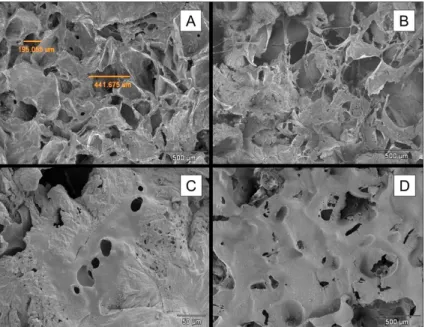
cytocompatibilty and biocompatibility of scaffolds with osteoblast-like cells because of the ease with which these scaffolds can be manufactured and the possibility of controlling pore size and structure26. Figure 4A shows SEM micrograph of the surface of a PLDLA scaffold.
The surface was characterized by homogeneously distributed irregular pores, with a range of pore sizes and surface topography that provided good interaction between cells and PLDLA scaffold surface during the irst 24h of culture (Figure 4B). Osteoblast-like cells adhered on scaffolds and had a lattened, elongated or polygonal morphology with cytoplasmic projections extending away from the central body to the top of adjacent wall pores.
From day 7 to 14 of culture, several non-speciic areas of the material surface were covered by a cellular layer as a result of osteoblast proliferation and migration (Figure 4 C,D). These indings conirmed the cytocompatibility of PLDLA scaffolds with osteoblast-like cells and their ability to support suitable cell growth that could be useful in bone tissue engineering applications.
Histological analysis of PLDLA scaffolds
implanted in rats
To evaluate the in vivo biocompatibility and inluence of the PLDLA scaffolds on bone healing, cell-seeded PLDLA scaffolds (constructs) were implanted in bilateral critical-size calvarial defects in Wistar rats and assessed histologically at 8 and 12 weeks after implantation. All rats recovered well during the post-surgical procedure, with no clinical signs of acute infection in the area of the defect and no systemic complications, which were only assessed macroscopically.
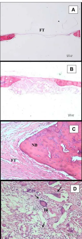
Figure 5 shows histological images of PLDLA constructs 8 weeks after implantation. In the control group (empty defect), the center of the defect area was illed with thin ibrous Figure 2. Viability of osteoblastic-like cells cultured on PLDLA
scaffolds for 2 h and 24 h . Cell viability was assessed by the MTT assay and expressed as the absorbance of the purple formazan product indicative of viable cells . The positive control consisted of cells grown in TCPS wells without any additions, while the negative
control consisted of cells exposed to 10% phenol. The columns are
the mean ± SD of ive independent experiments. *p<0.01 compared
to the control and PLDLA groups.
Figure 3.Proliferation of osteoblast-like cells grown on PLDLA scaffolds for 21 days. The points represent the mean ± SD of ive
independent experiments.
connective tissue while there was minimal bone formation at the borders of the defect (Figure 5A).
There was greater bone formation surround the margins of defect in the implants of the PLDLA constructs compared to the control group (Figure 5B), indicating that the 3D structure of the scaffolds was able to stimulate tissue ingrowth by providing mechanical assistance until the newly formed bone tissue assumed its biological functions. There was also an obvious belt of connective tissue ibers surrounding the space occupied by the material and the margins of the defect (Figure 5C).
Implantable biomaterials are recognized as foreign by the body and immediately after implantation, induce an initial host response to eliminate this foreign body. The magnitude and duration of the inlammatory process has a major inluence on the stability and biocompatibility of the device 27.
Figure 5D shows that a severe inlammatory response involving the iniltration of leukocytes, macrophages and giant cells was observed with all PLDLA implants. These reactions occurred primarily in the center of the defects, around the particle-polymer interface within the scaffold. The inlammatory response observed in the implants was not considered to be indicative of a toxic effect of the PLDLA constructs but was most likely related to the rapid degradation of the PLDLA polymer at this stage, which can result in a high concentration of polymer fragments and local acidiication of the implanted area 8,11,28,29.
The previous in vitro analysis of PLDLA degradation showed that material degradation starts after 8 weeks and is less pronounced after 16 weeks. It means that fewer polymer fragments and acidic degradation products are generated at the site of implant in the latter interval1,12. Besides, we have reported a similar PLDLA degradation rate and decrease in the inlammatory response along the time in the simulated internal ixation of PLDLA devices such as plates and screws in rabbit tibial implants30.
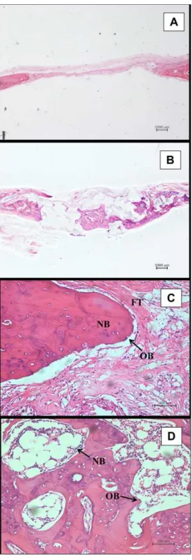
Figure 6 shows the histological appearance of PLDLA constructs after 12 weeks of implantation.
Panel A of this igure shows the presence of ibrous connective tissue in the control defect, although in two of four animals the control defect was also illed with a thin, uniform layer of newly formed bone (data not shown), indicating that the 5mm critical size defect used was not optimal for this study.
Calvarial critical size bone defects have been used in various studies to investigate the effectiveness of bone regenerative agents such as growth factors, biomaterials, cell or tissue implants, or any combination of these in repairing defective bone. However, the optimal critical size for bone calvarial defects within a given species remains controversial31,32.
In relation to the PLDLA scaffolds used in this work (Figure 6B), we observed active new bone formation in the central area of the implants that showed continuity with bone tissue growing inwards from the defect margins. In addition, a considerable amount of new thin bone trabeculae was observed surrounding the material implanted in the defect. However, in only one case the defect was completely illed with newly formed bone.
The inlammatory reaction within the implant decreased over time when compared with that seen after 8 weeks of implantation. The ibrous connective tissue became more disordered at the margins of control and implanted defects Figure 5. Histological alterations eight weeks after scaffold
implantation. Light microscopy (HE) of the critical size calvarial
defect at the control (empty defect) and scaffold-treated (PLDLA construct) sites. (A) The control site showed the presence of thin
ibrous tissue (FT). (B) Panoramic image of the implanted PLDLA construct. (C) Fibrous tissue (FT) formed at the interface between new bone (NB) and the PLDLA scaffold implantat the defect margin. (D) Severe inlammatory reaction (IR) showing lymphocytes, macrophages and foreign-body giant cells (arrows) inside the
(Figure 6C) and in the central area of the implant. A persistent tissue response was observed inside and around the remaining polymer fragments in the central area of newly formed bone (Figure 6D), thus conirming that the severe inlammatory response seen after 8 weeks was caused essentially by factors related to polymer degradation.
Careful histological analysis showed that the areas of newly formed bone gradually joined up to close the defect. During this process, the rate of PLDLA scaffold degradation was similar to that of bone formation since no signs of impaired bone growth, such as necrosis and ibrous capsule formation around the implanted material, were observed. These results indicated a good interaction at the interface between newly bone formed and the implanted PLDLA construct 33.
Although the histological analysis has revealed that the PLDLA constructs were biocompatible with bone tissue and were not cytotoxic during tissue repair, we assumed that some factors related to the porogen-leaching method used to manufacture the scaffolds may have affected the interactions between the PLDLA constructs and native tissue, thereby preventing better PLDLA scaffold-cell interaction and bone formation. Such factors include the lack of control of pore interconnectivity and pore size within the scaffold and the dificulty in obtaining scaffolds ~1mm thick (a thickness close to that of native calvarial). These factors may have inluenced the scaffold permeability and hindered luid diffusion through the polymeric network. This in turn would affect polymer degradation and hinder the transport of nutrients to the implanted area and to cells pre-seeded on scaffolds34-36.
Within the limits of this study, the contribution and inluence of transplanted osteoblast-like cells in bone replacement could not be assessed in terms of enhanced bone formation. However, histological results provided evidences on the beneits of using PLDLA scaffolds as synthetic temporary matrix in bone repair process, supporting a better growth and distribution of bone formation within the implanted area that was not seen in the control group. The latter generally showed only a uniform, thin bone layer covering the defect.
CONCLUSIONS
The results of this study have shown the potential usefulness of PLDLA copolymer as a scaffold for bone replacement procedures. The porogen-leached scaffolds were biocompatible with bone tissue and supported and stimulated osteoblast-like cell growth and migration, and still indicate the maintenance of cell morphology. The use of PLDLA cell-scaffold constructs in vivo showed that the material was mechanically stable, with a degradation rate similar to that of bone formation. The 3D structure of the constructs facilitated tissue growth into the implanted area and stimulated better distribution of the starting points of bone formation. More detailed studies of PLDLA polymer as a scaffold for bone regeneration are needed to conirm its usefulness for bone tissue engineering.
Acknowledgements
This work was supported by Fundação de Amparo à Pesquisa do Estado de São Paulo (FAPESP, grant no. 2012/12081-2) and the National Institute of Science and Technology in Biofabrication (INCT-Biofabris).
Figure 6. Histological alterations 12 weeks after scaffold implantation. Light microscopy (HE) of the critical size calvarial defect at
control (empty defect) and scaffold-treated (PLDLA construct)
implants. (A) Control site illed with thin ibrous connective tissue.
(B) Bone formation in the center of the implant. (C) Presence of
active osteoblasts (OB) around newly formed bone at the margin of a defect implanted with a PLDLA scaffold. Note the presence of ibrous tissue (FT) at the interface between new bone (NB) and space of implanted PLDLA. (D) New bone (NB) in a defect implanted with a PLDLA scaffold. Note the inlammatory reaction (arrow) and active osteoblasts (OB) surrounding the remaining polymeric
REFERENCES
1. Bitar KN, Zakhem E. Design strategies of biodegradable
scaffolds for tissue regeneration. Biomedical Engineering and Computational Biology. 2013;6:13-20. doi: 10.4137/BECB. S10961
2. Sharmas S, Srivastava D, Grover S, Sharma V. Biomaterials
in tooth tissue engineering: a review. Journal of clinical and diagnostic research: JCDR. 2014;8(1):309-315. doi: 10.7860/ JCDR/2014/7609.3937
3. Fröhlich M, Grayson WL, Wan LQ, Marolt D, Drobnic M, Vujnak-Novakovi G. Tissue engineered bone grafts: biological requirements, tissue culture and clinical relevance. Current Stem Cell Research & Therapy. 2008;3(4):254-264.
4. Shin, H. Fabrication methods of an engineered microenvironment for a nalysis of cell–biomaterial interactions. Biomaterials.
2007;28 (2):126-133. DOI: 10.1016/j.biomaterials.2006.08.007 5. Seyednejad H. Functional aliphatic polyesters for biomedical
and pharmaceutical applications. Journal of Controlled release. 2011; 152(1) :168-176. doi: 10.1016/j.jconrel.2010.12.016
6. Yu NY, Schindeler A, Little G, Ruys AJ. Biodegradable poly (α-hydroxy acid) polymer scaffolds for bone tissue engineering.
Journal of Biomedical Materials Research Part B: Applied Biomaterials. 2010;93(1):285-295. DOI: 10.1002/jbm.b.31588
7. Motta AC, Duek EA. Synthesis, characterization, and” in vitro” degradation of poly (L-lactic acid-co-glycolic acid), PLGA.
Matéria (Rio de Janeiro). 2006; 11 (3):340-350. http://dx.doi. org/10.1590/S1517-70762006000300024
8. Motta AC, Duek EA. Síntese e caracterização do copolímero poli (L-co-D, L Ácido Láctico). Polimeros: Ciencia e Tecnologia. 2007;17 (2):123-129.
9. Esposito AR, Moda M, Cattani SM, Santana M, Barbieri JÁ, Munhoz MM, et al. PLDLA/PCL-T Scaffold for Meniscus Tissue Engineering. BioResearch Open Access. 2013;2(2):138-147.
10. Mistura DV, Messias AD, Duek EA, Duarte MA. Development, characterization, and cellular adhesion of poly (l-Lactic Acid)/ Poly (caprolactone triol) membranes for potential application in bone tissue regeneration. Artiicial Organs. 2013;37(11):
978-984.
11. Ciambelli GS, Perez MO, Siqueira GV, Candella MA, Motta AC, Duarte MA, et al. Characterization of poly (L-co-D, L Lactic Acid) and a study of polymer-tissue interaction in subcutaneous
implants in wistar rats. Materials Research. 2013;16(1): 28-37.
http://dx.doi.org/10.1590/S151 6-14392012005000146 12. Pezzin AP, Cardoso TP, Rincón MC, Zavaglia CA, Duek EA.
Bioreabsorbable polymer scaffold as temporary meniscal prosthesis.
Artiicial Organs. 2003;27(5): 428-431. DOI: 10.1046/j.1525-1594.2003.07251.x
13. Sakata MM, Alberto-Rincon MC, Duek EA. Estudo da interação polímero/cartilagem/osso utilizando poli (Ácido Lático-co-Ácido Glicólico) e poli (p-Dioxanona) em condilo femural de coelhos.
Polímeros: Ciência e Tecnologia. 2004;14(3):176-180.
14. Pinto MR, Mariano EC, Alberto-Rincon MC, Duek EA. Análise histológica da Blenda PLLA/PCL como prótese de menisco.
Revista Matéria [Internet]. 2009 [citado 2015 Jul
27];14(4):1162-1171. Disponível em: http://www.materia.coppe.ufrj.br/sarra/
artigos/artigo11112
15. Minata MK, Motta AC, Brbo ML, Rincon MC, Duek EA. Biocompatibility study of poly (L-acid-lactic)/polycaprolactone triol blend. Polímeros. 2013;23(2): 242-247.http://dx.doi.
org/10.4322/polimeros.2013.074
16. Baraúna GS, Pierucci A, Oliveria A, Duarte MA, Duek EA. Estudo da degradação” in vivo” de poli (L-co-D, L-ácido láctico) aplicado como prótese para regeneração nervosa
periférica. Revista Matéria [Internet]. 2007 [citado 2015 Jun
21];12(2):298-306. Disponível em: http://www.materia.coppe. ufrj.br/sarra/artigos/artigo10851
17. Baraúna G, Coraça-Huber DC, Duek EA. In vitro degradation of Poly-L-co-D, L-lactic acid membranes. Materials Research. 2013;16(1):221-226.
http://dx.doi.org/10.1590/S1516-14392012005000154
18. Motta AC, Duek EA. Estudo inicial da degradação “in vitro” de poli (L-co-DL ácido lático) sintetizado em laboratório.
Matéria (Rio de Janeiro). 2008;13(3): 429-438.http://dx.doi.
org/10.1590/S1517-707620080003
19. Murphy CM, Matsiko A, Haugh M, Gleeson J, O’Brien FJ. Mesenchymal stem cell fate is regulated by the composition
and mechanical properties of collagen–glycosaminoglycan scaffolds. Journal of the Mechanical Behavior of Biomedical Materials. 2012;7:53-62. DOI: 10.1016/j.jmbbm.2011.11.009
20. Lucchesi C, Barbanti SH, Joazeiro PP, Duek EA. Cell culture on PCL/PLGA blends. Journal of Applied Polymer Science.2010;115 (5):2609-2615. DOI: 10.1002/app.29919
21. Más BA, Cattani SM, Rangel RC, Ribeiro GA, Cruz NC, Leite FL, et al. Surface characterization and osteoblast-like Cells
culture on collagen modified PLDLA scaffolds. Materials Research. 2014;17(6):1523-1534.
http://dx.doi.org/10.1590/1516-1439.269414
22. Blumenstein R, Dias M, Russo IH, Tahin Q, Russo J. DNA content and cell number determination in microdissected samples of breast carcinoma in situ. International Journal of Oncology. 2002; 21(2): 447-450.
23. Forsey RW, Chaudhuri JB. Validity of DNA analysis to determine cell numbers in tissue engineering scaffolds. Biotechnology Letters. 2009;31(6): 819-823. DOI 10.1007/s10529-009-9940-5
24. Santos AR, Ferreira BM, Duek EA, Dolder H, Wada RS, Wada
ML. Differentiation pattern of vero cells cultured on poly
(L-Lactic Acid)/Poly (Hydroxybutyrate-co-Hydroxyvalerate) blends. Artiicial Organs. 2004;28(4) : 381-389. doi:10.1089/
ten.2005.11.182
25. Ng KW, Leong DT, Hutmacher DW. The challenge to measure
cell proliferation in two and three dimensions. Tissue Engineering. 2005;11(1-2):182-191. doi:10.1089/ten.2005.11.182
26. Ke Y, Wang YJ, Ren L, Zhao QC, Huang W. Modified PHBV scaffolds by in situ UV polymerization: Structural characteristic, mechanical properties and bone mesenchymal stem cell compatibility. Acta Biomaterialia. 2010;6(4):1329-1336. DOI:
10.1016/j.actbio.2009.10.026
27. Nuss KM, Von Rechenberg B. Biocompatibility issues with modern implants in bone-a review for clinical orthopedics.
The Open Orthopaedics Journal. 2008; 2:66-78. doi:
10.2174/1874325000802010066
28. Cai K, Yao K, Yang Z, Li X. Surface modification of three-dimensional poly (d, l-lactic acid) scaffolds with baicalin: a
histological study. Acta Biomaterialia. 2007;3(4):597-605.
DOI: 10.1016/j.actbio.2006.12.005
29. Montjovent MO, Mathieu L,Schmoekel H, Mark S, Bourban PE, Zambelli PY, et al. Repair of critical size defects in the rat cranium using ceramic-reinforced PLA scaffolds obtained by
supercritical gas foaming. Journal of Biomedical Materials Research Part A, 2007;3(1):41-51.
30. Motta AC, Duek EA. Initial study of in vivo degradation of poly (L-lactic acid-co-D, L ácid láctic), PLDLA. Matéria (Rio de Janeiro). 2009;14(3):1070-1075. http://dx.doi.org/10.1590/
S1517-70762009000300018.
revisiting the concept of a critical-sized defect (CSD). Plastic and Reconstructive Surgery. 2010;125(6):1685-1692. doi:
10.1097/PRS.0b013e3181cb63a3
32. Horner EA, Kirkham J, Wood D, Curran S, Smith M, Thomson B, et al. Long bone defect models for tissue engineering
applications: criteria for choice. Tissue Engineering Part B: Reviews. 2010;16(2): 263-271. DOI: 10.1089=ten.teb.2009.0224
33. Nagata MJ, Messora M, Pola N, Campos N, Vieira R, Esper LA, et al. Influence of the ratio of particulate autogenous bone graft/platelet-rich plasma on bone healing in critical-size defects:
A histologic and histometric study in rat calvaria. Journal of Orthopaedic Research. 2010;28(4): 468-473. DOI: 10.1002/ jor.21027
34. Ho ST, Hutmacher DW. A comparison of micro CT with other techniques used in the characterization of scaffolds. Biomaterials. 2006;27(8):1362-1376.
35. Subia B, Kundu J, Kundu SC. Biomaterial scaffold fabrication
techniques for potential tissue engineering applications. In:
Eberli D, editor. Tissue Engineering. and regenerative medicine
[Internet] . 2010 . p.142-157. [cited 2015 Nov 21]. Available from: http://www.intechopen.com/books/tissue-engineering/
biomaterial-scaffold-fabrication-techniques-for-potential-tissue-engineering-applications
36. Suh SW, Shin JY, Kim J, Kim H, Beak CH, Kim DI, et al. Effect