The Brewed Rice Vinegar
Increases
HSPA1A Expression and Ameliorates
Cognitive Dysfunction in Aged P8 Mice
Hiroaki Kanouchi1*, Toshiaki Kakimoto1, Hideya Nakano1, Masahiro Suzuki1, Yuji Nakai2, Kazuhiro Shiozaki3, Kohei Akikoka4, Konosuke Otomaru5, Masanobu Nagano6,
Mitsuharu Matsumoto7
1Department of Veterinary Pathobiology, Joint Faculty of Veterinary Medicine, Kagoshima University, Korimoto, Kagoshima, Japan,2Institute for Food Sciences, Hirosaki University, Yanagawa, Aomori, Japan, 3Faculty of Fisheries, Kagoshima University, Shimoarata, Kagoshima, Japan,4Department of Veterinary Histopathology, Joint Faculty of Veterinary Medicine, Kagoshima University, Korimoto, Kagoshima, Japan, 5Veterinary Clinical Center, Joint Faculty of Veterinary Medicine, Kagoshima University, Korimoto, Kagoshima, Japan,6Sakamoto Kurozu, Inc., Uenosono-cho, Kagoshima, Japan,7Department of Veterinary Anatomy, Joint Faculty of Veterinary Medicine, Kagoshima University, Korimoto, Kagoshima, Japan
Abstract
Kurozuis a traditional Japanese rice vinegar. During fermentation and aging of theKurozu
liq-uid in an earthenware jar over 1 year, a solid residue calledKurozu Moromiis produced. In the present study, we evaluated whether concentratedKurozuorKurozu Moromicould ame-liorate cognitive dysfunction in the senescence-accelerated P8 mouse. Senescence-acceler-ated P8 mice were fed 0.25% (w/w) concentrSenescence-acceler-atedKurozuor 0.5% (w/w)Kurozu Moromifor 4 or 25 weeks.Kurozusuppressed cognitive dysfunction and amyloid accumulation in the brain, whileKurozu Moromishowed a tendency to ameliorate cognitive dysfunction, but the effect was not significant. We hypothesize that concentratedKurozuhas an antioxidant effect; however, the level of lipid peroxidation in the brain did not differ in senescence-accelerated P8 mice. DNA microarray analysis indicated that concentratedKurozuincreased HSPA1A mRNA expression, a protein that prevents protein misfolding and aggregation. The increase in HSPA1A expression byKurozuwas confirmed using quantitative real-time PCR and immu-noblotting methods. The suppression of amyloid accumulation by concentratedKurozumay be associated with HSPA1A induction. However, concentratedKurozucould not increase HSPA1A expression in mouse primary neurons, suggesting it may not directly affect neurons.
Introduction
Dementia is a common illness that affects the quality of life in the aging population. To date, there are no effective treatments; however, an early diagnosis and preventative measures, such as exercise, a healthy diet and social activity, have proven beneficial. Alzheimer’s disease (AD) and
OPEN ACCESS
Citation:Kanouchi H, Kakimoto T, Nakano H, Suzuki M, Nakai Y, Shiozaki K, et al. (2016) The Brewed Rice VinegarKurozuIncreases HSPA1A Expression and Ameliorates Cognitive Dysfunction in Aged P8 Mice. PLoS ONE 11(3): e0150796. doi:10.1371/ journal.pone.0150796
Editor:Masaki Mogi, Ehime University Graduate School of Medicine, JAPAN
Received:July 29, 2015
Accepted:February 10, 2016
Published:March 4, 2016
Copyright:© 2016 Kanouchi et al. This is an open access article distributed under the terms of the
Creative Commons Attribution License, which permits unrestricted use, distribution, and reproduction in any medium, provided the original author and source are credited.
Data Availability Statement:All relevant data are within the paper and its Supporting Information files.
Funding:The authors received no specific funding for this work.
cerebrovascular disease frequently co-exist and are part of a syndrome that may result in demen-tia. Excess oxidative stress has been suggested to contribute to dementia progression [1,2]. It has been reported that antioxidants from fruit, green tea or olive oil, which contain high concentra-tions of polyphenols, help to prevent cognitive dysfunction in animal studies [1,2]. However, further evaluation is needed on potential candidates that ameliorate cognitive dysfunction.
In this study, we focused on the traditional Japanese black vinegar calledKurozu.Kurozuis made from steamed rice. Saccharification, alcoholization, and acetification from starch to ace-tic acid occurs in the same earthenware jar and the produced vinegar is left to age for over 1 year. The liquid in the jar is filtered to produceKurozu, and the remaining extract is called Kur-ozu Moromi(KM). It has been reported thatKurozuandKMhave several health benefits. Kur-ozuprotects against colitis caused by dextran sulfate sodium [3], and suppresses proliferation of various cancer cell lines [4].Kurozualso has an antioxidant effect [5]. A 10-fold concen-trated form ofKurozu(CK) is prepared by vacuum distillation using a rotary evaporator. Using this process, acetic acid inCKis evaporated.
In the present study, we evaluated whetherCKorKMcould prevent cognitive dysfunction in senescence-accelerated P8 (P8) mice. The P8 mouse has been reported to be a good model for use in AD research [6–10]. The P8 mouse is one of nine senescence-prone strains of senes-cence-accelerated mice, which are originally generated from AKR/J mice. P8 mice exhibit a number of features that are known to occur in the pathogenesis of AD, such as increased oxida-tive stress, loss of neurons, gliosis,βamyloid alterations, and tau phosphorylation, as well as age-related deterioration in memory and learning. The senescence-resistance (R1) mouse is also generated from AKR/J mice at the same time. The R1 mouse shows normal aging and were used as the control mice in P8 mouse studies. The cognitive function of P8 mice following a diet ofCKorKMwas tested using the Morris water maze test.
Our aim was to identify a new candidate for the prevention of dementia, and we conclude thatCKcould ameliorate cognitive dysfunction.
Materials and Methods
Preparation of
CK
and
KM
TheCKdiet included 0.25% (w/w) CK in CE-2 basic rodent diet (Nihon CLEA, Tokyo, Japan). CKwas made fromKurozuliquid (Sakamoto Kurozu, Fukuyama, Kagoshima, Japan) by repeated vacuum distillation. TheKMdiet included 0.5% (w/w)KMpowder in CE-2 diet.KM powder (Sakamoto Kurozu) was made from the squeezed residue followingKurozu produc-tion. The squeezed residue was dried under a vacuum at 110°C. The chemical composition of CKwas 80% water, 9% crude protein (calculated as mineral nitrogen × 6.25), 2.5% organic acid, 5% ash, and 1% carbohydrate. The chemical composition ofKMwas 4% water, 12% crude protein (calculated as mineral nitrogen × 6.25), 23% organic acid, 1% ash, and 60% carbohydrate.
Animal experiments
R1 and P8 mice were purchased from Japan SLC (Shizuoka, Japan). Mice were housed at 25±2°C with 55±10% humidity on a 12-h light/dark cycle (lighting time 08:00–20:00). All mice were housed in independent cages and had free access to food and water. This study was carried out in strict accordance with the recommendations in the guide for the humane treatment and manage-ment of animals of the Japanese Law (No. 105) and Notification (No. 6). The protocol was approved by the Committee on the Ethics of Animal Experiments of the Kagoshima University Committee for Animal Experiment (Permit Number: A10030 and VM12018). All mice were killed by bleeding under isoflurane anesthesia, and all efforts were made to minimize suffering.
Experiment 1
Ten-week-old male R1 mice (n = 16) were fed a control CE2 diet. P8 mice were divided into three groups as follows: control CE2 diet group (n = 9);KMdiet group (n = 9); andCKdiet group (n = 9). Feeding of experimental diets started from 12 weeks of age until the mice were killed. The water maze test began when mice were 15 weeks of age and continued for 16 days. All mice were killed under anesthesia at 17 weeks old (4 months old). Serum was collected to measure the levels of thiobarbituric acid reactive substances (TBARS), alanine aminotransfer-ase (ALT), and aspartate aminotransferaminotransfer-ase (AST) using a commercial kit (Cayman Chemical, Ann Arbor, MI; Dry-chem Chemistry analyzer, Tokyo, Japan). Three mice from each group were fixed with neutralized 10% (v/v) formalin by perfusion fixation to obtain the brain.
Four-μm-thick tissue sections were prepared from paraffin-embedded brains and were used for the
detection of aggregated protein using the ProteoStat Amyloid Plaque Detection Kit (Enzo Life Sciences Inc., Farmingdale, NY) according to the manufacturer’s instructions. The detection reagent interacts with the cross-β-sheet quaternary structure of amyloid fibrils on the slides and is readily excited by an argon ion laser source, with an emission maximum of 600 nm. Fluorescent intensities in the cerebral cortex and hippocampus were detected using a confocal laser microscope (EZ-C1, Nikon, Tokyo, Japan). Brain homogenates were prepared to measure protein concentration (Pierce BCA Protein Assay Kit, Pierce, Rockford, IL) and TBARS. TBARS levels are expressed as MDA concentration. Heat shock 70 kDa protein 1A (HSPA1A) in brain homogenates was evaluated using the Hsp70 High-Sensitivity ELISA Kit (StressMarq Biosciences, British Columbia, Canada) according to the manufacturer’s instructions.
Experiment 2
Twelve 4-week-old male R1 mice and 36 4-week-old male P8 mice were purchased from Japan SLC. Mice were housed under normal conditions until 11 weeks of age, after which experimen-tal diets began. R1 mice were fed a control CE2 diet. P8 mice were divided into three groups as follows: control CE2 diet group (n = 12);KMdiet group (n = 12); andCKdiet group (n = 12). After 24 weeks (8.4 months old), the water maze test was started.
Morris water maze test
The standard Morris water maze test was used with minor modifications [11]. A circular pool (100 cm in diameter) was filled with water (17-cm-deep, 25°C) and divided into four quadrant zones: east, west, south, and north. A clear platform (10 cm in diameter and 16 cm in height) was hidden in the center of the right upper quadrant, submerged 1 cm below the water’s sur-face. Mice were trained to find the hidden platform in the water maze for 15 (Experiment 1) or 4 (Experiment 2) consecutive days, three trials per day. Mice were not allowed to search for the platform for more than 60 s, after which they were guided to the platform and allowed to stay on the platform for 15 s. On the last day, a probe trial test was performed for a period of 120 s without the platform. In each trial, the swimming path and escape time for locating the hidden platform was recorded using a web camera (Logicool HD Webcam C270, Logicool, Lausanne, Switzerland) and analyzed using a tracking system (TopScan Lite 2.0, Clever Sys, Reston, VA).
Antioxidant activities
of 9.1 mM salicylic acid, 25μL of 9.1 mM ferrous sulfate, and 500μL of 8.8 mM hydrogen
per-oxide. The reaction mixture was incubated for 10 min at room temperature, after which the absorbance of the mixture was measured at 510 nm using a UV/Vis spectrophotometer (Shi-madzu, Tokyo, Japan). The percentage of hydroxyl radical scavenging activity of the test sam-ple was determined in comparison with the negative control. Various concentrations of reduced ascorbic acid were used as positive controls. The negative control contained neither ascorbic acid norCK.
DNA microarray analysis and quantitative real-time PCR
The left side of the hippocampus region was excised from the brains of four mice in each group. Total RNA was extracted using the RNeasy Mini Kit (Qiagen, Valencia, CA). RNA quantity, purity, and concentration were determined using a nanodrop (Amersham Biosciences, Foster City, CA) and Experion RNA StdSens (BioRad Laboratories, Hercules, CA). Total RNA was used for microarray analysis and quantitative real-time PCR, as described below. DNA microar-ray analysis was performed according to the manufacturer’s instructions. We used GeneChip Mouse Gene 1.0 ST Array (Affymetrix, Santa Clara, CA). Fluorescent signals were scanned using the Affymetrix GeneChip System. Data analysis of the DNA microarray was carried out as described previously [13], except for normalization methods. Briefly, CEL files were normalized using the robust multi-array average method [14]. Hierarchical clustering was then performed using the pvclust() function in R. To identify differentially expressed genes, the rank products method [15] was applied to robust multi-array average normalized data. Probe sets with a false discovery rate<0.05 were regarded as having different expression levels between the two groups (R1 CE2vs. P8 CE2, or P8 CE2vs.CK). The annotation file for the Mouse Gene 1.0 ST Array was downloaded from the Affymetrix website. The data discussed in this publication have been deposited in NCBI’s Gene Expression Omnibus [16] and are accessible through GEO series accession number GSE70514 (http://www.ncbi.nlm.nih.gov/geo/query/acc.cgi?acc=GSE70514). Quantitative real-time PCR analyses were performed using a Lightcycler 1.5 (Roche Diagnostics, Tokyo, Japan) and SYBR Premix Ex Taq (Takara Bio, Shiga, Japan). The primer sets for
HSPA1A and glyceraldehyde 3-phosphate dehydrogenase (GAPD) were purchased from Takara Bio. Data are presented as relative values (HSPA1A/GAPD).
Mouse primary neuronal cultures
Pregnant C57BL/6N mice were killed by bleeding under isoflurane anesthesia. Brains were excised from embryonic (E18) mice. Neurons were collected from the cerebrum and hippo-campus using Nerve-Cell Culture System/Dissociation Solutions (Sumitomo Bakelite, Tokyo, Japan). Neurons (2.5 × 10⁵ cells/well) were plated on a 24-well plate (Celltight PL plate, Sumi-tomo) and cultured in Neuron Assay Medium (SumiSumi-tomo). After 4 days, media was replaced with fresh media containing various concentrations ofCK(0, 0.00125, 0.0025, 0.005, or 0.01% (v/v)). After 24 h, total RNA was extracted from neurons with TRIzol (Invitrogen Life Technol-ogies, Carlsbad, CA). Quantification of HSPA1A mRNA was carried out as described above.
Statistical analysis
Results
Effects of
KM
or
CK
feeding on cognitive function in P8 mice
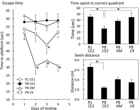
Experiment 1. In Experiment 1, mice were 15–17 weeks old at the time the Morris water maze test was performed. These mice were fedKMorCKfor 5 weeks. There were no signifi-cant differences in body weight gain and serum ALT and AST levels among the groups (S1 Table). The Morris water maze test showed that the escape time of the R1 CE2 group gradually shortened compared with that of the P8 CE2 group during training days, but there were no sig-nificant differences (Fig 1, left panel). Escape times for the P8KMand P8CKgroups were faster than that of the P8 CE2 group. Significant differences were observed at 12 to 15 days in the P8CKgroup compared with the P8 CE2 group, and 13 to 14 days in the P8KMgroup com-pared with the P8 CE2 group.
Following the completion of the Morris water maze test (day 15), the probe test was per-formed without the platform to evaluate working memory (Fig 1, upper right panel). As a result, the time spent in the correct quadrant in the P8CKgroup was slightly longer than that of the P8 CE2 group (p = 0.11). The time spent by the P8KMgroup was also slightly longer, but there were no significant differences when compared with the P8 CE2 group. During the probe test, mice did not stop swimming, therefore, there were no differences in swim distance among all groups.
We attempted to detect neuritic plaques in brain sections using a fluorescent dye that binds to aggregated protein, but no clear neuritic plaques could be detected for any group. This result could be because of the young age of the mice. However, the fluorescence intensity of
Fig 1. Evaluation of cognitive function and amyloid accumulation in 4-month-old senescence-accelerated mice.P8 mice were fed a normal diet (P8, n = 9),Kurozu Moromicontaining diet (KM, n = 9), or a concentratedKurozucontaining diet (CK, n = 9) for 5 weeks from 12 weeks old. R1 mice were fed a normal
diet (R1, n = 16). Cognitive function was evaluated using the Morris water maze test at 15 weeks of age. Left panel shows escape time during the training phase (R1 CE2, n = 16; P8 CE2, n = 9; P8KM, n = 9; P8CK, n = 9).§p<0.05, P8 CE2vs. P8KM;
*p<0.05, P8 CE2vs. P8CK. Right upper panel shows results of the probe test. Right lower panel shows amyloid accumulation (R1 CE2, n = 3; P8 CE2, n = 3; P8KM, n = 3; P8 CK, n = 3). Results are expressed as mean±SE.*p<0.05,vs. P8 CE2.
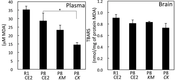
aggregated protein in the cerebral cortex and hippocampus was significantly lower in the P8 CKgroup compared with the P8 CE2 group (p<0.05). In serum, the MDA value in the P8CK group was significantly lower than that of the P8 CE2 group. However, in brain homogenates, the MDA values were not different among groups (Fig 2).
Experiment 2. In Experiment 2, mice were 7 months old at the time the Morris water maze test was performed. These mice were fed KM orCKfor 4 months. Body weight gains for P8 CE2, P8KM, and P8CKgroups were significantly lower than that of the R1 group.KMor CKfeeding did not affect the body weight of P8 mice. The difference observed in body weights between SAM R1 and SAM P8 is thought to be the result of aging. Serum ALT levels were not different among the groups (S2 Table). Escape time in the Morris water maze test revealed that the R1 group recognized the position of the platform, but that the P8 CE2 group and P8KM did not (Fig 3, left).
In the Morris water maze test performed without the platform to evaluate working memory, the time spent in the correct quadrant was significantly shorter in the P8 CE2 group compared with the R1 CE2 group (Fig 3, upper right). The time spent for the P8CKgroup, but not the P8 KMgroup, was significantly longer than that of the P8 CE2 group. Swim distance was signifi-cantly shorter for the P8 CE2 group compared with the R1 CE2 group. This result is thought to be a result of aging. As mice did not stop swimming during the probe test, the swim speed of P8 mice was slower than that of R1 mice.KMorCKfeeding did not affect the swim distance in P8 mice (Fig 3, lower right). Serum MDA values of P8 CE2 mice were significantly higher com-pared with the R1 group (Fig 4). Values of P8CKmice were significantly lower compared with P8 CE2 mice.
Antioxidant activity of
CK
The antioxidant activity ofCKwas evaluated using ferric reducing antioxidant assay, DPPH radical scavenging assay, superoxide radical scavenging assay, and hydroxyl radical scavenging assay (Table 1). SolidifiedCK, prepared by vacuum distillation, was dissolved in water. In the Fig 2. Level of TBARS in serum and brain homogenates in 4-month-old senescence-accelerated mice.Levels of TBARS in serum were measured (R1 CE2, n = 16; P8 CE2, n = 9; P8KM, n = 9; P8CK, n = 9) and brain homogenates (R1 CE2, n = 13; P8 CE2, n = 6; P8KM, n = 6; P8CK, n = 6). Results are expressed as mean±SE.*p<0.05,vs. P8.
Fig 4. Level of TBARS in serum in 7-month-old senescence-accelerated mice.TBARS levels in serum were measured (R1 CE2, n = 12; P8 CE2, n = 12; P8KM, n = 12; P8CK, n = 11). Results are expressed as
mean±SE.*p<0.05,vs. P8 CE2. doi:10.1371/journal.pone.0150796.g004
Fig 3. Evaluation of cognitive function in 8.4-month-old senescence-accelerated mice.P8 mice were fed a normal diet (P8 CE2, n = 12),Kurozu Moromicontaining diet (KM, n = 12), or concentratedKurozu
containing diet (CK, n = 12) for 24 weeks from 12 weeks old. Cognitive function was evaluated using the
Morris water maze test at 35 weeks of age. Left panel shows escape time during the training phase (R1 CE2, n = 12; P8 CE2, n = 12; P8KM, n = 12; P8CK, n = 11). Right upper panel shows the results of the probe test (R1 CE2, n = 12; P8 CE2, n = 12; P8KM, n = 12; P8CK, n = 11). Right lower panel shows swimming distance during the probe test (R1 CE2, n = 12; P8 CE2, n = 12; P8KM, n = 12; P8CK, n = 11). Results are expressed
present study, to evaluate antioxidant activities under the same conditions, the activities were compared with ascorbic acid. These experiments showed thatCKhad a free radical scavenging activity against superoxide and hydroxyl radicals. These activities were equivalent to 1.03–5.50 mM of ascorbic acid.
Effect of
CK
on gene expression in the brain-DNA microarray analysis
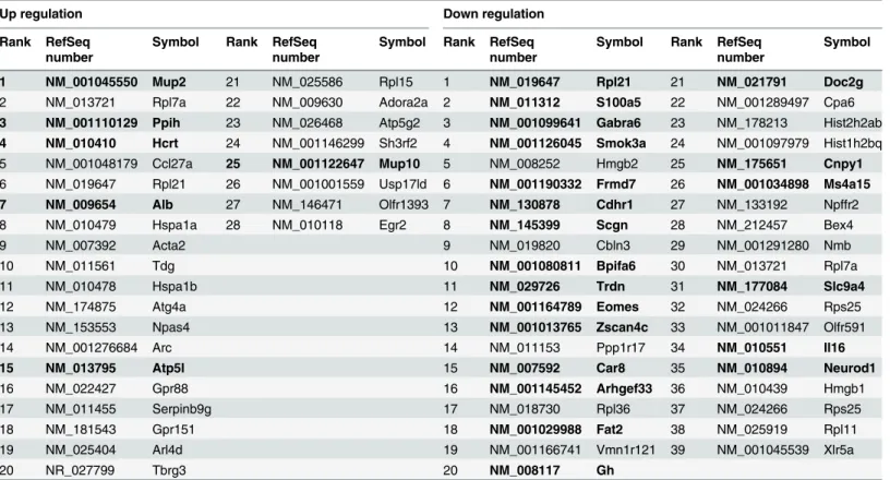
To identify changes in gene expression followingCKfeeding, DNA microarray analysis was carried out using cDNA libraries that were synthesized from total RNA of the R1 CE2 group, P8 CE2 group, and P8CKgroup. Differentially expressed genes were identified using the rank products method [15]. If the false discovery rate was<0.05, the gene was regarded as signifi-cantly down- or up-regulated. As a result, 116 and 191 genes were down-regulated and up-reg-ulated, respectively, in the P8 CE2 group compared with the R1 CE2 group (S3andS4Tables). Unfortunately, there were no genes related to AD or biological pathways known to improve cognitive function using the DAVID Bioinformatics Resources 6.7 (NIAID/NIH, Frederick, MD). Compared with the P8 CE2 group, 28 genes were up-regulated in the P8CKgroup, and six genes were down-regulated in the P8 CE2 group compared with the R1 CE2 group (Table 2, left column). Compared with the P8 CE2 group, 39 genes were down-regulated in theCK group, and 21 genes were up-regulated in the P8 CE2 group compared with the R1 CE2 group (Table 2, right column).
HSPA1A expression
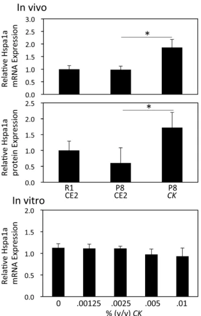
CKdecreased the amount of aggregated protein in the brain of P8 mice. DNA microarray anal-ysis showed that HSPA1A, a protein that aids in folding of misfolded proteins, was up-regu-lated byCK. HSPA1A mRNA levels and protein expression were confirmed using real-time PCR and ELISA, respectively.Fig 5shows that both mRNA and protein levels of HSPA1A sig-nificantly increased in the brains ofCK-fed mice (p<0.05). However, exposure of primary neu-ronal cultures toCKfor 24 h did not affect HSPA1A mRNA expression (Fig 5).
Discussion
In this study, we examined whetherKMorCKcould protect against cognitive dysfunction in P8 mice. In Experiment 1, escape times in the Morris water maze test for theKMandCK groups were faster than that for the P8 CE2 group at 4 months of age. In the probe trial test on the last day, there were no significant differences in time spent in the correct quadrant among the P8 CE2, P8KM, and P8CKgroups.
Table 1. Antioxidant Activities ofCK.
2.5%CKin working solution
Equivalent to ascorbic acid (undilutedCK)
Ferric reducing antioxidant power 0.67±1.2 (OD750) 1.47±0.10 mM
DPPH radical scavenging activity 63.7±1.2% 5.50±0.13 mM Superoxide radical scavenging
activity
22.4±2.4% 1.03±0.26 mM Hydroxyl radical scavenging
activity
62.3±0.9% 1.11±0.03 mM
Data are presented as the mean±SE (n = 3).
Mice fed aCKandKMdiet did show a slight amelioration in cognitive dysfunction. There-fore, in Experiment 2, we evaluated the cognitive function of P8 mice at 7 months of age. Body weights of 7-month-old P8 mice decreased compared with R1 mice of the same age. The total swim distance of the P8 CE2 group in the Morris water maze test was significantly shorter compared with the R1 CE2 group, suggesting that the exercise activity of 7-month-old P8 mice declined. Escape time decreased after each training day in the R1 CE2 group. In contrast, there was no change in the P8 CE2 group. The time spent in the correct quadrant for the P8 CE2 group was also significantly shorter than that of the R1 CE2 group. These results indicate that 7-month-old P8 mice lost not only exercise activity but also cognitive ability. Escape time of the P8CKgroup, but not theKMgroup, was slightly faster than that of the P8 CE2 group. There was a significant difference at day 3 (p<0.05). Time spent in the correct quadrant was significantly longer inCK-fed mice, but swimming distance did not change. These findings suggest thatCKfeeding ameliorates cognitive dysfunction, but not exercise ability, in older P8 mice.
Although we attempted to identify neuritic plaques and neurofibrillary tangles in the P8 CE2 mouse brain, none could be detected. However, aggregated protein increased in the P8 CE2 group and decreased in the P8CKgroup. A previous report suggests thatCKhas a scav-enging effect on DPPH and a suppressive effect on low-density lipoprotein oxidation [5]. Our in vitroexperiments also showed thatCKhad antioxidant properties. Although we expected thatCKwould suppress oxidation, we could not sufficiently evaluate oxidation levels in the brain. Effective molecules from food are generally modified in the digestive tract, and these Table 2. Genes Related toCKFeeding.
Up regulation Down regulation
Rank RefSeq number
Symbol Rank RefSeq number
Symbol Rank RefSeq number
Symbol Rank RefSeq number
Symbol
1 NM_001045550 Mup2 21 NM_025586 Rpl15 1 NM_019647 Rpl21 21 NM_021791 Doc2g
2 NM_013721 Rpl7a 22 NM_009630 Adora2a 2 NM_011312 S100a5 22 NM_001289497 Cpa6
3 NM_001110129 Ppih 23 NM_026468 Atp5g2 3 NM_001099641 Gabra6 23 NM_178213 Hist2h2ab 4 NM_010410 Hcrt 24 NM_001146299 Sh3rf2 4 NM_001126045 Smok3a 24 NM_001097979 Hist1h2bq
5 NM_001048179 Ccl27a 25 NM_001122647 Mup10 5 NM_008252 Hmgb2 25 NM_175651 Cnpy1
6 NM_019647 Rpl21 26 NM_001001559 Usp17ld 6 NM_001190332 Frmd7 26 NM_001034898 Ms4a15
7 NM_009654 Alb 27 NM_146471 Olfr1393 7 NM_130878 Cdhr1 27 NM_133192 Npffr2
8 NM_010479 Hspa1a 28 NM_010118 Egr2 8 NM_145399 Scgn 28 NM_212457 Bex4
9 NM_007392 Acta2 9 NM_019820 Cbln3 29 NM_001291280 Nmb
10 NM_011561 Tdg 10 NM_001080811 Bpifa6 30 NM_013721 Rpl7a
11 NM_010478 Hspa1b 11 NM_029726 Trdn 31 NM_177084 Slc9a4
12 NM_174875 Atg4a 12 NM_001164789 Eomes 32 NM_024266 Rps25
13 NM_153553 Npas4 13 NM_001013765 Zscan4c 33 NM_001011847 Olfr591
14 NM_001276684 Arc 14 NM_011153 Ppp1r17 34 NM_010551 Il16
15 NM_013795 Atp5l 15 NM_007592 Car8 35 NM_010894 Neurod1
16 NM_022427 Gpr88 16 NM_001145452 Arhgef33 36 NM_010439 Hmgb1
17 NM_011455 Serpinb9g 17 NM_018730 Rpl36 37 NM_024266 Rps25
18 NM_181543 Gpr151 18 NM_001029988 Fat2 38 NM_025919 Rpl11
19 NM_025404 Arl4d 19 NM_001166741 Vmn1r121 39 NM_001045539 Xlr5a
20 NR_027799 Tbrg3 20 NM_008117 Gh
Bolded genes showed opposite expression pattern in P8 CE2 compared with R1 CE2.
modified molecules may have different effects from the original molecules. Further experi-ments are required to determine whetherCKfeeding has effects on excessive oxidation in the brain.
CKameliorated both cognitive dysfunction and the accumulation of aggregated protein in the brain more effectively thanKM. We therefore focused on the effect ofCKon cognitive dys-function. To further understand the mechanism behind the effect ofCK, we measured the change in gene expression in brains from R1 CE2, P8 CE2, and P8CKgroups. DNA microarray analysis showed that expression levels of many genes were altered in 4-month-old P8 mice Fig 5. Effect ofCKon the expression of HSPA1A mRNA and protein.Total RNA was prepared from brains of R1 CE2, P8 CE2, and P8CKmice from Experiment 1 (n = 4). Expression levels of HSPA1A and GAPD mRNA were evaluated using quantitative real-time PCR. Results are presented as the relative HSPA1A mRNA level (HSPA1A/GAPD). HSPA1A protein expression in brain homogenates was evaluated using a commercial ELISA kit (R1 CE2, n = 11; P8 CE2, n = 4; P8CK, n = 4). Total RNA was prepared from mouse primary neurons treated withCKfor 24 h (n = 4). Expression levels of HSPA1A and GAPD mRNA were evaluated using quantitative real-time PCR. Results are presented as the relative HSPA1A mRNA level (HSPA1A/GAPD). Results are expressed as mean±SE.*p<0.05,vs. P8 CE2.
compared with R1 mice of the same age. Unfortunately, we could not find genes directly related to brain function. Interestingly, changes in gene expression accompanied by aging were reversed in the P8CKgroup. Six of the genes down-regulated in the P8 CE2 group were up-regulated in the P8CKgroup, and 15 up-regulated genes in P8 CE2 mice were down-regulated in the P8CKgroup. Moreover,CKfeeding increased major urinary protein-2 (α2u-globulin), which dramatically decreases in the liver during aging [17]. However, its function in the brain has not been elucidated. Overall,CKameliorates senescing of P8 mice.
It has been suggested that accumulation of tau and/or amyloidβoligomers in the brain initi-ates a cascade of pathological events resulting in neurodegeneration and cognitive decline [18]. Heat shock proteins (HSP) are known chaperones and play a major role in preventing protein misfolding and aggregation. HSPs may also prevent aggregation and oligomerization of amy-loidβand tau protein [19]. Leak reviewed the relationship between HSPA1A expression and AD [20]. He concluded that although there is generally an increase in HSPA1A expression seen with mild cognitive decline, some authors have observed age-related reduction in HSP70 in studies using older male mice. Therefore, it is difficult to form conclusions about the expres-sion level in AD. Bobkovaet al. revealed that exogenous HSP70 inhibited the accumulation of amyloidβand cognitive abnormalities in a mouse model of AD [21]. DNA microarray analysis indicated that HSPA1A (also known as HSP70) was up-regulated byCK. This finding was con-firmed using quantitative real-time PCR and ELISA. The decreased accumulation of aggregated protein in the P8CKgroup may be related to the activation of HSPA1A.In vitroexperiments using primary neurons revealed that the addition ofCKto culture medium had no effect on HSPA1A mRNA expression levels. This result suggests thatCKdoes not have a direct effect on neurons. Further studies are needed to clarify the induction mechanism of HSPA1A byCK.
Conclusion
We evaluated the effect ofKMorCKfeeding on cognitive function in P8 mice.CKfeeding ameliorated cognitive dysfunction and suppressed the accumulation of aggregated protein in the brain.CKincreased HSPA1A expression in the brain. The activation of HSPA1A may be associated with the attenuated accumulation of aggregated protein in the brain.
Supporting Information
S1 Table. Body Weight and Serum ALT and AST Level at 4 Months Old. (TIF)
S2 Table. Body Weight Gain and Serum ALT at 8 Months Old. (TIF)
S3 Table. Down-regulated Genes in P8 Mice. (TIF)
S4 Table. Up-regulated Genes in P8 Mice. (TIF)
Author Contributions
References
1. Abdel Moneim AE. Oxidant/antioxidant imbalance and the risk of Alzheimer’s disease. Curr Alzheimer Res. 2015; 12: 335–349. PMID:25817254
2. Rege SD, Geetha T, Griffin GD, Broderick TL, Babu JR. Neuroprotective effects of resveratrol in Alzhei-mer disease pathology. Front Aging Neurosci. 2014 Sep 11. doi:10.3389/fnagi.2014.00218
3. Shizuma T, Ishiwata K, Nagano M, Mori H, Fukuyama N. Protective effects of Kurozu and Kurozu Moro-mimatsu on dextran sulfate sodium-induced experimental colitis. Dig Dis Sci. 2011; 56: 1387–1392. doi:10.1007/s10620-010-1432-xPMID:20936352
4. Nanda K, Miyoshi N, Nakamura Y, Shimoji Y, Tamura Y, Nishikawa Y, et al. Extract of vinegar "Kurosu" from unpolished rice inhibits the proliferation of human cancer cells. J Exp Clin Cancer Res. 2004; 23: 69–75. PMID:15149153
5. Nishidai S, Nakamura Y, Torikai K, Yamamoto M, Ishihara N, Mori H, et al. Kurosu, a traditional vinegar produced from unpolished rice, suppresses lipid peroxidation in vitro and in mouse skin. Biosci Biotech-nol Biochem. 2000; 64: 1909–1914. PMID:11055395
6. Pallàs M. Senescence-Accelerated Mice P8. A Tool to Study Brain Aging and Alzheimer’s Disease in a Mouse Model. ISRN Cell Biology. Article ID 917167, 2012.
7. Markowska AL, Spangler EL, Ingram DK. Behavioral assessment of the senescence-accelerated mouse (SAM P8 and R1). Physiol Behav. 1998; 64: 15–26. PMID:9661977
8. Gang B, Yue C, Han N, Xue H, Li B, Sun L, et al. Limited hippocampal neurogenesis in SAMP8 mouse model of Alzheimer’s disease. Brain Res. 2011; 1389: 183–193. doi:10.1016/j.brainres.2011.03.039 PMID:21439270
9. Chang X, Rong C, Chen Y, Yang C, Hu Q, Mo Y, et al. (-)-Epigallocatechin-3-gallate attenuates cogni-tive deterioration in Alzheimer’s disease model mice by upregulating neprilysin expression. Exp Cell Res. 2015; 334: 136–145. doi:10.1016/j.yexcr.2015.04.004PMID:25882496
10. Zheng JD, Hei AL, Zuo PP, Dong YL, Song XN, Takagi Y, et al. Age-related alterations in the expres-sion of MTH2 in the hippocampus of the SAMP8 mouse with learning and memory deterioration. J Neu-rol Sci. 2009; 287: 188–196. doi:10.1016/j.jns.2009.07.027PMID:19735921
11. Morris R. Developments of a water-maze procedure for studying spatial learning in the rat. J Neurosci Methods. 1984; 11: 47–60. PMID:6471907
12. Ikeda A, Ichino H, Kiguchiya S, Chigwechokha P, Komatsu M, Shiozaki K. Evaluation and identification of potent angiotensin-I converting enzyme inhibitory peptide derived from dwarf gulper shark (Centro-phorus atromarginatus). J Food Process Pres. 2015; 39: 107–115.
13. Suyama T, Okada S, Ishijima T, Iida K, Abe K, Nakai Y. High phosphorus diet-induced changes in NaPi-IIb phosphate transporter expression in the rat kidney: DNA microarray analysis. PLoS One. 2012 Jan 3. e29483. doi:10.1371/journal.pone.0029483PMID:22235299
14. Irizarry RA, Hobbs B, Collin F, Beazer-Barclay YD, Antonellis KJ, Scherf U, et al. Exploration, normali-zation, and summaries of high density oligonucleotide array probe level data. Biostatistics. 2003; 4: 249–264. PMID:12925520
15. Breitling R, Armengaud P, Amtmann A, Herzyk P. Rank products: a simple, yet powerful, new method to detect differentially regulated genes in replicated microarray experiments. FEBS Lett. 2004; 573: 83– 92. PMID:15327980
16. Edgar R, Domrachev M, Lash AE. Gene Expression Omnibus: NCBI gene expression and hybridization array data repository. Nucleic Acids Res. 2002; 30: 207–210. PMID:11752295
17. Roy AK, Nath TS, Motwani NM, Chatterjee B. Age-dependent regulation of the polymorphic forms of alpha 2u-globulin. J Biol Chem. 1983; 258: 10123–10127. PMID:6193107
18. Hardy J, Selkoe DJ. The amyloid hypothesis of Alzheimer’s disease: progress and problems on the road to therapeutics. Science. 2002; 297: 353–356. PMID:12130773
19. Repalli J, Meruelo D. Screening strategies to identify HSP70 modulators to treat Alzheimer’s disease. Drug Des Devel Ther. 2015; 9: 321–331. doi:10.2147/DDDT.S72165PMID:25609918
20. Leak RK. Heat shock proteins in neurodegenerative disorders and aging. J Cell Commun Signal. 2014; 8: 293–310. doi:10.1007/s12079-014-0243-9PMID:25208934