Variants in the Carboxy-Terminal Region of BRCA1
Renato S. Carvalho1,2¤, Renata B. V. Abreu2, Aneliya Velkova1¤, Sylvia Marsillac1, Renato S. Rodarte3, Guilherme Suarez-Kurtz2, Edwin S. Iversen4, Alvaro N. A. Monteiro1*, Marcelo A. Carvalho2,5*
1Cancer Epidemiology Program, H. Lee Moffitt Cancer Center & Research Institute, Tampa, Florida, United States of America,2Programa de Farmacologia, Instituto Nacional de Caˆncer, Rio de Janeiro, RJ, Brazil,3Universidade Federal de Alagoas, Maceio´, AL, Brazil,4Department of Statistical Science, Duke University, Durham, North Carolina, United States of America,5Instituto Federal de Educac¸a˜o, Cieˆncia e Tecnologia do Rio de Janeiro, Rio de Janeiro, RJ, Brazil
Abstract
Germline inactivating variants inBRCA1lead to a significantly increased risk of breast and ovarian cancers in carriers. While the functional effect of many variants can be inferred from the DNA sequence, determining the effect of missense variants present a significant challenge. A series of biochemical and cell biological assays have been successfully used to explore the impact of these variants on the function of BRCA1, which contribute to assessing their likelihood of pathogenicity. It has been determined that variants that co-localize with structural or functional motifs are more likely to disrupt the stability and function of BRCA1. Here we assess the functional impact of 37 variants chosen to probe the functional impact of variants in phosphorylation sites and in the BRCT domains. In addition, we perform a meta-analysis of 170 unique variants tested by the transcription activation assays in the carboxy-terminal domain of BRCA1 using a recently developed computation model to provide assessment for functional impact and their likelihood of pathogenicity.
Citation:Carvalho RS, Abreu RBV, Velkova A, Marsillac S, Rodarte RS, et al. (2014) Probing Structure-Function Relationships in Missense Variants in the Carboxy-Terminal Region of BRCA1. PLoS ONE 9(5): e97766. doi:10.1371/journal.pone.0097766
Editor:Alvaro Galli, CNR, Italy
ReceivedJanuary 17, 2014;AcceptedApril 23, 2014;PublishedMay 20, 2014
Copyright:ß2014 Carvalho et al. This is an open-access article distributed under the terms of the Creative Commons Attribution License, which permits unrestricted use, distribution, and reproduction in any medium, provided the original author and source are credited.
Funding:This work was supported by a National Institutes of Health award CA116167, and grants from the Florida Breast Cancer Foundation, Fundac¸a˜o de Amparo a` Pesquisa do Estado do Rio de Janeiro (FAPERJ), and INCT para o Controle do Caˆncer (CNPq – Conselho Nacional de Desenvolvimento Cientı´fico e Tecnolo´gico). A.V. was the recipient of a predoctoral fellowship from the Department of Defense Breast Cancer Research Program (W81XWH-08-1-0509). The funders had no role in study design, data collection and analysis, decision to publish, or preparation of the manuscript.
Competing Interests:The authors have delcared that no competing interests exist.
* E-mail: marcelo.carvalho@ifrj.edu.br (MAC); alvaro.monteiro@moffitt.org (ANAM)
¤ Current address: Faculdade de Farma´cia – Universidade Federal do Rio de Janeiro, Rio de Janeiro, RJ, Brazil
Introduction
InheritedBRCA1inactivating mutations are major determinants of breast and ovarian cancer risk, accounting for 46–68% of cases with a family history of breast cancer cases [1,2,3,4]. Since 1996 genetic testing to identify mutations in BRCA1and BRCA2 has been offered to women with a family history of breast and ovarian cancer [5,6]. Presently several different assay platforms are used to investigate BRCA alterations, including amplicon-based Sanger sequencing, target capture followed by next-generation sequenc-ing, and methods to detect large genomic rearrangements [5,7,8]. Variations found during sequencing include nonsense, frameshift, missense, splicing, and small insertions and deletions. Variants in BRCA1that lead to functional inactivation, either by compromis-ing gene expression, correct spliccompromis-ing, or protein structure and stability are associated with an increased risk for cancer [9]. In many instances, inactivation can be inferred from the DNA sequence alone (e.g.nonsense or frameshift changes). However, in cases such as missense or splicing variants the resulting impact on function cannot be directly inferred. While many variants have been evaluated using functional assays and multifactorial statistical models [10,11], cancer association has not been determined for several variants, referred to as Variants of Uncertain Clinical Significance (VUS).
An array of functional tests and computation prediction tools have been developed to aid in the determination of the functional
impact of sequence variants of BRCA1, in particular,in vitroassays that assess the integrity and functionality of the N-terminal RING finger and the C-terminal BRCT tandem domains (tBRCT) of BRCA1 [11,12]. Variants in these domains are more likely to have a functional impact [13,14]. Analysis of these variants fulfills a double purpose: they provide information to aid in the classifica-tion of variants, and inform the biology of BRCA1 by pinpointing specific regions on the protein critical for different biochemical activities.
VarCall [18], to assess the likelihood of pathogenicity given their functional impact.
Materials and Methods
Rationale for Choice of Variants
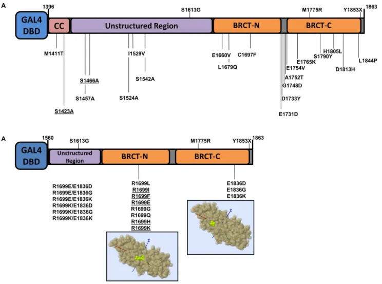
In total we analyzed thirty seven BRCA1 missense variants (Table 1, Figure 1). These variants represent three distinct groups: variants of uncertain significance in BRCA1, phosphorylation site variants, and salt-bridge variants in the BRCT domains. With the exception of R1699W, no other variant was found in the NHBLI Exome Sequencing Project (data release ESP6500 SI-V2).
Variants of uncertain significance. Five variants (M1411T, S1457A, S1524A, I1529V and S1542A) were chosen to increase coverage of the unstructured region (aa 1396–1648) of BRCA1 C-terminus (aa 1396–1863, corresponding to exons 13 to 24 coding sequence, encompassing the two BRCT tandem domains) [19]. Three of these naturally-occurring variants (S1457A, S1524A and S1542A) are located in BRCA1 phosphor-ylation sites. Several phosphorphosphor-ylation sites have been identified on the C-terminal of BRCA1 that are involved in biological functions such as cell cycle checkpoint and caspase activation [20]. We also generated artificial (cannot result from a single nucleotide change
in the natural codon) alanine variants to probe the role of phosphorylation at position S1423 and S1466 to be consistent with the other phosphorylation sites. Alanine was choosen to substitute serine residues in order to impede phosphorylation. The remaining 13 variants (E1660V, L1679Q, C1697F, E1731D, D1733Y, G1748D, A1752T, E1754V, E1765K, S1790Y, H1805L, D1813H and L1844P) are missense variants in the tBRCT domains (Table 1, Figure 1).
BRCT salt-bridge variants. In previous studies we identi-fied and analyzed a naturally occurringBRCA1allele, R1699W, which showed a temperature-sensitive behavior [16,21]. R1699 and E1836 are critical residues for a salt-bridge that stabilizes the tBRCT [15,16]. We reasoned that variants in residues involved in the salt-bridge could generate useful structure-function informa-tion and potential temperature-sensitive proteins [21]. Thus, we generated a panel of eight variants of residue 1699 and three variants of residue 1836. We also generated four double mutants combining changes in residues R1699 and E1836 (Table 1, Figure 1).
Figure 1. BRCA1 carboxy-terminal variants. (A) Natural and artificial (underlined) BRCA1 variants in the context of the analyzed region (comprising amino acids residues 1396–1863).(B)BRCA1 R1699 and E1836 variants in the context of the analyzed region (comprising amino acids residues 1560–1863). Structural models were obtained with Mupit tool (http://mupit.icm.jhu.edu/) using the 1jnx PDB structure (amino acid residues R1699 and E1936 are depicted in green).
Constructs
BRCA1 expression constructs (controls and variants) were generated in 2 different construct contexts: for VUS and phosphorylation sites variants analysis we used the region comprising amino acid residues 1396–1863, corresponding to
exon 13–24 coding sequence (Figure 1A); for salt-bridge variants analysis we used the region comprising amino acid residues 1560– 1863, corresponding to exon 16–24 coding sequence (Figure 1B). All variants were confirmed by sequencing.
Table 1.BRCA1 variants analyzed in this study.
Exon Protein Variant (HGVS) DNA variant (HGVS) DNA variant (BIC) Notes
13 p.Met1411Thr c.4232T.C T4351C VUS; Coiled-coil domain
13 p.Ser1423Ala c.4268G.A G4386A Disordered region; Phosphorylation site; artificial variant
14 p.Ser1457Ala c.4369T.G T4488G Disordered region; Phosphorylation site
14 p.Ser1466Ala c.4398C.A C4517A Disordered region; Phosphorylation site; artificial variant
15 p.Ser1524Ala c.4570T.G T4689G Disordered region; Phosphorylation site
15 p.Ile1529Val c.4585A.G A4704G Disordered region
15 p.Ser1542Ala c.4624T.G T4743G Disordered region; Phosphorylation site
16 p.Glu1660Val c.4979A.T A5098T BRCT;
17 p.Leu1679Gln c.5036T.A T5155A BRCT
18 p.Cys1697Phe c.5090G.T G5209T BRCT
19 p.Glu1731Asp c.5193G.C G5312C BRCT
20 p.Asp1733Tyr c.5197G.T G5316T BRCT
20 p.Gly1748Asp c.5243G.A G5362A BRCT
20 p.Ala1752Thr c.5254G.A G5373A BRCT
20 p.Glu1754Val c.5261A.T A5380T BRCT
21 p.Glu1765Lys c.5293G.A G5412A BRCT
22 p.Ser1790Tyr c.5369C.A C5488A BRCT
23 p.His1805Leu c.5414A.T A5533T BRCT
23 p.Asp1813His c.5437G.C G5556C BRCT
24 p.Leu1844Pro c.5531T.C T5650C BRCT
18 p.Arg1699Gly c.5095C.G 5214 Salt bridge variants
18 p.Arg1699Leu c.5096G.T 5215 Salt bridge variants
18 p.Arg1699Gln c.5096G.A 5215 Salt bridge variants
18 p.Arg1699Glu N/A N/A Salt bridge variants
18 p. Arg1699Phe N/A N/A Salt bridge variants
18 p. Arg1699His N/A N/A Salt bridge variants
18 p.Arg1699Ile N/A N/A Salt bridge variants
18 p.Arg1699Lys N/A N/A Salt bridge variants
24 p.Glu1836Ly c.5506G.A 5625 Salt bridge variants
24 p.Glu1836Gly c.5507A.G 5626 Salt bridge variants
24 p.Glu1836Asp c.5508G.C 5627 Salt bridge variants
18/24 p.Arg1699Lys Salt bridge variants (double mutants)
p.Glu1836Lys
18/24 p. Arg1699Lys Salt bridge variants (double mutants)
p.Glu1836Gly
18/24 p. Arg1699Lys Salt bridge variants (double mutants)
p.Glu1836Asp
18/24 p.Arg1699Glu Salt bridge variants (double mutants)
p.Glu1836Lys
18/24 p.Arg1699Glu Salt bridge variants (double mutants)
p.Glu1836Gly
18/24 p.Arg1699Glu Salt bridge variants (double mutants)
p.Glu1836Asp
VUS and phosphorylation sites variants. Control con-structs (amino acid context 1396–1863) containing the wt BRCA1, S1613G, M1775R, and Y1853X were generated previously (Figure 1A) [22]. Mutations in the cDNA sequence were introduced by site-directed mutagenesis using plasmid p385-BRCA1 as template, as previously described [23]. Primers sequences are available upon request. For each variant, both products (59and 39regions) were combined and used as a template for a final round of PCR using 24ENDT and UX13 primers [23]. The final PCR products were then digested with BamH1 and EcoR1 and ligated to pGBT9 vector. To obtain the heterologous GAL4 DNA binding domain (GAL4-DBD) fusions in a mamma-lian expression vector, pGTB9 constructs were digested with HindIII andBamH1, a 1.8 Kb band was isolated and ligated into equally digested pCDNA3 vector.
BRCT salt-bridge variants. Control constructs (amino acid context 1560–1863, Figure 1B) containing the wt BRCA1, M1775R, and Y1853X were previously described [16,24]. Mutations in the cDNA sequence were introduced as described above. To obtain the double mutants the same procedure and primers described were used but instead of wild-type cDNA as templates we used the individual constructs containing the single mutants at position 1699. The final PCR products were then digested and cloned in pGBT9 vector, then subcloned in pCDNA3 as described above.
Transcriptional Assay
The transcriptional assays were performed in mammalian cells as described [12,19,22]. Briefly, we used pG5Luc as a reporter and transfections were normalized with an internal control phGR-TK (Promega), which contains a Renilla luciferase gene under a constitutive TK basal promoter. Transfections were conducted in human HEK293T, HCC1937 [25] or SUM1315 cells in triplicate using Fugene 6 (Roche), harvested 24 h post-transfection, and luciferase activity was measured using the Dual Luciferase Reporter Assay System (Promega). Results were plotted as a percentage of the wild-type activity. We inferred disease relevance for each missense variant using a computational tool, VarCall, based on a Bayesian hierarchical model. [18].
VarCall is a tool that uses functional data, quantitative or categorical, as input and accounts for sources of experimental heterogeneity. Specifically, here we used non-normalized ratios of Firefly luciferase/Renilla luciferase indexed by batch as the specific input data and each batch also records the ratio for the wild type control. VarCall generates a likelihood of pathogenicity for each variant given the input data in the form of a posterior probability of being damaging. In addition, VarCall also generates a Bayesian integrated likelihood ratio statistic that measures the degree to which the data support the hypothesis that a variant is protein damaging. To generate accurate probabilities VarCall uses large datasets and re-assesses previously anazide variants given the new data. Therefore, we included data for all (n = 176) variants previously tested under controlled conditions (see Results for further details). We used this model to infer disease relevance for all the naturally occurring variants described in this study, an additional set of 119 variants previously assayed for transcriptional activation [17] and provide a reanalysis of the 82 variants previously assessed by this model [18]. This combined set includes 2,436 measurements of transcriptional activity for 176 unique variants in the region analyzed in the assay: amino acid residues context 1396–1863 (Tables S1 and S2). Single nucleotide changes in this region can generate 2,740 unique variants (1,244 in the tBRCT). Of those, 219 have been documented in the population (BIC Database as of this writing) with 129 unique variants in the
tBRCT domains. The present dataset represents 61% and 89.9% coverage of all variants documented in the amino acid region limited by residues 1398–1863 and the tBRCT domains, respectively. VarCall can also assess results from different construct contexts (aa 1396–1863 or aa 1560–1863) because each variant is assessed relative to the wild type in the same context. We have extensively tested several contexts for the TA assay including aa 1560–1863 [26], aa 1396–1863 [22], and aa 1646–1859 [17]. While they show different absolute activities the relative activities of variants (for example M1775R) are comparable in terms of the percentage of activity of the corresponding wild type. Thus, data from different contexts can be directly compared using VarCall since each batch is compared against its own wild-type construct.
Results
BRCA1 C-terminus VUS
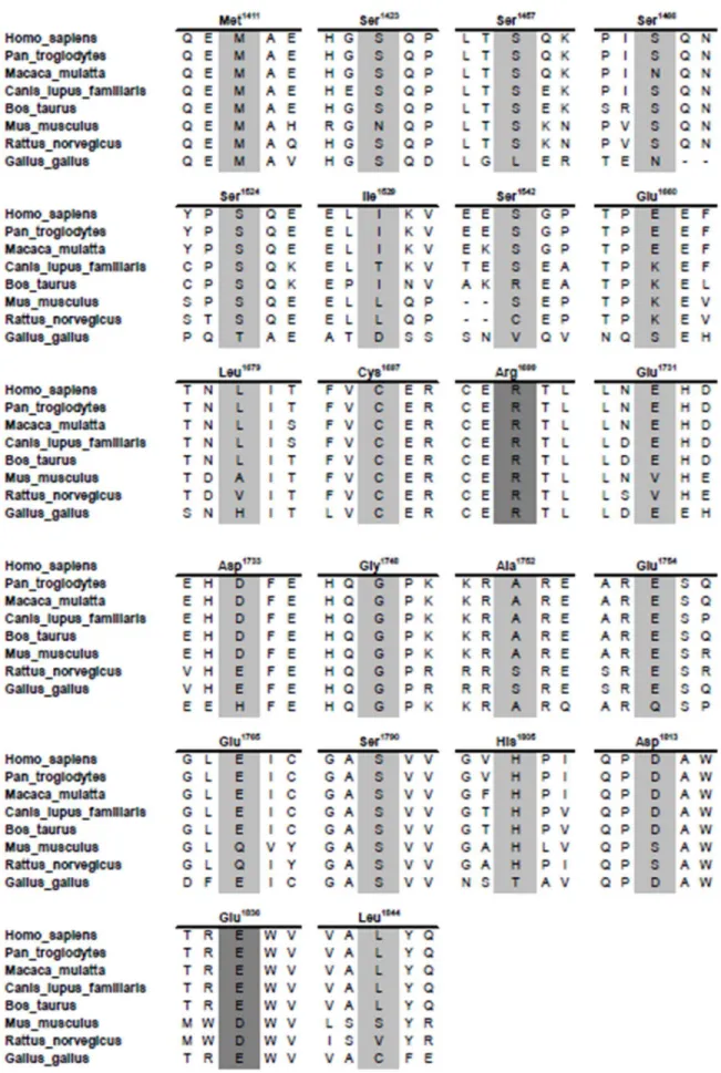
In order to increase the coverage of variants located at carboxy-terminus of BRCA1 we selected a set of 18 naturally occuring missense variants (Table 1). Six are located outside the tandem BRCT (tBRCT) domains: M1411T, located in the coiled-coil region; S1457A, S1524A, I1529V, and S1542A, located in the unstructured region of BRCA; and L1844P located in the carboxy-terminal tail of the molecule. Twelve are located in tBRCT domains: E1660V, L1679Q, C1697F, E1731D, D1733Y, G1748D, A1752T, E1754V, E1765K, S1790Y, H1805L and D1813H (Figure 1A). To comprehensively probe the role of the phosphorylation sites we also included two artificial variants located outside the tBRCT that are ATM phosphorylation target sites: S1423A and S1466A [20] (Table 1, Figure 1A). All variants are located at amino acid residues strongly conserved in the vertebrate lineage (Figure 2).
Among the naturally occuring variants, I1529V, E1660V, L1679Q, D1733Y, E1754V, E1765K, S1790Y, H1805L and L1844P showed transcriptional activity values corresponding to the wild-type reference (Figure 3A) indicating that they have no detectable functional impact. Similarly, the naturally-occurring phosphorylation variants S1457A, S1524A, and S1542A, and the artificial phosphorylation site variants, S1423A, S1466A, also displayed transcriptional activity.75% of the wild-type activity (Figure 3B) suggesting that phosphorylation at these sites is not required for the transcriptional activity of BRCA1 in normal growing conditions.
The remaining six variants exhibited reduced functional activity. Variants M1411T, C1697F, G1748D and A1752T showed significantly reduced (,30% of wt) levels of activity (Figure 3A). Two variants, E1731D and D1813H presented activty levels around 50% of the wild type reference (55610% and 55611% respectively, Figure 3A) suggesting that they might have a mild to moderate impact on the function of BRCA1.
BRCT Salt-bridge Variants
dissect the role of these residues in this molecular interaction we generated a series of BRCA1 tBRCT single amino acid variants at positions 1699 and 1836 (Figure 1B).
We investigated these variants using the transcription activation assay in three different temperatures to assess temperature-sensitive behaviors (Figures 4A and 4B). Variants R1699I, R1699F, R1699E, R1699G, R1699Q and R1699H showed low activity across different temperatures. Interestingly, variant R1699L displayed an activity comparable to the wild type reference consistent across different temperatures (Figure 4A). One variant, a conservative change from an arginine residue to a lysine, R1699K, displayed a temperature dependent behavior, presenting transcriptional activation ,80% of wt at 30uC and progressively decreasing with temperature. At 37uC this variant showed a significantly reduced activity (38% of wt, Figure 4B). This behavior was consistent in two different cell lines, HEK293T and HCC1937.
We also tested three variants at position 1836, where the original glutamic acid residue was replaced by an aspartic acid, a glycine or a lysine. E1836D exhibited a somewhat reduced transcriptional activation values that were comparable at 33uC and 37uC (Figure 4C). Variants E1836G and E1836K, on the other hand exhibited a temperature-sensitive behavior (Figure 4C). Then, we combined the R1699K (temperature-sensitive) or the R1699E (not temperature-sensitive) with variations at the 1836 site and tested the double mutants at 33uC and 37uC. All R1699K double mutants retained the temperature-sensitive behavior but only the conservative E1836D mutations retained normal activity at the permissive temperature (33uC) highlighting the role of both residues (1699 and 1836) on the stability of the BRCT domains (Figure 4D). Moreover, changes in one residue can compensate for changes in the other as demonstrated by the R1699E double
mutants, which show levels of activity comparable to wt at the permissive temperature but dramatically reduced levels when cells are shifted to 37uC. In particular, the R1699E/E1836G showed the largest difference of activity between the two temperatures and might constitute a useful tool to probe the function of BRCA1 (Figure 3E).
VarCall
Next, we evaluated the activity of missense variants using the VarCall computational model [18]. Results from this model reflect the likelihood of pathogenicity given the functional impact data. VarCall results are depicted in Figure 5 where each variant’s activity is represented by a boxplot summarizing the marginal posterior distribution of its random effect. A point estimate of the mixture model is plotted on the right margin. Its top component corresponds to variants with no functional impact, whereas its bottom component corresponds to variants with functional impact. VarCall data indicate that C1697F, G1748D and A1752T have significant impact on function and are likely to represent pathogenic variants (Figure 5). M1411T variant also showed reduced activity (Figure 5), resulting in a reduced but still significant probability of being pathogenic (0.35, Table S1).
Discussion
A significant percentage of genetic tests conducted for breast and ovarian cancer susceptibility results in findings of a variant of uncertain clinical significance (VUS). While some VUS are located in intronic or other putative regulatory regions, many are missense variants. These individual VUS alleles usually are very rare in the population precluding family-based or population-based genetic analysis to determine their disease association. Functional assay M. mulatta(NP_001108421.1),C.lupus(NP_001013434.1),B.taurus(NP_848668.1),M. musculus(NP_033894.3), R. norvegicus(NP_036646.1) andG. gallus(NP_989500.1). Target amino acids residues are depicted in light grey and salt-bridge involved residues in dark grey.
doi:10.1371/journal.pone.0097766.g002
Figure 3. Functional analysis of missense variants in BRCA1 C-terminal region.Transcriptional activity of BRCA1 variants were evaluated in HEK293T cells using a GAL4-responsive firefly luciferase reporter gene (shown above the graphs) at 37uC. Cells were harvested 24h after transfections and the lysate was used to assess transcriptional activation ability by luciferase activity measurement. Activity is depicted as % of the wild-type activity.(A)Natural missense variants and(B)natural and artificial (underlined) variants located on phosphorylation sites. S1613G, M1775R and Y1853X variants were used as controls.
have been used to determine whether specific amino acid changes lead to detectable functional impact in a number of biochemical and biological processes that have been associated with BRCA1 ([11] and references therein).
In this study we focused on the transcriptional activation assays for BRCA1. The method is based on the ectopic expression of BRCA1 C-terminal fragments fused to GAL4-DBD and the ability of the resulting chimeric protein to activate transcription of a reporter gene [27,28]. Interestingly, there is a strong correlation of transcriptional activation results and other biochemical activities assigned to BRCA1 such as the specific recognition of phosphor-ylated peptides [17] indicating that the transcriptional assay
functions as a monitor of the structural integrity of the BRCA1 C-terminal region. Importantly, the TA displays a strong correlation with pathogenicity indicating that the assay is specific and sensitive for BRCA1 missense variants in the C-terminus [17].
We analyzed 18 naturally occurring BRCA1 VUS (Figure 1A) using the TA assay [11,19]. We also tested two artificial missense variants targeting phosphorylation sites in BRCA1 (Figure 1A). Transcriptional activity data was assessed by VarCall, a recently developed computational tool to infer disease association from functional data [18].
M1411T, C1697F, G1748D, and A1752T displayed signifi-cantly decreased transcriptional activity compared to the wt Figure 4. Functional analysis of BRCA1 R1699 and E1836 variants at different temperatures.Transcriptional activity of BRCA1 variants were evaluated using a GAL4-responsive firefly luciferase reporter gene (shown above the graphs). Cells were harvested 24h after transfections and the lysate was used to assess transcriptional activation ability by luciferase activity measurement. Activity is depicted as % of the wild-type activity.(A)
Transcriptional activity of R1699L, R1699I, R1699F, R1699E, R1699G, R1699Q and R1699H variants and the controls M1775R and Y1853X at different temperatures evaluated in HEK293T cells; (B) transcriptional activity of the R1699K variant and the controls M1775R and Y1853X at different temperatures evaluated in HEK293T and HCC1937 cells and(C)transcriptional activity of the E1836D, E1836G and E1836K variants and the control M1775R at different temperatures evaluated in HEK293T. Transcriptional activity of double variants(D)R1699K/E1836D, R1699K/E1836G and R1699K/ E1836K(E)R1699E/E1836D, R1699E/E1836G and R1699E/E1836K and the control M1775R at different temperatures evaluated in HEK293T at different temperatures.
reference and were, with the exception of M1411T, determined to be likely to be pathogenic by VarCall (Figure 5, Table S1). Different variants on the same positions (A752V, A1752P, and C1697R) lead to severe protein folding defects inferred by increased protease sensitivity and impaired transcriptional activity [17]. Taken together these data highlight the relevance of these amino acid residues (C1697 and A1752) for the integrity of the BRCA1 tBRCT structure. Assessment of M1411T by VarCall was inconclusive. This variant, first reported in a Swedish ovarian cancer patient with a family history of breast cancer and other malignancies [29], lies on the BRCA1 coiled-coil domain and was reported to disrupt the interaction between BRCA1 and PALB2 [30].
All other natural variants tested (S1457A, S1524A, I1529V, S1542A, E1660V, L1679Q, E1731D, D1733Y, E1754V, E1765K, S1790Y, H1805L, D1813H and L1844P) displayed transcriptional activity comparable to the wt reference, as did the two artificial variants analyzed (S1423A and S1466A). These natural variants were determined to be unlikely to be pathogenic by VarClass (Figure 5, Table S1). Because unphosphorylatable (alanine) variants at phosphorylation sites did not have impact on transcriptional activity we conclude that phosphorylation of these residues is not required for transcription activation under normal conditions. They also provide a demonstration that serine to alanine substitutions in these residues do not induce dramatic structural changes to the tBRCT domains. Our data do not address the relevance of these variants following DNA damage, but it is clear that these sites are critical for DNA injury induced ATM phosphorylation of BRCA1, and the overall repair response [13,20,31,32].
Next, we probed the role of the salt bridge that stabilizes the tandem BRCT interaction between residues R1699, E1836, and D1840 [15]. The temperature-dependent effects of the R1699W variant on transcriptional activity were previously reported [21]. We performed site-directed mutagenesis generating a series of
eight variants of this residue (Figure 1B). Except for R1699L that behaved as reference in all tested conditions, and R1699K, discussed below, all other variants showed low TA values in all ranges of temperature tested (30uC to 37uC, Figure 4). Interest-ingly, the R1699L variant displayed impaired phosphopeptide binding activity [17] suggesting that this variant can be used to uncouple transcriptional activation from phosphopeptide binding. In addition to its role in the salt bridge, R1699L and R1699W, are predicted to abolish the interaction of the T1700 residue with PRKCD, PRKC, PRKCQ, PRKCZ, PRKCA, PRKCG and MST2 [33,34]. Interestingly, R1699K was found to have a temperature-dependent behavior, showing about 80% of wild-type activity at 30uC and,40% activity at 37uC. These results were confirmed in theBRCA1-deficient HCC1937 cells [25] (Figure 4). We also generated aspartic acid, glycine and lysine variants at position 1836 (Figure 1B). While the conservative change E1836D showed no temperature-dependent behavior, the E1836G and E1836K have significantly lower transcriptional activity at 37uC. E1836K was also reported to have modest effects on protein folding and transcriptional activation but significant decrease in peptide binding and specificity [17] in a pattern reminiscent of the R1699L variant. Then, we combined variants for R1699K and R1699E, which had temperature dependent and independent behaviors, respectively (Figure 1B and 4) with variants in E1836. As expected, for double variants of R1699K, the substitution of a glutamic acid by an aspartic acid (E1836D) did not change the pattern observed for the single R1699K. R1699K double variants containing a non-charged (E1836G) or a positively charged amino acid residue (E1836K) resulted in reduced transcriptional activities even at low temperatures (Figure 4). R1699E double variants showed the largest difference in activities from the permissive to the non-permissive temperature of all variants, suggesting that these double variants can be used as genetic tools to investigate the role of different biochemical processes mediated by the BRCT domains.
Figure 5. Estimated Variant Specific Effects (Bayesian statistical model, VarCall, graphical summary).Variants are depicted in order of amino acid from residues 1396 to 1863. The top panel shows secondary structures in the C-terminal region. Coiled-coil region anda-helixes are depicted as pink and blue boxes, respectively.b-sheets are shown as gray arrows. The shaded area on the graph corresponds to different structures of similar color. The linker region is indicated with green shading. Each variant’s activity is represented by a boxplot summarizing the marginal posterior distribution of its random effect. A point estimate of the mixture model is plotted on the right margin. Its top component corresponds to variants with no functional impact, whereas its bottom component corresponds to variants with functional impact. The mean of the benign/ damaging component is plotted as a green/red dotted horizontal line. Yellow box represents wild-type reference, green and red boxes represent low (class 1 and 2) and high (class 4 and 5) risk variants respectively, classified according to the International Agency for Research on Cancer (IARC), purple boxes represent VUS previously analyzed by our group (used to feed VarClass algorithm), and blue boxes represent the VUS analyzed by the first time in this study.
The study of temperature influence on BRCA1 structure/ function is especially relevant because temperature-sensitive variants may have different clinical presentations or penetrances. The evaluated variants could, judged by the behavior of the R1699W allele be potential intermediate risk variants. [21].
Finally, combined with the natural variants in this study (Table S1) we examined 170 unique variants using VarCall and generated likelihoods of pathogenicity. Note that because this tool uses a Bayesian approach every variant, even the 82 variants assessed previously [18] are re-evaluated given the new data. This analysis (Figure 5) reveals interesting insights about the architec-ture of the C-terminus. Some variants in the coiled-coil region displayed functional impact albeit a moderate one. The disordered region, located N-terminally to the BRCT domains is tolerant to changes. Similarly, a1 helixes on both BRCTs seem also to be tolerant to changes. Otherwise, most secondary structures and the linker region tend to be sensitive to changes. A note of caution is warranted here as these results, obtained in a research environ-ment, are not meant to guide clinical decisions. Although the VarCall tool infers the likelihood of a variant being pathogenic, the results provided here are derived from a single data source (activity of a functional assay). Only determination of pathogenic-ity by a multifactorial likelihood model using independent data sources (e.g.segregation analysis, allele frequency, tumor pathology markers, co-occurrence, and co-observation with BRCA2 patho-genic variants) should be considered clinically [35,36,37].
In summary, in this paper we directly assessed the transcrip-tional assay of several BRCA1 VUS and conducted a compre-hensive analysis of 170 unique variants using the VarCall computational tool. We also report data on several natural and artificial variants with temperature dependent behavior that can be utilized as reagents to dissect the functions of BRCA1.
Supporting Information
Table S1 Posterior probabilities and log Bayes Factor from VarCall.
(XLSX)
Table S2 Variant Dataset for VarCall. (XLSX)
Acknowledgments
Additional support for this work was provided by the Molecular Genomics core at the H.L. Moffitt Cancer Center.
Author Contributions
Conceived and designed the experiments: RSC ANAM MAC. Performed the experiments: RSC RBVA AV SM RSR. Analyzed the data: RSC GSK ESI ANAM MAC. Contributed reagents/materials/analysis tools: ANAM MAC. Wrote the paper: RSC ANAM MAC.
References
1. Mavaddat N, Antoniou AC, Easton DF, Garcia-Closas M (2010) Genetic susceptibility to breast cancer. Mol Oncol 4: 174–191.
2. Couch FJ, Nathanson KL, Offit K (2014) Two decades after BRCA: setting paradigms in personalized cancer care and prevention. Science 343: 1466–1470. 3. Evans DG, Shenton A, Woodward E, Lalloo F, Howell A, et al. (2008) Penetrance estimates for BRCA1 and BRCA2 based on genetic testing in a Clinical Cancer Genetics service setting: risks of breast/ovarian cancer quoted should reflect the cancer burden in the family. BMC Cancer 8: 155. 4. Chen S, Iversen ES, Friebel T, Finkelstein D, Weber BL, et al. (2006)
Characterization of BRCA1 and BRCA2 mutations in a large United States sample. J Clin Oncol 24: 863–871.
5. Frank TS, Deffenbaugh AM, Reid JE, Hulick M, Ward BE, et al. (2002) Clinical characteristics of individuals with germline mutations in BRCA1 and BRCA2: analysis of 10,000 individuals. Journal of Clinical Oncology 20: 1480–1490. 6. Frank TS, Manley SA, Olopade OI, Cummings S, Garber JE, et al. (1998)
Sequence analysis of BRCA1 and BRCA2: correlation of mutations with family history and ovarian cancer risk. J Clin Oncol 16: 2417–2425.
7. Jackson SA, Davis AA, Li J, Yi N, McCormick SR, et al. (2014) Characteristics of individuals with breast cancer rearrangements in BRCA1 and BRCA2. Cancer doi: 10.1002/cncr.28577.
8. Concolino P, Mello E, Minucci A, Santonocito C, Scambia G, et al. (2014) Advanced tools for BRCA1/2 mutational screening: comparison between two methods for large genomic rearrangements (LGRs) detection. Clin Chem Lab Med doi: 10.1515/cclm-2013-1114.
9. Smith SA, Easton DF, Evans DG, Ponder BA (1992) Allele losses in the region 17q12–21 in familial breast and ovarian cancer involve the wild-type chromosome. Nat Genet 2: 128–131.
10. Spurdle AB, Healey S, Devereau A, Hogervorst FB, Monteiro AN, et al. (2011) ENIGMA-Evidence-based network for the interpretation of germline mutant alleles: An international initiative to evaluate risk and clinical significance associated with sequence variation in BRCA1 and BRCA2 genes. Hum Mutat. 11. Millot GA, Carvalho MA, Caputo SM, Vreeswijk MP, Brown MA, et al. (2012) A guide for functional analysis of BRCA1 variants of uncertain significance. Hum Mutat 33: 1526–1537.
12. Carvalho MA, Couch FJ, Monteiro AN (2007) Functional assays for BRCA1 and BRCA2. Int J Biochem Cell Biol 39: 298–310.
13. Abkevich V, Zharkikh A, Deffenbaugh AM, Frank D, Chen Y, et al. (2004) Analysis of missense variation in human BRCA1 in the context of interspecific sequence variation. J Med Genet 41: 492–507.
14. Tavtigian SV, Byrnes GB, Goldgar DE, Thomas A (2008) Classification of rare missense substitutions, using risk surfaces, with genetic- and molecular-epidemiology applications. Human Mutation 29: 1342–1354.
15. Williams RS, Green R, Glover JN (2001) Crystal structure of the BRCT repeat region from the breast cancer- associated protein BRCA1. Nat StructBiol 8: 838–842.
16. Vallon-Christersson J, Cayanan C, Haraldsson K, Loman N, Bergthorsson JT, et al. (2001) Functional analysis of BRCA1 C-terminal missense mutations identified in breast and ovarian cancer families. Human Molecular Genetics 10: 353–360.
17. Lee MS, Green R, Marsillac SM, Coquelle N, Williams RS, et al. (2010) Comprehensive analysis of missense variations in the BRCT domain of BRCA1 by structural and functional assays. Cancer Res 70: 4880–4890.
18. Iversen ES, Couch FJ, Goldgar DE, Tavtigian SV, Monteiro ANA (2011) A Computational Method to Classify Variants of Uncertain Significance Using Functional Assay Data with Application to BRCA1. Cancer Epidemiology Biomarkers & Prevention 20: 1078–1088.
19. Carvalho MA, Marsillac SM, Karchin R, Manoukian S, Grist S, et al. (2007) Determination of cancer risk associated with germ line BRCA1 missense variants by functional analysis. Cancer Res 67: 1494–1501.
20. Ouchi T (2006) BRCA1 phosphorylation: biological consequences. Cancer Biol Ther 5: 470–475.
21. Worley T, Vallon-Christersson J, Billack B, Borg A, Monteiro AN (2002) A naturally occurring allele of BRCA1 coding for a temperature-sensitive mutant protein. Cancer Biol Ther 1: 497–501.
22. Phelan CM, Dapic V, Tice B, Favis R, Kwan E, et al. (2005) Classification of BRCA1 missense variants of unknown clinical significance. J Med Genet 42: 138–146.
23. Johnson D (2003) PCR: A practical approach; Press I, editor. Oxford: IRL Press. 24. Hayes F, Cayanan C, Barilla D, Monteiro AN (2000) Functional assay for BRCA1: mutagenesis of the COOH-terminal region reveals critical residues for transcription activation. Cancer Res 60: 2411–2418.
25. Tomlinson GE, Chen TT, Stastny VA, Virmani AK, Spillman MA, et al. (1998) Characterization of a breast cancer cell line derived from a germ-line BRCA1 mutation carrier. Cancer Res 58: 3237–3242.
26. Mirkovic N, Marti-Renom MA, Weber BL, Sali A, Monteiro AN (2004) Structure-based assessment of missense mutations in human BRCA1: implica-tions for breast and ovarian cancer predisposition. Cancer Res 64: 3790–3797. 27. Monteiro AN, August A, Hanafusa H (1996) Evidence for a transcriptional activation function of BRCA1 C-terminal region. ProcNatlAcadSciUSA 93: 13595–13599.
28. Chapman MS, Verma IM (1996) Transcriptional activation by BRCA1. Nature 382: 678–679.
29. Malander S, Ridderheim M, Masback A, Loman N, Kristoffersson U, et al. (2004) One in 10 ovarian cancer patients carry germ line BRCA1 or BRCA2 mutations: results of a prospective study in Southern Sweden. Eur J Cancer 40: 422–428.
31. Gatei M, Scott SP, Filippovitch I, Soronika N, Lavin MF, et al. (2000) Role for ATM in DNA damage-induced phosphorylation of BRCA1. Cancer Res 60: 3299–3304.
32. Okada S, Ouchi T (2003) Cell cycle differences in DNA damage-induced BRCA1 phosphorylation affect its subcellular localization. J Biol Chem 278: 2015–2020.
33. Matsuoka S, Ballif BA, Smogorzewska A, McDonald ER, III, Hurov KE, et al. (2007) ATM and ATR substrate analysis reveals extensive protein networks responsive to DNA damage. Science 316: 1160–1166.
34. Tram E, Savas S, Ozcelik H (2013) Missense Variants of Uncertain Significance (VUS) Altering the Phosphorylation Patterns of BRCA1 and BRCA2. PLoS One 8: e62468.
35. Plon SE, Eccles DM, Easton D, Foulkes WD, Genuardi M, et al. (2008) Sequence variant classification and reporting: recommendations for improving the interpretation of cancer susceptibility genetic test results. Hum Mutat 29: 1282–1291.
36. Easton DF, Deffenbaugh AM, Pruss D, Frye C, Wenstrup RJ, et al. (2007) A systematic genetic assessment of 1,433 sequence variants of unknown clinical significance in the BRCA1 and BRCA2 breast cancer-predisposition genes. AmJ HumGenet 81: 873–883.


