Diagnostic yield of endobronchial ultrasound-guided
transbronchial needle aspiration for mediastinal
staging in lung cancer*
Rendimiento diagnóstico de la ultrasonografía endobronquial con aspiración transbronquial por aguja fina en el estudio de etapificación
mediastínica en pacientes con cáncer pulmonar
Sebastián Fernández-Bussy1, Gonzalo Labarca2, Sofia Canals3,
Iván Caviedes4, Erik Folch5, Adnan Majid6
Abstract
Objective: Endobronchial ultrasound-guided transbronchial needle aspiration (EBUS-TBNA) is a minimally invasive diagnostic test with a high diagnostic yield for suspicious central pulmonary lesions and for mediastinal lymph node staging. The main objective of this study was to describe the diagnostic yield of EBUS-TBNA for mediastinal lymph node staging in patients with suspected lung cancer. Methods: Prospective study of patients undergoing EBUS-TBNA for diagnosis. Patients ≥ 18 years of age were recruited between July of 2010 and August of 2013. We recorded demographic variables, radiological characteristics provided by axial CT of the chest, location of the lesion in the mediastinum as per the International Association for the Study of Lung Cancer classification, and definitive diagnostic result (EBUS with a diagnostic biopsy or a definitive diagnostic method). Results: Our analysis included 354 biopsies, from 145 patients. Of those 145 patients, 54.48% were male. The mean age was 63.75 years. The mean lymph node size was 15.03 mm, and 90 lymph nodes were smaller than 10.0 mm. The EBUS-TBNA method showed a sensitivity of 91.17%, a specificity of 100.0%, and a negative predictive value of 92.9%. The most common histological diagnosis was adenocarcinoma. Conclusions:
EBUS-TBNA is a diagnostic tool that yields satisfactory results in the staging of neoplastic mediastinal lesions.
Keywords: Lung neoplasms; Bronchoscopy; Endosonography; Neoplasm staging.
1. Physician. Division of Interventional Pulmonology, Clinica Alemana of Santiago, Universidad del Desarrollo, Santiago, Chile. 2. Resident in Internal Medicine. Pontifical Catholic University of Chile School of Medicine, Santiago, Chile.
3. Physician. Clinica Alemana of Santiago, Universidad del Desarrollo, Santiago, Chile.
4. Physician. Division of Pulmonology, Clinica Alemana of Santiago, Universidad del Desarrollo, Santiago, Chile.
5. Staff Physician. Division of Thoracic Surgery and Interventional Pulmonology, Beth Israel Deaconess Medical Center, Harvard Medical School, Boston, MA, USA.
6. Director. Division of Thoracic Surgery and Interventional Pulmonology, Beth Israel Deaconess Medical Center, Harvard Medical School, Boston, MA, USA.
*Study carried out at the Clinica Alemana of Santiago, Universidad del Desarrollo, Santiago, Chile. Correspondence to: Sebastián Fernández-Bussy. Avenida Manquehue Norte, 1410, Vitacura, Santiago, Chile. Tel. 56 2 2210-1111. Fax: 56 2 575-4972. E-mail: [email protected]
Financial support: None.
Submitted: 31 October 2014. Accepted, after review: 20 April 2015.
Introduction
Early diagnosis and staging of pulmonary lesions are central to the treatment and survival of patients with suspected lung cancer, especially of those with suspected non-small cell lung cancer. (1) Among the
available alternatives, surgery by mediastinoscopy, video-assisted thoracoscopy, or other techniques remains the reference standard for diagnosis and mediastinal staging. However, assessment by minimally invasive techniques, such as flexible
bronchoscopy with bronchial or transbronchial biopsy, endobronchial ultrasound (EBUS) with transbronchial needle aspiration (TBNA), virtual navigation, and electromagnetic navigation bronchoscopy, has made it possible to obtain representative histologic and cytologic samples with greater speed and a lower rate of complications.(1,2)
Alemana of Santiago, Universidad del Desarrollo, Santiago, Chile.
All procedures were performed with a flexible video bronchoscope (BF-Q180; Olympus Corp., Miami, FL, USA) and a flexible ultrasound bronchoscope (BF-UC180F; Olympus Corp.) by an endoscopist. The procedures were performed in accordance with standard recommendations, with monitoring and sedation under anesthesia. (2)
All patients had to sign a written informed consent before undergoing the procedure. The airway and mediastinal lymph node stations were thoroughly inspected with a flexible bronchoscope. Subsequently, the lymph nodes were identified by radiological imaging study and linear transducer ultrasound. Mediastinal lymph nodes with suspected neoplastic involvement—defined as enlargement (10 mm in size), irregular borders, and irregular shape—that was not confirmed by imaging was aspirated at least six times using a transbronchial needle.
We recorded demographic variables such as age and gender, as well as lesion characteristics on axial CT of the chest, classifying them in accordance with the affected lymph node; location of the lesion in the mediastinum as per the IASLC lymph node map(6); lymph node
morphology (round, oval, triangular, or other); lesion borders (defined or irregular); lesion size; and complications associated with the procedure. The anatomopathological study of the samples was carried out at the Department of Pathology by an operator who was blinded to the clinical history and to the diagnostic result of the previous procedure. A sample that was positive for lung cancer was considered to be diagnostic, dispensing with another surgical biopsy. In cases in which bronchoscopy was nondiagnostic, the definitive diagnostic procedure that is defined as the reference standard (surgery by video-assisted thoracoscopy or by mediastinoscopy) was resorted to within 2 months, whereas in cases in which the lesion sample was nondiagnostic by all (bronchoscopic or surgical) methods, a 12-month or longer, chest CT follow-up was defined as the reference standard.
Data were recorded in a database designed in Microsoft Office Excel 2010. The working definitions used for this study were as follows: end to confirm the location of the lesion in
real time, which allows fine-needle aspiration of the lesion contents (i.e., TBNA), significantly increasing the diagnostic yield. This method was first used in early 2000 by Herth et al.(3,4)
The diagnostic yield of EBUS-TBNA has been studied in different types of airway lesions, as well as in central and peripheral lesions. The procedure has proven to be useful in lesions adjacent to central airway structures, reportedly yielding a sensitivity of 90% and a specificity of 100%. A systematic review and meta-analysis,(5)
which included 11 studies involving 1,299 patients referred for EBUS for mediastinal staging in non-small cell lung cancer, found a sensitivity of 93% (95% CI, 91%-94%) and a specificity of 100% (95% CI, 99%-100%). However, this diagnostic method is not sufficiently accurate to locate all mediastinal lymph node stations, especially stations 5, 6, 8, and 9 of the mediastinal lymph node map developed by the International Association for the Study of Lung Cancer (IASLC).(3,6)
The objective of the present study was to describe the diagnostic yield of EBUS-TBNA as a diagnostic and staging method for secondary mediastinal nodal involvement in patients with non-small cell lung cancer.
Methods
This was a prospective descriptive study of patients undergoing EBUS-TBNA for diagnosis of operable non-small cell lung cancer and referred for mediastinal nodal staging. All procedures performed in outpatients and inpatients of the Clinica Alemana of Santiago, Santiago, Chile, between July of 2010 and August of 2013 were included. Patients ≥ 18 years of age with a presumptive diagnosis of lung cancer or non-small cell lung cancer who met criteria for surgery were consecutively selected on the basis of clinical history and imaging studies (chest X-ray and CT scans). Lung cancer patients with distant metastasis (M1, as per the tumor-node-metastasis classification)(7) were excluded, as were patients
undergoing restaging after chemotherapy and those who refused the procedure.
was 93.9, whereas the likelihood ratio for a negative diagnostic result was 0.06.
Analysis of the diagnostic yield of EBUS according to lesion size revealed that the result of 1/90 biopsies was false negative in those lesions smaller than 10 mm, with sensitivity being 90.0% and specificity being 100.0% for this group, whereas, in those lesions greater than 10 mm, the yield was 91.25%.
A definitive diagnosis was established by EBUS-TBNA in 164 cases, by surgical techniques in 15 cases, and by radiological follow-up in 175 cases. Of the EBUS-TBNA samples that were negative, 15 corresponded to false negatives (pulmonary adenocarcinoma, in 11; squamous neoplasia, in 4).
Of all pulmonary lesions that were diagnosed by any method, 170 were found to be consistent with lung cancer, whereas 9 were of benign etiology. The prevalence of lung cancer in our series was 48.02%. The histologic results are summarized in Table 4.
Finally, in our series, there was one episode of pneumomediastinum that was considered related to the procedure and occurred after biopsy sampling of the pretracheal region, which was performed as part of mediastinal lymph node assessment. Management of this complication was medical, without major complications, with a 2-day hospital stay.
Discussion
Assessment of pulmonary lesions by minimally invasive techniques plays an increasingly important true positive (TP) = an EBUS-TBNA sample
positive for lung cancer; true negative (TN) = an EBUS-TBNA sample negative for lung cancer and a surgical sample negative for lung cancer, or axial chest CT follow-up showing no significant changes; false negative (FN) = an EBUS-TBNA sample negative for lung cancer and a surgical sample positive for lung cancer.
In addition, diagnostic yield was defined as the sum of EBUS-TBNA results that were diagnostically positive for cancer and EBUS-TBNA results that were negative for cancer and were confirmed by the reference standard (TP + TN).
On the basis of the results obtained, we assessed sensitivity—defined as TP/(TP + FN)— specificity—defined as TN/(TN + FP)—positive predictive value—defined as TP/(TP + FP)—and negative predictive value—defined as TN/(TN + FN). Once the diagnostic yield results were obtained, we calculated the positive and negative likelihood ratios for EBUS-TBNA.
Results
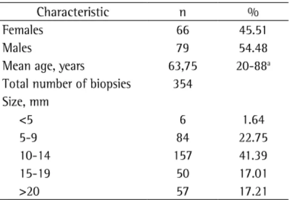
Our analysis included 354 mediastinal lymph node biopsies, from 145 consecutive patients. Of those 145 patients, 54.48% were male. The mean age was 63.75 years (range, 20 to 88 years). The mean lymph node size was 15.03 mm, and 90 lymph nodes were smaller than 10.0 mm. The demographic and lesion characteristics of the patients included in this study are summarized in Table 1.
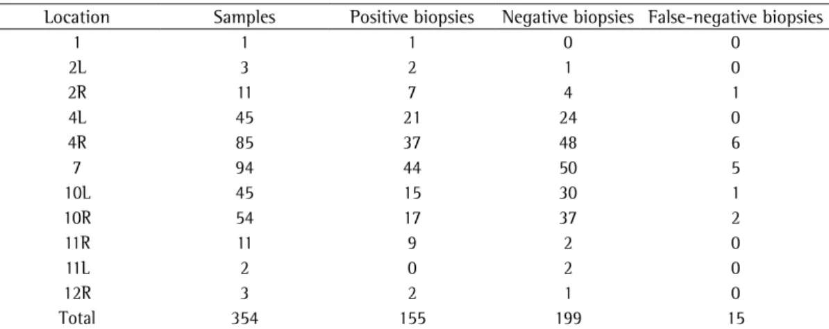
Lesions were located as follows: 40.96% in the lower or upper paratracheal region; 27.96% in the hilar region; 26.55% in the subcarinal region; and 4.53% in the interlobar, lobar, and subsegmental regions. Regarding the ultrasound characteristics of the lymph nodes, 35.0% were oval, 16.6% were triangular, and 51.41% had regular borders, whereas the location of the lesion in the mediastinum as per the IASLC lymph node map and biopsy diagnostic yield is summarized in Tables 2 and 3. The EBUS-TBNA method showed a sensitivity of 91.17%, a specificity of 100.0%, a positive predictive value of 100.0%, and a negative predictive value of 92.9%. The likelihood ratio for a positive diagnostic result
Table 1 - Demographic characteristics of and lymph node size in the patients included in the study (N = 145).
Characteristic n %
Females 66 45.51
Males 79 54.48
Mean age, years 63,75 20-88a
Total number of biopsies 354 Size, mm
<5 6 1.64
5-9 84 22.75
10-14 157 41.39
15-19 50 17.01
>20 57 17.21
diagnostic yield of pulmonary lesions suggestive of cancer do not have the reference standard applied to all positive histologic results. The main reason for this is that it is complicated to perform another diagnostic surgical procedure in patients diagnosed with lung cancer.
Another application of EBUS-TBNA is its ability to stage lung cancer in the mediastinum, mainly because the technique allows easy access to most lymph node stations, except for stations 5, 6, 8, and 9 (IASLC classification), from which it is not possible to obtain histologic or cytologic samples by using this technique; in such cases, EBUS-TBNA can be complemented with ultrasound-guided endoscopy.(8) However, the yield of EBUS-TBNA
is significantly decreased in peripherally located pulmonary lesions.(3,8)
The diagnostic yield of mediastinal staging by EBUS-TBNA is high, with a reported sensitivity of approximately 90% and a specificity of 100%. In a recently published randomized clinical trial,(9) the
yield of EBUS-TBNA as the first-choice minimally invasive method for the staging and diagnosis of lung cancer in lesions adjacent to the central airways was compared with that of commonly used techniques. The group randomized to EBUS-TBNA had a decrease in the number of unnecessary thoracotomies and complications, as well as a decrease in the time required to confirm or rule out the diagnosis of lung cancer and make therapeutic decisions. In that study, EBUS-TBNA had a sensitivity of 92% and a specificity of 100%.
role as diagnostic yields are increased. EBUS has been used as a method that allows TBNA of pulmonary lesions to be performed reliably and safely. Methodologically, studies analyzing the
Table 2 - Location of the lymph nodes in the mediastinum as per the IASLC classification and biopsy results. Location Samples Positive biopsies Negative biopsies False-negative biopsies
1 1 1 0 0
2L 3 2 1 0
2R 11 7 4 1
4L 45 21 24 0
4R 85 37 48 6
7 94 44 50 5
10L 45 15 30 1
10R 54 17 37 2
11R 11 9 2 0
11L 2 0 2 0
12R 3 2 1 0
Total 354 155 199 15
IASLC: International Association for the Study of Lung Cancer.
Table 3 - Ultrasound characteristics of the lymph nodes aspirated (N = 354) under EBUS guidance for mediastinal staging.
Characteristic n %
Shape
Oval 124 35.02
Round 148 41.8
Triangular 59 16.66
Other 23 6.49
Borders
Regular 182 51.41
Irregular 172 48.58
EBUS: endobronchial ultrasound.
Table 4 - Definitive histologic results for the EBUS-TBNA samples.
Histologic result %
Neoplasia (n = 170)
Adenocarcinoma 69.41
Squamous disease 18.82
Metastasis 1.76
Small cell disease 7.64
Neuroendocrine disease 2.35
Non-Hodgkin’s lymphoma
-Benign lesion (n = 9)
Sarcoidosis granuloma 4.70 Tuberculosis granuloma 11.1
this study was conducted in a single center, with the same operator. Diagnostic yield is a learning curve-dependent variable, and this can reduce result applicability to other centers with less experience in EBUS-TBNA.
In conclusion, EBUS-TBNA is a diagnostic tool with a high diagnostic yield, has few associated complications, and should be considered an option for mediastinal nodal staging in patients with lung cancer, especially in those with non-small cell lung cancer.
References
1. Rivera MP, Mehta AC, Wahidi MM. Establishing the diagnosis of lung cancer: Diagnosis and management of lung cancer, 3rd ed: American College of Chest Physicians evidence-based clinical practice guidelines. Chest. 2013;143(5 Suppl):e142S-65S.
2. Ernst A, Silvestri GA, Johnstone D; American College of Chest Physicians. Interventional pulmonary procedures: Guidelines from the American College of Chest Physicians. Chest. 2003;123(5):1693-717. http://dx.doi.org/10.1378/ chest.123.5.1693
3. Anantham D, Koh MS, Ernst A. Endobronchial ultrasound. Respir Med. 2009;103(10):1406-14. http://dx.doi. org/10.1016/j.rmed.2009.04.010
4. Dincer HE. Linear EBUS in staging non-small cell lung cancer and diagnosing benign diseases. J Bronchology Interv Pulmonol. 2013;20(1):66-76. http://dx.doi. org/10.1097/LBR.0b013e31827d1514
5. Gu P, Zhao YZ, Jiang LY, Zhang W, Xin Y, Han BH. Endobronchial ultrasound-guided transbronchial needle aspiration for staging of lung cancer: a systematic review and meta-analysis. Eur J Cancer. 2009;45(8):1389-96. http://dx.doi.org/10.1016/j.ejca.2008.11.043 6. Rusch VW, Asamura H, Watanabe H, Giroux DJ,
Rami-Porta R, Goldstraw P, et al. The IASLC lung cancer staging project: a proposal for a new international lymph node map in the forthcoming seventh edition of the TNM classification for lung cancer. J Thorac Oncol. 2009;4(5):568-77. http://dx.doi.org/10.1097/ JTO.0b013e3181a0d82e
7. Tanoue LT, Detterbeck FC. New TNM classification for non-small-cell lung cancer. Expert Rev Anticancer Ther. 2009;9(4):413-23. http://dx.doi.org/10.1586/era.09.11 8. Vilmann P, Puri R. The complete ‘’medical’’ mediastinoscopy
(EUS-FNA + EBUS-TBNA). Minerva Med. 2007;98(4):331-8. 9. Navani N, Nankivell M, Lawrence DR, Lock S, Makker
H, Baldwin DR, et al.; Lung-BOOST trial investigators. Lung cancer diagnosis and staging with endobronchial ultrasound-guided transbronchial needle aspiration compared with conventional approaches: an open-label, pragmatic, randomised controlled trial. Lancet Respir Med. 2015;3(4):282-9. http://dx.doi.org/10.1016/ S2213-2600(15)00029-6
10. Tedde ML, Figueiredo VR, Terra RM, Minamoto H, Jatene FB. Endobronchial ultrasound-guided transbronchial needle aspiration in the diagnosis and staging of mediastinal lymphadenopathy: initial experience in Brazil. J Bras Pneumol. 2012;38(1):33-40.
In our series, the diagnostic yield of EBUS-TBNA was high, especially because all lesions were located in the central airway, with lesions visualized by EBUS. We evaluated the yield of EBUS-TBNA and found that, in small lesions (those smaller than 10 mm), it remained satisfactory. This finding is a contribution to the assessment of solitary pulmonary nodules and lymph nodes between 5 and 10 mm, given that lesions smaller than 7 mm are not seen on imaging studies such as positron emission tomography-CT, whereas, by using ultrasound, those lesions can be found and aspirated transbronchially, with a diagnostic yield of 90% or more.
Tedde et al.,(10) the only available reference
regarding EBUS-TBNA in South America, included 50 patients who underwent a total of 201 EBUS-TBNA biopsies of 81 lymph nodes or mediastinal masses for diagnosis and staging. In that series, the diagnosis of cancer was confirmed in 57% of the patients in whom EBUS-TBNA was diagnostic. A negative point in that study was that, in 13 of the 50 patients, the cytologic material was not optimal for anatomopathological examination.
Complications associated with EBUS-TBNA are rare and usually minor. In our series, there was one case of pneumomediastinum—a complication with a reported rate of less than 1%—that was managed medically, without other major complications. In the literature, complications are infrequent, with self-limited bleeding being reported in less than 5% of the procedures, whereas the rate of pneumothorax is 1% with no fatal events,(8,11)
similar to what was found in our series. This study has some limitations. First, the study design: the protocol for studies of diagnostic yield should be in accordance with the recommendations of the STARD statement.(12) In our study, those
12. Bossuyt PM, Reitsma JB, Bruns DE, Gatsonis CA, Glasziou PP, Irwig LM, et al. The STARD statement for reporting studies of diagnostic accuracy: explanation and elaboration. Ann Intern Med. 2003;138(1):W1-12. http://dx.doi. org/10.7326/0003-4819-138-1-200301070-00010 11. von Bartheld MB, van Breda A, Annema JT.