Metriorhynchid Crocodylomorph Genera
Dakosaurus
and
Plesiosuchus
from the Late Jurassic of Europe
Mark T. Young1*, Stephen L. Brusatte2,3, Marco Brandalise de Andrade4, Julia B. Desojo5, Brian L. Beatty6, Lorna Steel7, Marta S. Ferna´ndez8, Manabu Sakamoto9, Jose Ignacio Ruiz-Omen˜aca10,11,
Rainer R. Schoch12
1School of Geosciences, University of Edinburgh, Edinburgh, United Kingdom,2Division of Paleontology, American Museum of Natural History, New York City, New York, United States of America,3Department of Earth and Environmental Sciences, Columbia University, New York City, New York, United States of America,4Departamento de Paleontologia e Estratigrafia, Universidade Federal do Rio Grande do Sul, Porto Alegre, Brazil,5Divisio´n de Paleontologı´a, Museo Argentino de Ciencias Naturales ‘Bernardino Rivadavia’, Buenos Aires, Argentina,6Department of Anatomy, New York College of Osteopathic Medicine, Old Westbury, New York, United States of America,
7Department of Earth Sciences, Natural History Museum, London, United Kingdom,8Departamento Paleontologı´a de Vertebrados, Museo de La Plata, La Plata, Argentina,9School of Earth Sciences, University of Bristol, Bristol, United Kingdom,10Museo del Jura´sico de Asturias (MUJA), Colunga, Spain,11Departamento de Geologı´a, Universidad de Oviedo, Oviedo, Spain,12Staatliches Museum fu¨r Naturkunde, Stuttgart, Germany
Abstract
Background:DakosaurusandPlesiosuchusare characteristic genera of aquatic, large-bodied, macrophagous metriorhynchid crocodylomorphs. Recent studies show that these genera were apex predators in marine ecosystems during the latter part of the Late Jurassic, with robust skulls and strong bite forces optimized for feeding on large prey.
Methodology/Principal Findings:Here we present comprehensive osteological descriptions and systematic revisions of the type species of both genera, and in doing so we resurrect the genusPlesiosuchusfor the speciesDakosaurus manselii. Both species are diagnosed with numerous autapomorphies. Dakosaurus maximus has premaxillary ‘lateral plates’; strongly ornamented maxillae; macroziphodont dentition; tightly fitting tooth-to-tooth occlusion; and extensive macrowear on the mesial and distal margins. Plesiosuchus manselii is distinct in having: non-amblygnathous rostrum; long mandibular symphysis; microziphodont teeth; tooth-crown apices that lack spalled surfaces or breaks; and no evidence for occlusal wear facets. Our phylogenetic analysis finds Dakosaurus maximusto be the sister taxon of the South American Dakosaurus andiniensis, and Plesiosuchus manselii in a polytomy at the base of Geosaurini (the subclade of macrophagous metriorhynchids that includesDakosaurus,GeosaurusandTorvoneustes).
Conclusions/Significance:The sympatry ofDakosaurusandPlesiosuchusis curiously similar to North Atlantic killer whales, which have one larger ‘type’ that lacks tooth-crown breakage being sympatric with a smaller ‘type’ that has extensive crown breakage. Assuming this morphofunctional complex is indicative of diet, thenPlesiosuchuswould be a specialist feeding on other marine reptiles whileDakosauruswould be a generalist and possible suction-feeder. This hypothesis is supported by Plesiosuchus manseliihaving a very large optimum gape (gape at which multiple teeth come into contact with a prey-item), while Dakosaurus maximuspossesses craniomandibular characteristics observed in extant suction-feeding odontocetes: shortened tooth-row, amblygnathous rostrum and a very short mandibular symphysis. We hypothesise that trophic specialisation enabled these two large-bodied species to coexist in the same ecosystem.
Citation:Young MT, Brusatte SL, de Andrade MB, Desojo JB, Beatty BL, et al. (2012) The Cranial Osteology and Feeding Ecology of the Metriorhynchid Crocodylomorph GeneraDakosaurusandPlesiosuchusfrom the Late Jurassic of Europe. PLoS ONE 7(9): e44985. doi:10.1371/journal.pone.0044985
Editor:Richard J Butler, Ludwig-Maximilians-Universita¨t Mu¨nchen, Germany
ReceivedApril 24, 2012;AcceptedAugust 16, 2012;PublishedSeptember 18, 2012
Copyright:ß2012 Young et al. This is an open-access article distributed under the terms of the Creative Commons Attribution License, which permits unrestricted use, distribution, and reproduction in any medium, provided the original author and source are credited.
Funding:The authors have no support or funding to report.
Competing Interests:The authors have declared that no competing interests exist. * E-mail: zoologika@gmail.com
Introduction
The evolution and diversification of metriorhynchid crocodylo-morphs in the Mesozoic seas is a classic example of an evolutionary radiation in the fossil record [1], [2]. Metriorhynch-ids are highly aberrant compared to other crocodylomorphs (which are terrestrial or semi-aquatic), and evolved numerous adaptations to their pelagic lifestyle, including a complete loss of their osteoderm armour, hydrofoil-like forelimbs, a hypocercal tail,
sizes, skull shapes, biting behaviours, and dental morphologies during their evolutionary history [1], [2], [11], [13–17].
One of the major metriorhynchid subclades, Geosaurinae, includes large-bodied taxa such as ‘‘Mr Leeds’ specimen’’ (GLAHM V972, the generic and specific name for this taxon is currently in press [2]),Torvoneustes,Geosaurus, andDakosaurus, which had skulls and teeth well suited for feeding on large prey [1], [2], [11], [13], [14], [17]. There seems to have been a temporal and phylogenetic trend towards increasing super-predatory behaviour within this group, as progressively more derived and younger taxa had skulls that were better optimized for enduring strong bite forces [1], [13], [16]. Furthermore, because of the high diversity of tooth crown and serration morphologies among geosaurines [11], Younget al. [13] hypothesised that contemporaneous geosaurines were limiting competition through ecological specialisation and niche partitioning. Indeed, Late Jurassic marine ecosystems frequently had two sympatric geosaurine genera that were either apex-predators or second tier super-predators [1], [11], [18].
The geosaurine genusDakosaurushas been of particular interest, especially due to its unusual cranial morphology. Its skull and mandible were the most robust and powerful within
Metrior-hynchidae, as shown by biomechanical analyses [1], [16]. Furthermore, it had a brevirostrine and oreinirostral snout and a robust dentition, with the largest apicobasal crown lengths of any metriorhynchid and serrated carinae composed of a keel and true denticles [10], [11], [17], [19], [20] (Fig. 1). As has been hinted at in previous studies, and as we argue more fully in this monograph, it is likely thatDakosauruswas macrophagous: an animal that could feed upon prey items of similar body size. The larger body size of
Dakosauruscompared to other metriorhynchids would be beneficial
for such a feeding style, as it would allow this taxon to target larger prey, and would allow for a reduction in the time taken to process prey, making larger organisms more energetically feasible prey items [21].
The genusDakosaurushas been known for over 150 years, and it was among the handful of large marine reptiles discovered in early–mid 19th century Europe that helped reveal a hitherto unknown ancient fauna of peculiar, predatory reptiles from the Mesozoic. Since that time numerous new species have been placed in the genus. The recent phylogenetic analysis of Young & Andrade [10] and the taxonomic changes necessitated by that analysis indicated that the genusDakosaurushad four valid species
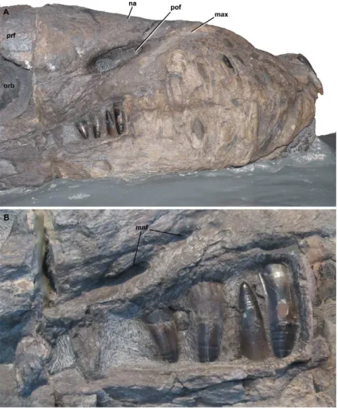
Figure 1.Dakosaurus andiniensis, referred specimen MOZ 6146P.Skull and mandible in, (A) lateral (right) view of the snout and (B) close-up on the posterior teeth, showing the interlocking dentition. Note the robust teeth and snout. Abbreviations: max, maxilla; mnf, maxillary neurovascular foramen; na, nasal; orb, orbit; pof, preorbital fenestra; prf, prefrontal.
[22–25]:D. andiniensis(Fig. 1),D. maximus[23] (Figs. 2, 3, 4, 5, 6, 7, 8),D. manselii[24] (Figs. 9, 10, 11, 12, 13, 14, 15, 16, 17, 18, 19, 20, 21, 22, 23) and D. nicaeensis [25]. Furthermore, two species originally assigned to Dakosaurus were referred by Young & Andrade [10] to the genusGeosaurus:D. lapparentiandD. carpenteri. However, the latter was recently given its own genus,Torvoneustes [11]. Most of the Callovian–Oxfordian teeth from England, France and Poland that previously were referred toDakosaurusare now considered as belonging to a new genus (still in press [2]), whereas another intriguing specimen, NHMUK PV R486, is considered Geosaurinae indeterminate [2]. Furthermore, incom-plete material from the Kimmeridgian of Mexico may represent a fifth species ofDakosaurus[26], [27] but this is currently unclear (see discussion below). As is clear, Dakosaurus had a wide geographic range, with specimens known from Argentina, England, France, Germany and Switzerland [1], and possibly also Spain [28]. It may have had a worldwide distribution during the Mesozoic.
Recently, however, it has been suggested thatDakosaurus manselii may also not belong within the genusDakosaurus. This contention was first suggested by Young et al. [14], based on a subsidiary phylogenetic analysis (presented in their supplementary material and differing from their primary analysis in the use of some ordered characters) that foundD. manseliito be the sister taxon of the cladeGeosaurus+Dakosaurus. This change in position was solely
based on dental characters. Young et al. [2] re-iterated in their discussion of metriorhynchid denticle evolution that the taxonomic affinities ofD. manselii are currently unclear. They noted thatD.
manseliihas microscopic denticles (whereas bothD. maximusandD.
andiniensishave macroscopic denticles), and apicobasally aligned
ridges on both the labial and lingual surfaces (whichD. maximus
and D. andiniensislack). Updated anatomical information,
there-fore, is necessary for resolving the affinities of D. manselii. Furthermore, the systematic placement of D. manselii also has bearing on the systematics, especially the generic placement, ofD. nicaeensis, a poorly-understood species that shares an unusual large dentition with bothD.manseliiandD.maximus. IfD.manseliidoes not belong to the same subclade asD.maximus, then this distinctive dentition would be homoplastic and insufficient for assigningD. nicaeensistoDakosaurus. Resolving the phylogenetic position of D. manselii, therefore, is currently one of the most pressing issues in metriorhynchid systematics and a keystone upon which rests many wider issues of metriorhynchid classification and phylogeny.
Despite the recent upsurge of interest in metriorhynchid phylogeny and evolution, the original specimens of Dakosaurus maximusand ‘‘Dakosaurus’’manseliifrom Europe have received little attention. In fact, they have been only briefly described in the literature (with Fraas’ [4] monograph of Geosaurus [Cricosaurus]
suevicus and Dakosaurus maximus being the only exception), which
makes it difficult to incorporate them into phylogenetic analyses
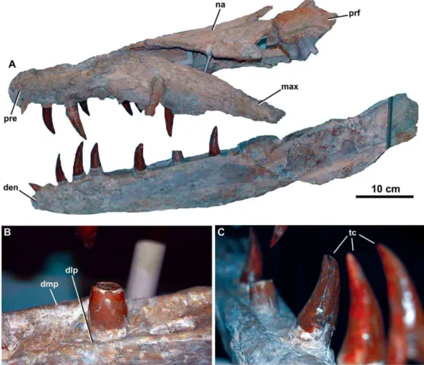
Figure 2.Dakosaurus maximus, neotype SMNS 8203.(A) General view of the skull and mandible, (B) close-up on the dentary alveoli and raised lateral and medial margins, and (C) oblique forward view of the dentary tooth row. Abbreviations: den, dentary; dlp, dentary lateral plate; dmp, dentary medial plate; max, maxilla; na, nasal; pre, premaxilla; prf, prefrontal; tc, tooth crowns.
Figure 3.Dakosaurus maximus, neotype SMNS 8203.(A) Anterior view of the skull and mandible, note that level to the fourth dentary tooth the mandibular ramus deflects laterally (i.e. short symphysis), and (B) oblique dorsal view of the skull, emphasising the blunt ‘‘bullet-shaped’’ snout (i.e. amblygnathy). Abbreviations: den, dentary; max, maxilla; na, nasal; pre, premaxilla; prf, prefrontal.
doi:10.1371/journal.pone.0044985.g003
Figure 4.Dakosaurus maximus, neotype SMNS 8203.(A) Close-up on the premaxillary ‘lateral plates’, (B) close-up on the maxillary alveoli in oblique ventral view and (C) close-up on the maxillary teeth showing tooth crown wear. Abbreviations: plp, premaxillary lateral plate; rp, reception pit.
and compare them with newly discovered specimens. This is especially surprising given that several new species and specimens have been assigned to the genus Dakosaurus in recent years, including spectacularly preserved material that has revealed the strong bite forces and theropod-like skulls characteristic of the genus [19], [20] (Fig. 1). Here we redescribe the type specimens of Dakosaurus maximusand ‘‘D.’’manselii, as well as a large partial skull
and mandible assigned to ‘‘Pliosaurus trochanterius’’ (and later
Machimosaurus mosae), which we conclusively demonstrate is a
metriorhynchid and, for the first time, refer to ‘‘D.’’manselii. These redescriptions reveal a number of characters unique to each species, and allow us to present a comprehensive osteology for these important historical taxa. Furthermore, these redescriptions highlight significant craniodental differences betweenD. maximus
Figure 5.Dakosaurus maximus, referred specimens SMNS 10819a and SMNS 10819b.Snout (SMNS 10819a) in: (A) dorsal view, (B) ventral (palatal) view, and (C) close-up on the maxillary tooth-row in ventral view. Note that several teeth exhibit carinal wear, broken apices and spalling of the enamel at the apex. (D) Left-half of the posterior region of the skull (SMNS 10819b) in dorsal view. Abbreviations: en, external nares; fr, frontal; max, maxilla; pa, parietal; po, postorbital; pre, premaxilla; prf, prefrontal; rp, reception pit; sq, squamosal; stf, supratemporal fenetra.
and ‘‘D.’’manselii, and show that there are numerous autapomor-phies shared by D. maximusand D. andiniensis that are absent in
‘‘D.’’manselii. We then incorporate new information gleaned from
these redescriptions into a revised phylogenetic analysis of Thalattosuchia. Most importantly, this analysis does not recover a distinct grouping ofD.maximus,D.andiniensis, and ‘‘D.’’manselii, which supports the removal of the latter species into its own genus, the resurrectedPlesiosuchus. Based on our greater understanding of Geosaurini craniodental form we also revise the generic diagnoses
for Geosaurusand Torvoneustes. Finally, based on our craniodental
descriptions ofD. maximusand ‘‘D.’’manseliiwe outline hypothet-ical feeding behaviours for both species, and hypothesise that trophic specialisation enabled these two species to co-exist in the same ecosystem.
Institutional Abbreviations
BRSMG, Bristol City Museum and Art Gallery, Bristol, UK; BSPG, Bayerische Staatssammlung fu¨r Pala¨ontologie und Geolo-gie, Mu¨nchen, Germany; CAMSM, Sedgwick Museum, Cam-bridge, UK; CM, Carnegie Museum of Natural History, Pittsburgh, PA, USA; GLAHM, Hunterian Museum, Glasgow, UK; JME, Jura Museum, Eichsta¨tt, Germany; MOZ, Museo Profesor J. Olsacher, Zapala, Argentina; MUJA, Museo del Jura´sico de Asturias, Colunga, Spain; NHMUK, Natural History Museum, London, UK; NMS, Naturmuseum Solothurn, Switzer-land; OUMNH, Oxford University Museum of Natural History, Oxford, UK; SMNS, Staatliches Museum fu¨r Naturkunde Stuttgart, Germany.
Historical Overview ofDakosaurus maximus
The first species assigned to the genus Dakosaurus was D.
maximus, which was first erected asGeosaurus maximusby Plieninger
Figure 6.Dakosaurus maximus, referred specimen SMNS 82043.Left mandibular ramus in lithographic limestone in: (A) general view, (B) close-up on the anterior teeth, and (C) close-close-up on the dentary dorsal margin at the first preserved tooth crown, showing the various foramina and a reception pit and (D) close-up on the dentary dorsal margin, slightly further back along the tooth row. Abbreviations: an, angular; art, articular; cp, coronoid process on the surangular; den, dentary; rp, reception pit; sdg, surangulodentary groove; sur, surangular.
Figure 7.Dakosaurus maximus, referred specimen SMNS 56999.Left isolated maxilla in: (A) lateral view, (B) dorsal view and (C) ventral view (showing the tooth row). Abbreviations: al, alveolus; mnf, maxillary neurovascular foramen; pof, preorbital fenestra; tc, tooth crown.
doi:10.1371/journal.pone.0044985.g007
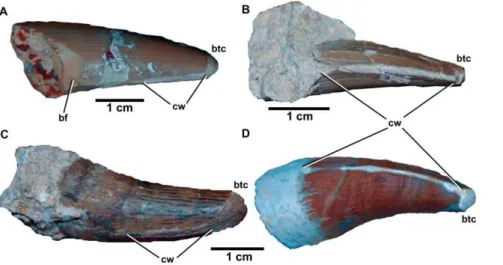
Figure 8.Dakosaurus maximus, referred specimens SMNS 91425.Numerous isolated teeth ofD. maximusshowing the occlusion wear patterns and apical breakage. Top left scale bar for (A), top right scale bar for (B), and bottom scale bar for images (C)–(D). Abbreviations: bf, basal facet; btc, broken tip; cw, carinal wear.
[23], for an isolated tooth found at Schnaitheim, near Heiden-heim, Baden-Wu¨rttemberg, Germany (upper Kimmeridgian;
Hybonoticeras beckeriSub-Mediterranean ammonite Zone).
Plienin-ger [29] later referred a partial dentary with six in situ teeth preserved in lithographic limestone from Ulm toGeosaurus maximus. Quenstedt [30], [31] initially considered isolated teeth from Schnaitheim, very similar to those of figured by Plieninger, as belonging to the theropod dinosaur genusMegalosaurus. However, later he erected the nameDakosaurusfor the Schnaitheim teeth and Plieninger’s species [32], [33]. Furthermore, Quenstedt [33] referred a dentigerous bone (probably a partial maxilla) from Schnaitheim with threein situ crowns toD. maximus. As the type material ofD.maximusis missing, Young & Andrade [10] suggested a skull and mandible described by Fraas [4] should be the neotype of the species. This specimen (SMNS 8203; Figs. 2, 3, 4) was found at Staufen, Baden-Wu¨rttemberg, Germany, and was also from the upper Kimmeridgian H. beckeri Sub-Mediterranean ammonite Zone. Fraas [4] described a second D. maximus skull (SMNS 10819a, b; Fig. 5) from the upper Kimmeridgian of Sontheim an der Brenz, Baden-Wu¨rttemberg, Germany.
During the 19thcentury there were numerous species assigned
toDakosaurus, most of which were erected for isolated teeth. Fraas
[4] synonymised allDakosaurus species with the exceptions of D. manselii(he consideredD.manseliito be either a junior synonym of, or closely related to,D. maximus) and the tooth taxonD. paradoxus
withD. maximus. Central to this argument, Fraas [4] demonstrated
that the various morphological differences used to erect these numerous tooth taxa were actually part of a continuum of variation that was normal for a single species. This argument followed an earlier, but long neglected, study by Mason [34], who discussed the variation in mediolateral compression and symmetry
inDakosaurus teeth as being related to position in the tooth row,
and to which bone the teeth belonged.
Two historic tooth taxa now considered as synonymous withD. maximus,Liodon paradoxus[35] andTeleosaurus suprajurensis[36], were erected for isolated teeth discovered in the lower TithonianDiceras Limestones near Kelheim, Bavaria, Germany.Teleosaurus
suprajur-ensis was considered to be a subjective junior synonym of D.
maximus by Lydekker [3], von Zittel [37] and Fraas [4]. Liodon
paradoxus was referred to the early Tithonian speciesCricosaurus
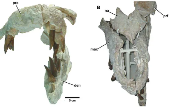
grandis[38] by von Zittel [37], while Fraas [4] referred the species Figure 9.Plesiosuchus manselii, holotype NHMUK PV OR40103.Dorsal view of the skull, (A) hypothetical skull reconstruction (grey lines represent elements that are missing) and (B) photograph of what is preserved. Abbreviations: en, external nares; max, maxilla; na, nasal; orb, orbit; pa, parietal; pre, premaxilla; prf, prefrontal; sq, squamosal; stf, supratemporal fenestra.
toDakosaurus, based on a mandible within situteeth and isolated teeth from Schnaitheim. All teeth referred toD. paradoxusare less robust than those that had been referred toD. maximusduring the mid 1800s, with a narrower labiolingual cross-section (see Fraas
[4]: Plate 2, Figs 1, 12, 13, and 14). This is the primary reason why Fraas [4] retainedD. paradoxusas a separate species. Interestingly, however, the mandible Fraas [4] referred to D. paradoxus only preserves the anterior-most dentition. One specimen ofDakosaurus, SMNS 10819a, preserves the premaxilla and maxilla within situ crowns (Fig. 5A). It demonstrates that the premaxillary and newly erupted maxillary crowns of D. maximusare notably less robust (narrower labiolingual cross-section) than fully-erupted maxillary crowns (confirming Mason [34]). In other words, Dakosaurus exhibits heterodonty across the tooth row, and not all of its teeth possess the ‘characteristic’ robust morphology that was assumed by early workers. As such, there are no grounds to retainD. paradoxus as a separate species.
Another historic tooth taxon now considered as D. maximus,
Dakosaurus gracilisis known from small isolated teeth discovered in
lower Tithonian deposits near Steinheim, Baden-Wu¨rttemberg, Germany [39]. Fraas [4] did not considerD. gracilisas a separate species, but regarded it to be at most a variety ofD. maximus. An incomplete skeleton discovered from an unnamed Lower Creta-ceous (early Hauterivian in age) formation in De´partement du Var, Provence-Alpes-Coˆte d’Azur, France was referred to as
Dacosaurus maximus var. gracilis (sic) [40]. Subsequently [41],
another incomplete skeleton from a nearby locality (late Valanginian in age) was namedDacosaurus lapparenti (sic), and is now known asGeosaurus lapparenti[10].
Yet another historic taxon, Dakosaurus lissocephalus, is known from a poorly preserved and dorsoventrally crushed skull (CAMSM J29419) discovered in the upper Kimmeridgian
(Aulacostephanus eudoxusSub-Boreal ammonite Zone) Lower
Kim-meridge Clay Formation of Ely, Cambridgeshire, England [42]. Lydekker [3] considered this species to be a subjective junior synonym ofD. maximus, while Fraas [4] provisionally synonymised the two. Young & Andrade ([10]: appendix) considered D. lissocephalusand‘‘Dakosaurus’’ manseliinot to be conspecific, due to the shape of the supratemporal fenestra, squamosal, and parietal
inD. lissocephalus being more reminiscent ofD. andiniensisand D.
Figure 10.Plesiosuchus manselii, holotype NHMUK PV OR40103. Snout in dorsal view, (A) line drawing (grey squares represent the premaxilla-maxilla suture, the exact nature of which we are unsure) and (B) photograph. Abbreviations: en, external nares; fr, frontal; max, maxilla; na, nasal; pre, premaxilla; prf, prefrontal.
doi:10.1371/journal.pone.0044985.g010
Figure 11.Plesiosuchus manselii, holotype NHMUK PV OR40103.Snout in lateral (right) view, (A) photograph and (B) line drawing (grey shaded area represents the orbital cavity, grey lines represents sutures we are unsure of). Abbreviations: al, alveolus; max, maxilla; na, nasal; orb, orbit; pal, palatine; pre, premaxilla; prf, prefrontal; sut?, suture?; tc, tooth crown.
maximus. Furthermore, they considered the synonymy betweenD.
maximus and D. lissocephalus provisional. Here, we formally
synonymise D. lissocephalus and D. maximus. This is due to two similarities that they share, unique to other Dakosaurus species. First, D. lissocephalus like other metriorhynchids has a quadrate distal articular surface separated into two protuberances (condyles) by a sulcus [5].Plesiosuchus manselii, on the other hand, lacks this sulcus (Fig. 15). Secondly, isolated teeth from the D. lissocephalus type locality (NHMUK PV OR20283) share the same suite of characters as the German D. maximus teeth (no apicobasally aligned ridges, large conspicuous denticles; Fig. 8). TheP. manselii teeth, however, possess apicobasal ridges (of low-relief) and small denticles (Figs. 22, 23). As the type locality (a quarry in Ely) has subsequently been flooded, discovering more material to confirm these observations is difficult.
Finally, another historical taxon, Leiodon primaevum is known from isolated teeth discovered in the upper Kimmeridgian (A.
autissiodorensis Sub-Boreal ammonite Zone) Argiles de Chaˆtillon
Formation of Boulogne-sur-Mer, Pas-de-Calais, France [43]. Sauvage [44] later placed L. primaevum in Dacosaurus (sic) as D.
primaevus. Lydekker [3], von Zittel [37] and Fraas [4] all considered this species to be a subjective junior synonym ofD. maximus. We agree, as isolated teeth from the type locality (NHMUK PV OR32414; SMNS 57210) share the same suite of characters as the GermanD. maximusand the English teeth from Ely (see above). A partial left maxilla (SMNS 56999) from the locality also shares the same distinctive maxillary ornamentation as D. maximus (see description below; Fig. 7).
Historical Overview ofPlesiosuchus manselii
The holotype ofPlesiosuchus manseliiis a broken and incomplete skull (NHMUK PV OR40103; Figs. 9, 10, 11, 12, 13) with a mandible and isolated post-cranial remains (NHMUK PV OR40103a; Figs. 17, 18, 22, 23) from a large individual discovered in the upper Kimmeridgian (A. autissiodorensisSub-Boreal ammon-ite Zone) Lower Kimmeridge Clay Formation of Kimmeridge Bay, Dorset, England. The specific epithetmanselii is frequently misspelt in the literature, generally as manseli or mansellii [3], [45–47]. The type and referred specimens were given to the Figure 12.Plesiosuchus manselii, holotype NHMUK PV OR40103.Snout in ventral (palatal) view, (A) line drawing (grey lines represent the sutures we are unsure of) and (B) photograph. Abbreviations: al, alveolus; max, maxilla; pal, palatine; pre, premaxilla; sof, suborbital fenestra; sut?, suture?; tc, tooth crown.
British Museum (now in the Natural History Museum, London) by John Clavell Pleydell in 1866. During the 1860s Mansel-Pleydell discovered the remains of several large-bodied marine reptiles along the coast of Dorset, most especially at Kimmeridge Bay. He discovered the remains of P. manselii in a reef at Kimmeridge Bay, exposed at low tide [48].
The holotype ofPlesiosuchus manselii was described by Hulke in two papers. In the first, Hulke [48] described the right mandibular ramus, isolated vertebrae, an isolated premaxilla, a femur and a dentigerous bone that he tentatively referred to as the ‘‘upper maxilla’’ (these specimens are now curated as NHMUK PV OR40103a, although the isolated premaxilla is now part of NHMUK PV OR40103). He referred the specimens to Geoffroy’s
[49]Steneosaurus rostro-minor. Hulke [48] posited that NHMUK PV
OR40103a was probably identical to Cuvier’s [50] second Honfleur gavial ‘‘teˆte a` museau plus court’’, that the dentition of NHMUK PV OR40103a was identical toDakosaurus maximus, and that all these species/specimens could be referred to Steneosaurus
rostro-minor. It must be noted that metriorhynchids were poorly
known at this time, with Dakosaurus maximus known only from isolated teeth and a fragments of dentigerous bones within situ crowns, while Cuvier’s [50] ‘‘teˆte a` museau plus court’’ was a chimera of two metriorhynchid species (Metriorhynchus superciliosus
andM. geoffroyii[1], [51]). The characteristics Hulke [48] used to
unite these specimens are now known to be either metriorhynchid apomorphies (e.g. oval, ‘‘spoon-shaped’’ external nares; three Figure 13. Plesiosuchus manselii, holotype NHMUK PV OR40103. Braincase: in (A) dorsal view, (B) ventral view and (C) occipital view. Abbreviations: bt, basal tubera; eo, exocciptial; fm, foramen magnum; pa, parietal; qu, quadrate; soc, supraoccipital; sq, squamosal; stf, supratemporal fenestra.
premaxillary alveoli; absence of external mandibular fenestrae; distinct coronoid process on the mandible) or Geosaurini apomorphies (e.g. bicarinate serrated dentition). Other character-istics used were subsequently found to have been widespread in Mesozoic crocodylomorphs (e.g. teeth with apicobasal ‘striations’; teeth that are unequally convex, that have some degree of mediolateral compression and recurve lingually; amphicoelous vertebrae). A note was added to the end of the first publication stating a ‘‘considerable part of the skull’’ had been discovered through further examination of the material presented to the British Museum.
In the second paper, Hulke [24] described a skull, which is preserved in two sections: the rostrum and the occiput (the latter
with partial supratemporal arches preserved; now curated as NHMUK PV OR40103; Fig. 9). Initially, the then matrix-encased skull was believed to pliosaurian and set aside; it was the preparator Mr Davies that realised the skull material was in fact crocodylian in nature [24]. It was here that the specific epithet
manselii was erected as Steneosaurus Manselii (sic). The ‘‘head’’
(NHMUK PV OR40103) and the ‘‘lower jaw and associated post-crania’’ (NHMUK PV OR40103a, the specimen described by Hulke [48]) have been considered to be from the same individual. Hulke ([24]:167) stated: ‘‘The general agreement of their dimensions, and their discovery near together (in a reef exposed at low water in Kimmeridge Bay), make it highly probable that this head and the lower jaw both belonged to one individual’’. We agree that the two specimens are most likely from the same individual, especially as the size of the two specimens is comparable.
Interestingly, the isolated bone fragment referred to as the ‘‘upper maxilla’’ has never been figured and cannot be located. Hulke [48] describes the specimen as being a fragment of 14 cm in length, containing five alveoli of which four still have portions of teeth remainingin situ. Furthermore, the bone is not mentioned in the latter publication (unlike the mandible and premaxilla). There is a possibility as to why this specimen cannot be located. A note in the NHMUK specimen register beside NHMUK PV OR40103 states that some of the material was destroyed, with a date of 1 August 1931. Unfortunately, what was destroyed is not stated. As both NHMUK PV OR40103 and NHMUK PV OR40103a suffer pyrite decay and require periodic conservation, it is possible that the ‘‘upper maxilla’’ was destroyed after extensive decay. A box of NHMUK PV OR40103 fragments was discovered by one of us (LS), and it could represent some of the destroyed material.
Owen [52] erected the genusPlesiosuchusforSteneosaurus manselii as he considered it to be more similar, in a morphological sense, to extant crocodylians than toSteneosaurus. Woodward [53] referred the species to the genus Dakosaurus. Lydekker ([3]:92) saw no reason to separate P. manselii from D. maximus, considering the former to be a subjective junior synonym of the latter. It appears as if this decision was based on dental characteristics, as previous authors noticed the similarity between the dentition ofP. manselii
andD. maximus[24], [48], [52], [53]. Woodward’s [53] taxonomic
decision could not have been based on craniomandibular morphology, as the firstD. maximusskull was not described until several years later [4]. Fraas [4] regardedP. manseliieither as a junior synonym of, or closely related to,D. maximus; interestingly, however, he did not includeP. manseliiin his synonymy list ofD.
maximus. The phylogenetic analysis of Young & Andrade [10]
supports the hypothesis that the two are separate species. As stated above, there has been a growing realisation that Dakosaurus/
Plesiosuchus manseliimay not belong within Dakosaurusand that its
taxonomic affinities are unclear [2], [14].
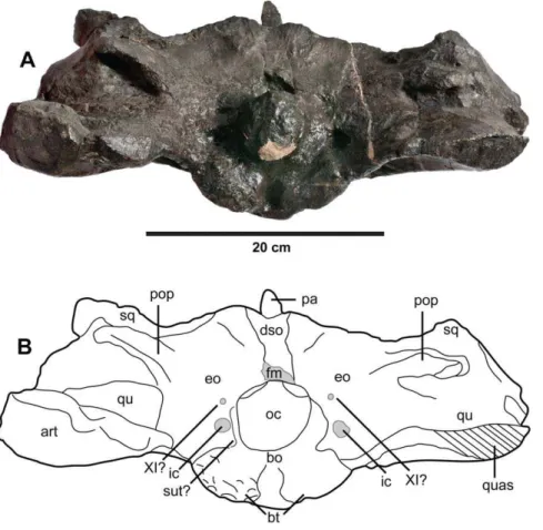
The Lost ‘Pliosaurus trochanterius’ Skull and Mandible In 1866, Mansel-Pleydell presented numerous marine reptile fossils he discovered at Kimmeridge Bay to the British Museum. One of these specimens, a mandible and an incomplete skull (braincase with part of the supratemporal arches), was from an individual even larger than the ‘‘D.’’manseliiholotype (NHMUK PV OR40103). As with NHMUK PV OR40103, this specimen (NHMUK PV R1089; Figs. 14, 15, 16, 19, 20, 21) was initially considered to belong to a pliosaurid plesiosaur. It was originally described by Owen [54] within his speciesPliosaurus trochanterius. However, Owen never provided evidence to show that NHMUK PV R1089 belonged within the species. There are no overlapping elements, as Owen [55], [56] erectedPliosaurus trochanteriusbased Figure 14.Plesiosuchus manselii, referred specimen NHMUK PV
R1089. Braincase and intertemporal bar in: (A) dorsal view and (B) ventral view. Abbreviations: bt, basal tubera; crB, crest B; fr, frontal; ic, foramen for the internal carotid artery; oc, occipital condyle; pa, parietal; pop, paroccipital process; qu, quadrate; quas, quadrate articular surface; sq, squamosal.
Figure 15.Plesiosuchus manselii, referred specimen NHMUK PV R1089.Close-up on the articular surface of the right quadrate. Abbreviations: bt, basal tubera; crB, crest B; oc, occipital condyle; qu, quadrate; quas, quadrate articular surface; sq, squamosal.
doi:10.1371/journal.pone.0044985.g015
Figure 16.Plesiosuchus manselii, referred specimen NHMUK PV R1089.Braincase in occipital view, (A) photograph and (B) line drawing (filled grey areas represent foramina). Abbreviations: XI?, foramen for cranial nerve XI?; art, articular; bo, basioccipital; bt, basal tubera; dso, depression for the supraoccipital; eo, exoccipital; fm, foramen magnum; ic, foramen for the internal carotid artery; oc, occipital condyle; pa, parietal; pop, paroccipital process; qu, quadrate; quas, quadrate articular surface; sq, squamosal; sut?, suture?.
on an isolated femur (according to Brown [57] it is actually a humerus). Additionally, they come from different localities: Pliosaurus trochanteriusis from the early Tithonian of Shotover Hill, Oxfordshire, England, whereas NHMUK PV R1089 is from the early Tithonian of Kimmeridge Bay, Dorset. Eudes-Deslong-champs ([58]:329) quickly demonstrated the crocodylian affinities of NHMUK PV R1089, and referred it to the metriorhynchid genus Metriorhynchus, as did Woodward ([53]:502). Lydekker ([3]:104), however, believed the specimen belonged to the teleosaurid speciesMachimosaurus mosae, although he noted that it lacked the anterior transverse expansion of the mandibular symphysis seen in teleosaurids. Tarlo [59] considered the specimen to be crocodylian, while Buffetaut [60] considered it to be a large metriorhynchid, probably Dakosaurus. More recently, Benton & Spencer [61] figured NHMUK PV R1089 as the plesiosaur
Colymbosaurus trochanterius, while Vignaud [51] referred the
speci-men to the teleosaurid speciesMachimosaurus mosae.
Following Hua et al. [62], there are two valid species of
Machimosaurus in the Late Jurassic of Europe: the type species
Machimosaurus hugii(early–late Kimmeridgian of France, Portugal
and Switzerland) andM. mosae(latest Kimmeridgian of France). Comparing NHMUK PV R1089 to the mandibles ofM. hugii[63]
and M. mosae [62] clearly shows it does not belong to
Machimosaurus. Both species of Machimosaurus possess external
mandibular fenestrae and an anterior transverse expansion of
the mandibular symphysis, whereas NHMUK PV R1089 lacks both features. Additionally, both species ofMachimosauruslack the prearticular, which is present in NHMUK PV R1089. The absence of the external mandibular fenestrae is a metriorhynchid apomorphy, while the anterior transverse expansion of the mandibular symphysis and loss of the prearticular are teleosaurid apomorphies [5], [10] (Hua pers. comm. 2011). In addition, NHMUK PV R1089 has far fewer dentary alveoli than either
Machimosaurusspecies: 13 compared to their 19–25. This extreme
reduction in dentition is observed in geosaurine metriorhynchids (Table 1).
Furthermore, the dentary interalveolar spaces of NHMUK PV R1089 are very small, far smaller than in both Machimosaurus species, and most thalattosuchians. In teleosaurids [5], [62–64], metriorhynchine metriorhynchids [5], [64], [65] and basal geosaurine metriorhynchids (‘‘Metriorhynchus’’ brachyrhynchus and ‘‘Mr Leeds’ specimen’’ [2], [5]) the dentary symphyseal interal-veolar spaces are variable in size, ranging from being larger than the proceeding and preceding alveolus, to being half the size. The extreme reduction in dentary symphyseal interalveolar distances in NHMUK PV R1089 (always being less than a quarter the length of the immediate alveoli, and usually even smaller; Fig. 21) is characteristic of Geosaurini metriorhynchids (Dakosaurus maximus: Figs. 2, 3;Torvoneustes carpenteri[18]). This reduction in symphyseal interalveolar spaces means the typical thalattosuchian ‘diastema’ between dentary alveoli 4 and 5 is absent [2], [5], [58], [62], [64], [65]. Curiously, both the extreme reduction in symphyseal interalveolar spaces and the absence of the D4–D5 ‘diastema’ are observed in the holotype of the geosaurine metriorhynchid
Suchodus durobrivensis(NHMUK PV R1994: a mandibular
symphy-sis). These unusual two characteristics were first highlighted by Lydekker ([66]:287); moreover, he noted there was ‘‘marked resemblance between’’ the Suchodus durobrivensis holotype and NHMUK PV R1089. This has led two of us (MTY and LS) to begin re-examining the NHMUK Callovian metriorhynchids to determine whether the synonymy of Suchodus durobrivensis and ‘‘Metriorhynchus’’brachyrhynchusis valid [2], [14]; as such, herein we do not follow Young et al. [1] in referring ‘‘Metriorhynchus’’ brachyrhynchusto the genusSuchodus.
The occiput/braincase of NHMUK PV R1089 exhibits two metriorhynchid autapomorphies: 1) enlarged carotid artery foramina ventrolateral to the foramen magnum (apomorphy was confirmed through computed tomography scanning of an Oxfordian braincase [67]); and 2) the trigeminal fossa is developed mainly posterior to the trigeminal foramen [67]. As such, we can conclusively refer NHMUK PV R1089 to Metriorhynchidae, and by extension remove it fromMachimosaurus mosae.
Although we can refer NHMUK PV R1089 to Metriorhynch-idae, can we refer it to Dakosaurus/Plesiosuchus manselii? The surangulodentary groove in NHMUK PV R1089 and NHMUK PV OR40103a is deeply excavated and strongly developed on both elements (Figs. 17, 18, 19, 20). This morphology is only observed inD. maximus(SMNS 8203, SMNS 82043; Figs. 2, 6) and
D. andiniensis [19], [20] among metriorhynchids (and to some
extent in the more poorly preserved specimens ofGeosaurus giganteus [10]). This therefore allows us to assign it to Geosaurini. We can exclude both the holotype of D./P. manselii and NHMUK PV R1089 from D. maximus because they lack the sharp dorsal inclination of the ventral margin of dentary and the raised alveolar margins of the posterior dentary alveoli that are characteristic of this species (see below; compare Fig. 2 with Figs. 17, 18, 21). Furthermore, both the holotype ofD./P. manseliiand NHMUK PV R1089 share a cranial apomorphy: distal articular surface of the quadrate is not divided into two condyles by a sulcus (Fig. 15), Figure 17. Plesiosuchus manselii, holotype NHMUK PV
OR40103a.Mandible, with the left ramus is lateral view and the right ramus in medial view.
doi:10.1371/journal.pone.0044985.g017
Figure 18. Plesiosuchus manselii, holotype NHMUK PV OR40103a.Mandible, right ramus in lateral view.
which is an unusual feature among archosaurs. This suite of shared derived characters (see Table 2) allows us to refer NHMUK PV R1089 toDakosaurus/Plesiosuchus manselii.
Methods Ethics Statement
We had permission to look at, and photograph, the relevant collections in the MUJA, NHMUK and SMNS. The curators whose remit includes fossil crocodylians from the MUJA (JIR-O), NHMUK (LS) and SMNS (RS) are co-authors on this manuscript. None of these specimens were purchased, donated or loaned as part of this study.
Phylogenetic Analyses
We undertook two phylogenetic analyses to assess the evolu-tionary relationships ofDakosaurus andPlesiosuchuswithin Thalat-tosuchia. This analysis is the latest in a series of iterative analyses, beginning with the publication of Young & Andrade [10], in which our research group (led by MTY) has added new character data and newly-described taxa to a growing discrete character dataset. The analysis presented here is a revised version of the most recent analysis by our group, that published by Younget al. [2]. See the online supplementary material for sources of character coding and the character list (Text S1) and the character scores (Text S2). Here, craniomandibular and dental characters make up 73% (175/240) of the character list, while the post-cranial characters contribute 27% (65/240). The analysis presented here differs from that of Younget al. [2] in that:
1. 39 new or revised characters have been added.
2. We have revised the character codings ofDakosaurus maximus
and Plesiosuchus manselii based on first-hand examination and
the anatomical revisions presented in this monograph. 3.Erpetosuchus grantiis no longer included in the analysis, with the
outgroup taxon now being Postosuchus kirkpatricki. Recent comprehensive phylogenetic work on the relationships of basal archosaurs [68], [69] strongly supports the close relationship of
Postosuchus(and related rauisuchians) with crocodylomorphs but
does not corroborate previous hypotheses [70], [71] that
Erpetosuchus, a taxon known only from highly incomplete
material, is a close crocodylomorph outgroup. Additionally, the fragmentary nature of knownErpetosuchusspecimens results in a high amount of missing data when this taxon is scored in phylogenetic analyses, which is not a desired characteristic of an outgroup taxon used to root phylogenetic trees.
4. We have expanded the non-metriorhynchid taxon selection substantially, with eight more teleosaurids and 16 non-thalattosuchians, resulting in 73 total taxa.
5. The putativeDakosaurusspecimens from Mexico were removed due to their poor preservation and the fact that we cannot be sure they belong to the same taxon.
6. The metriorhynchidPurranisaurus potenswas removed, as its type specimen is currently under re-description by one of us (MF) with colleagues. This redescription will result in a more confident set of character scores for this taxon.
7. Finally, we recoded ‘‘Metriorhynchus’’ brachyrhynchus due to the uncertainty of whether the Suchodus durobrivensisholotype is a junior synonym of the former (see above discussion regarding dentary interalveolar spaces). This is currently being investi-gated by two of us (MTY and LS).
The two phylogenetic analyses were carried out using TNT v1.1 (Willi Hennig Society Edition) [72]. They differed in that: 1) the first analysis had all characters treated as unordered, while 2) in the second analysis 40 multi-state characters were treated as ordered (transformational sequences). The 1st, 7th, 8th, 10th, 13th, 25th, 38th, 39th, 42nd, 43rd, 47th, 50th, 56th, 58th, 69th, 86th, 87th, 96th, 126th, 132nd, 133rd, 151st, 152nd, 154th, 156th, 166th, 179th, 181st, 182nd, 183rd, 184th, 198th, 202nd, 214th, 218th, 225th, 228th, 230th, 231st and 237th characters are ordered in the second analysis. Other than the ordering of those 40 characters the analyses were identical.
Tree-space was searched using the advanced search methods in TNT, namely: sectorial search, tree fusion, ratchet and drift, for 1,000 random addition replicates. The default settings for the advanced search methods were changed to increase the iterations of each method per replicate: now 100 sectorial search drifting cycles, 100 ratchet iterations, 100 drift cycles and 100 rounds of tree fusion per replicate. This tree-space search procedure was repeated for five different random start seeds. All characters were treated with equal weight. Character polarity was determined with reference to a pre-defined non-crocodylomorph outgroup taxon
(Postosuchus kirkpatricki). Nodal support was evaluated using
non-parametric bootstrapping [73] with 1000 replicates, using TBR searching.
Results
Systematic Palaeontology
Superorder Crocodylomorpha Hay, 1930 [74] (sensu Walker, 1970) [75].
Infraorder Thalattosuchia Fraas, 1901 [76] (sensu Young & Andrade, 2009) [10].
Family Metriorhynchidae Fitzinger, 1843 [77] (sensuYoung & Andrade, 2009) [10].
Subfamily Geosaurinae Lydekker, 1889 [78] (sensu Young & Andrade, 2009) [10].
Tribe Geosaurini Lydekker, 1889 [78] (sensuCau & Fanti, 2011) [12].
Type genus. GeosaurusCuvier, 1824 [50].
Diagnosis. Metriorhynchid crocodylomorphs with the fol-lowing unique combination of characters (autapomorphic charac-ters are indicated by an asterisk): high absolute tooth-crown apicobasal length (in some species exceeding 12 centimetres)*; contiguous row of true denticles along the mesial and distal carinae of the teeth*; bicarinate serrated dentition; the inflexion point of the prefrontal lateral margin (in dorsal view) is directed posteriorly at an angle less of 70 degrees or less from the anteroposterior axis of the skull*; acute angle between the medial and the posterolateral processes of the frontal; supratemporal fenestrae enlarged, in dorsal view the posterolateral corner extends further posterior to the intertemporal bar*; all dentary interalveolar distances are very small (always less than a quarter the length of the immediate alveoli)*; dentary tooth-row is ventrally displaced relative to the jaw joint; humerus shaft greatly reduced, contributing less than 25% of total humeral length.
Phylogenetic definition. The least inclusive clade consisting of Geosaurus giganteus, Dakosaurus maximus and Torvoneustes carpenteri (sensuCau & Fanti [12]).
DakosaurusQuenstedt, 1856 [32].
Type species. Geosaurus maximus Plieninger, 1846 [23] (fol-lowing Recommendation 67B of the ICZN Code).
Etymology. ‘‘Biter lizard’’. Dakos (da´xoz from Quenstedt [33]:785) is derived from the Ancient Greek ‘to bite’ (da´xuv). Furthermore, Quenstedt ([39]:182) places da´xoz in parentheses
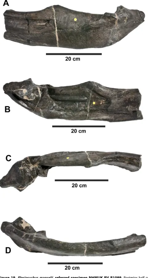
beside Beisser, the German for biter. While –saurusis the latinised version ofsauros, the Ancient Greek for lizard. Note that Wilkinson et al. [18] incorrectly translatedDakosas meaning ‘‘tearing’’. Figure 19.Plesiosuchus manselii, referred specimen NHMUK PV R1089.Posterior half of the left mandibular ramus in: (A) lateral view, (B) medial view, (C) dorsal view and (D) ventral view.
Geological range. Upper Kimmeridgian to lower Berriasian. Geographical range. Europe (England, France, Germany and Switzerland) and South America (Argentina).
Geographical note. Possible Dakosaurus remains have been found at Khoroshevskii Island, in the Volga region of Russia [79]. They consist of a vertebra and metatarsal from upper Tithonian or lower Berriasian deposits. The possible referral to Dakosaurus is presumably due to their large size. However, as there are currently no vertebral or metatarsalDakosaurusautapomorphies this referral cannot be substantiated. The taxonomic affinities of the two fragmentary skull specimens from the Kimmeridgian of Mexico are in question due to newly discovered metriorhynchine specimens from the early Tithonian of Mexico (see discussion; [80]).
Spelling. Quenstedt [31], [32] used the spelling, Dakosaurus for the genus. However, there has been a question around the transliteration of the Greek letterxinto the Latin letterscandk. During the latter half of the 19thcentury and the first half of the 20thcenturyDacosauruswas the predominant spelling [3–5], [36], [40], [41], [44], [81], [82]. From the latter half of the 20thcentury onwards the original spelling, Dakosaurus, became dominant [1], [2], [10–20], [22], [26–28], [46], [60]. The first use of the ‘‘c’’ spelling was by Sauvage ([44]:380), while Lydekker ([3]:92) was the first to explicitly state that the original spelling had been amended
toDacosaurus. However, under the ICZN Code (Article 32.5) an
incorrect original spelling cannot be corrected solely on the grounds that it was incorrectly transliterated or latinized. As such, the genus is properly speltDakosaurus.
Emended diagnosis. Metriorhynchid crocodylomorph with the following unique combination of characters (autapomorphic characters are indicated by an asterisk): large robust teeth, with moderate to strong mediolateral compression; carinae formed by a keel and true macroscopic denticles (macroziphodonty, all dimensions exceed 300mm)*; tooth enamel ornamentation is inconspicuous, visible under SEM and comprising an anastomosed pattern; rostrum proportionately short (brevirostrine, less than 55% of basicranial length), dorsoventrally tall with a convex dorsal margin (oreinirostral)*, and in dorsal view has a distinctly wide and blunt, ‘‘bullet’’ shape (amblygnathous)*; separation between premaxilla and nasal half, or less than half, the midline length of the premaxilla; aligned set of large neurovascular foramina on the maxilla extending posteroventrally from the preorbital fossa (not homologous to the archosaurian antorbital fenestra [8], [9]) *; in dorsal view, the lateral margins of the prefrontals have an inflexion point directed posteriorly at an angle of approximately 50 degrees from the anteroposterior axis of the skull*; acute angle (between 60 and 45 degrees depending on species) between the medial and the posterolateral processes of the frontal; the supratemporal fossae (intratemporal flange) reach the minimum interorbital distance; ventral margin of dentary sharply rises dorsally at the anterior tip*; very short mandibular symphysis (only one third of dentary tooth-row adjacent to the symphysis)*; surangulodentary groove has a well-developed foramen at the dentary terminus*; surangular anteroposteriorly short, terminates posterior to the anterior margin of the orbit.
Dakosaurus maximus(Plieninger, 1846) [23] Quenstedt, 1856 [32].
1843Megalosaurussp. –Quenstedt, p. 493. [30]
Figure 20.Plesiosuchus manselii, referred specimen NHMUK PV R1089.Posterior half of the right mandibular ramus in: (A) lateral view, (B) medial view, (C) dorsal view and (D) ventral view.
doi:10.1371/journal.pone.0044985.g020
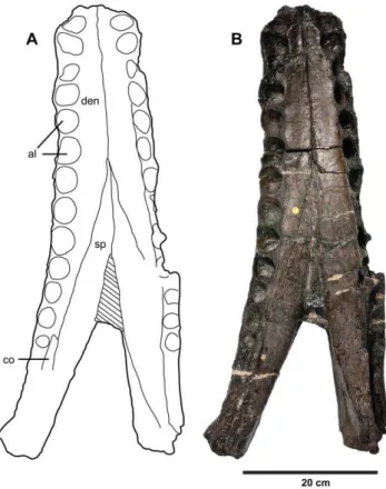
Figure 21.Plesiosuchus manselii, referred specimen NHMUK PV R1089.Mandibular symphysis in dorsal view, (A) line drawing and (B) photograph. Abbreviations: al, alveolus; co, coronoid, den, dentary; sp, splenial.
v* 1846Geosaurus maximussp. nov. – Plieninger, p. 150, Taf. 3 Figure 2. [23]
v 1849Geosaurus maximusPlieninger – Plieninger, p. 252, Taf. 1 Figure 7. [29]
v 1852Megalosaurussp. –Quenstedt, p. 112, Taf. 8 Figure 4 [31] v* 1853 Liodon paradoxus sp. nov. – Wagner, p.263, Taf. 3 Figures 9–13. [35]
v 1856 Dakosaurus maximus (Plieninger) gen. et comb. nov. – Quenstedt, p. 131. [32]
v 1858Dakosaurus maximus(Plieninger) –Quenstedt, p. 785. [33] v* 1869Dakosaurus lissocephalussp. nov. – Seeley, p. 92. [42] * 1871Leiodon primaevumsp. nov. – Sauvage, p. 141. [43]
1873Dacosaurus primaevus(Sauvage) comb. nov. et unjust.emend.
– Sauvage, p. 380, pl. 7 Fig. 3–5. (sic) [44]
* 1881Teleosaurus suprajurensissp. nov. – Schlosser. [36] v 1885Dakosaurus maximus(Plieninger) –Quenstedt, p. 182. [39] v* 1885Dakosaurus gracilissp. nov. –Quenstedt, p. 184. [39] v 1888 Dacosaurus maximus (Plieninger) – Lydekker, p. 92–94, Figure 13. (sic) [3]
v 1902Dacosaurus maximus (Plieninger) – Fraas, p. 7, Fig. 1–2 Taf. 1, Taf. 2 Fig. 2–11, Taf 3–4 (sic) [4]
v 1902Dacosaurus paradoxus(Wagner) comb. nov. – Fraas, Tafel 2, Fig. 1, 12–13 (sic) [4]
v 1902Dacosaurus suprajurensis(Schlosser) comb. nov. – Fraas, p. 20 (sic) [4]
1925Dakosaurus maximus(Plieninger) – Huene, p. 600, plate 26
Figure 57. [83]
v 1973Dakosaurus maximus(Plieninger) – Steel, p. 42, Figure 18 (6, 11). [46]
v 2009Dakosaurus maximus(Plieninger) – Young & Andrade, p. 555, Fig. 5. [10]
v2009 Dakosaurus lissocephalus (Seeley) – Young & Andrade, p.
579. [10]
v 2010Dakosaurus maximus (Plieninger) – Young et al., p. 804, Fig. 4, 6. [1]
v2010 Dakosaurus lissocephalus(Seeley) – Younget al., p. 859. [1] Figure 22. Plesiosuchus manselii, holotype NHMUK PV
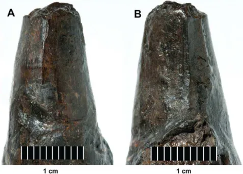
OR40103a. Isolated tooth crown in: (A) right lateral view, (B) left lateral view and (C) dorsal (apical) view.
Figure 23.Plesiosuchus manselii, holotype NHMUK PV OR40103a.Close-up on the carinae of an isolated tooth crown, (A) anterior carina and (B) posterior carina.
Torvoneustes: BRSMG Cd7203, BRSMG Ce17365
Geosaurus: BSPG AS-VI-1, NHMUK
PV OR37020, NHMUK PV R1229, NHMUK PV R1230
Plesiosuchus: NHMUK PV OR40103,
NHMUK PV OR40103a, NHMUK PV R1089
Dakosaurus: CAMSM J29419,
NHMUK PV OR20283, SMNS 8203, SMNS 82043, SMNS 10819
Dentition
Premaxillary alveoli count 3 3 3 3
Maxillary alveoli count 14a 14 14 13
Dentary alveoli count ? 13a 13 12
Dentary alveoli adjacent to symphysis
? 8a 9 4
Enamel ornamentation Intense ornamentation on both labial and
lingual surfaces. Basal half has apicobasally aligned ridges, apical half has pronounced anastomosed pattern
No conspicuous enamel ornamentation Ornamentation largely inconspicuous,
apicobasal ridges on labial and lingual surfaces but of low relief
No conspicuous enamel ornamentation
Facetting on labial surface No Yes, three apicobasal facets No No
Crown shape Conical Laminar/strongly compressed Robust, some compression Robust, some compression
Wear facets on mesial and distal margins
No No No Yes
Denticles Microscopic, but in apical half the
denticles are hard to distinguish from enamel ornamentation
Microscopic Microscopic Macroscopic
Cranium
Premaxillary lateral plates No No No Yes
Distance between premaxilla and nasal
,Equal to the midline length of the
premaxilla
Less than half the midline length of the premaxilla
,Equal to the midline length of the
premaxilla
Less than half the midline length of the premaxilla
Rostrum-to-skull roof dorsal margin
Concave Concave Concave Convex
Prefrontals in dorsal view: angle between the inflexion point on the lateral margin and the anteroposterior axis of the skull
,70 degrees ,70 degrees ,70 degrees ,50 degrees
Intratemporal flange anterior development
Does not reach minimum interorbital distance
Does not reach minimum interorbital distance
? Reaches minimum interorbital
distance
Palatine anterior development along the
midline
Maxillopalatine suture reaches as far anteriorly as the 9th maxillary alveoli
Comparison between the specimens suggests palatine reached level to either the 8th or 9th maxillary alveoli
Level to 4th maxillary alveoli Palatine not preserved, maxillae form
the secondary palate up to at least the 8th maxillary alveoli
Mandible
Dentary lateral and medial plates
? No No Two ridges either side of the dentary
alveoli (prominent between D6 to D12)
Mandibular lateral groove ( = surangulodentary groove)
No large foramen at dentary terminus No large foramen at dentary terminus No large foramen at dentary terminus Large foramen present at dentary
terminus
Re-Descriptio
n
of
Dakosaur
us
and
Plesiosuchus
ONE
|
www.ploson
e.org
19
September
2012
|
Volume
7
|
Issue
9
|
v 2010Dakosaurus maximus(Plieninger) – Andradeet al., p., Fig. 4. [11]
Holotype. Isolated tooth, the location of which is unknown and is presumed lost.
Etymology. ‘Greatest biter lizard’. From the Latin maximus, meaning largest/greatest.
Holotype locality and horizon. Schnaitheim, Baden-Wu¨rt-temberg, Germany. Mergelsta¨tten Formation.Hybonoticeras beckeri Sub-Mediterranean ammonite Zone, upper Kimmeridgian, Up-per Jurassic.
Neotype. SMNS 8203– incomplete skull and mandible (first suggested by Young & Andrade [10]).
Neotype locality and horizon. Staufen bei Giengen, Baden-Wu¨rttemberg, Germany. Mergelsta¨tten Formation. Hybonoticeras
beckeriSub-Mediterranean ammonite Zone, upper Kimmeridgian,
Upper Jurassic.
Designation of neotype. Herein we formally designate SMNS 8203 as the neotype ofDakosaurus maximus. In order to be in full accordance of Article 75 of the ICZN Code, in particular Article 75.3, we make the following statements:
1. This designation is made with the express purpose of clarifying the taxonomic status ofDakosaurus maximus.
2. Our statement of the characters that we regard as differenti-atingDakosaurus maximusfrom other taxa is given by the species diagnosis below.
3. The neotype can be recognised through both the description below and Figs. 2, 3, and 4.
4. The holotype (an isolated tooth) cannot be located and is presumed lost. The type was described in the 1846 [23], and there is no known documentation to suggest which institution the holotype was given to, assuming the specimen was curated in a scientific institution.
5. The holotype is an isolated tooth, from the description and figure given by Plieninger [23] show it was both robust and macroziphodont. As such, the neotype is consistent with what is known of the former name-bearing type.
6. While the neotype is not from the same locality as the holotype, both are from the same Sub-Mediterranean ammonite Zone. The two localities are little over 10 km from one another. 7. The neotype is the property of a recognized scientific
institution, SMNS, which maintains a research collection with proper facilities for preserving name-bearing types, and is accessible for study.
Geological range. Upper Kimmeridgian (A. eudoxus Sub-Boreal ammonite Zone) to lower Tithonian (H. hybonotum Sub-Mediterranean ammonite Zone).
Geographical range. Cambridgeshire, England; Pas-de-Ca-lais, France; Baden-Wu¨rttemberg & Bayern, Germany; Canton Solothurn, Switzerland.
Referred specimens. NHMUK PV OR33186, NHMUK PV OR35766, NHMUK PV OR35835, NHMUK PV OR35836, NHMUK PV OR35837, SMNS 51494, SMNS 55420, SMNS 80148: isolated teeth from Schnaitheim (H. beckeri Sub-Mediter-ranean Zone); SMNS 81793: isolated tooth from Nusplingen, Baden-Wu¨rttemberg, Germany (H. beckeri Sub-Mediterranean Zone); SMNS 10819a, b: broken and dorsoventrally compressed skull from Sontheim an der Brenz, Baden-Wu¨rttemberg, Ger-many; SMNS 82043: right mandibular ramus in lithographic limestone, from Painten, Bayern, Germany; CAMSM J29419: incomplete dorsoventrally crushed skull (holotype ofD. lissocepha-lus) from Ely, Cambridgeshire, England, Lower Kimmeridge Clay Formation (A. eudoxusSub-Boreal Zone); NHMUK PV OR20283:
Table 1. Cont. Torvoneustes : BRSMG Cd7203, BRSMG Ce17365 Geosaurus : BSPG AS-VI-1, NHMUK PV OR37020, NHMUK PV R1229, NHMUK PV R1230 Plesiosuchus : NHMUK PV OR40103, NHMUK PV OR40103a, NHMUK PV R1089 Dakosaurus : CAMSM J29419, NHMUK PV OR20283, SMNS 8203, SMNS 82043, SMNS 10819 valign="top"> Reception pits along the dentigerous bones No Yes, on the lateral margin of the dentary and m edial surface of maxilla No Yes, between the alveoli o f the upper and lower jaw dentigerous bones Postcrania Humerus d eltopectoral crest shape Prominent convex curve that is continuous with the p roximal articular surface ? P rominent and subtriangular, easily distinguished from proximal articular surface Prominent and subtriangular, easily distinguished from p roximal articular surface Femur posteromedial tuber enlargement Does not encompass the entire medial femoral surface in dorsal view Does not encompass the entire medial femoral surface in dorsal view Encompasses the entire medial femoral surface in dorsal view, creating a pronounced medial deflection o f femoral h ead ? Ischium b lade Posterior-edge is a convex curve Posterior-edge is a convex curve ? Posterior-edge is subrectangular The hunmerus of Geosaurus in the Kimmeridgian-Tithonian is unknown, but the Valanginian-Hauterivian species Geosaurus lapparenti has a peculiar humerus morphology that does not conform with either the Torvoneustes , Plesiosuchus or Dakosaurus humeral shapes [40], [41].
aEstimate. doi:10.1371/journal.pone.
0
an isolated tooth, also from Ely, England; NHMUK PV OR32414, SMNS 57210: isolated teeth from Boulogne-sur-Mer, Pas-de-Calais, France, Argiles de Chaˆtillon Formation (A.
autissiodorensis Sub-Boreal Zone); SMNS 56999: partial maxilla
also from Boulogne-sur-Mer, France; NMS 7009: isolated tooth from Canton Solothurn, Switzerland, Reuchenette Formation (upper Kimmeridgian); JME-SOS4577, JME-SOS2535: isolated teeth from Schernfeld, Bayern, Germany, Solnhofen Formation (lower Tithonian;H. hybonotumSub-Mediterranean Zone).
Diagnosis. Metriorhynchid crocodylomorph within the
ge-nusDakosauruswith four autapomorphic characters: 1) wear facets
on the mesial and distal edges of the crown that obliterate the carinae; 2) thin lamina of bone projecting from the lateral alveolar margin of the premaxilla (‘‘premaxillary lateral plates’’); 3) maxilla is strongly ornamented, with most of the element covered in long deep grooves and long raised ridges orientated to the long axis of the skull, but with the alveolar margin largely smooth; 4) in the posterior half of the dentary, there are laminae of bone projecting from the lateral and medial dentary alveolar margins (‘‘dentary lateral and medial plates’’). Note that the preservation of
Dakosaurus andiniensis makes it difficult to assess whether it also
possesses characteristics one and two.
Body length estimate. The largest known specimen of
Dakosaurus maximusis the isolated mandible SMNS 82043 (Fig. 6),
which is 87.5 cm in length. Using the ratio of basicranial length to mandibular length in ‘‘Metriorhynchus’’ brachyrhynchus as a guide (NHMUK PV R3804: the most three-dimensionally preserved NHMUK specimen with a complete skull and mandible; mandible length = 80.9 cm, basicranial length = 76.8 cm) and assuming that the basicranium and mandibles of D. maximus scale in the same
proportions, SMNS 82043 is estimated as having a basicranial length of 83.1 cm. This gives a total body length estimate of 4.49 m, using the Young et al. [14] equations. This is slightly greater than the body length given in Younget al. [14], however that was based on an estimated length of the neotype SMNS 8203, which they found to be 4.28 m long.
Description and Comparisons
Skull: general comments. Many cranial and mandibular bones are preserved in the neotype (SMNS 8203; Figs. 2, 3, 4), referred cranial elements (SMNS 10819, Fig. 5; SMNS 56999, Fig. 7) and the referred mandible (SMNS 82043, Fig. 6), but several other bones are not preserved and are thus unknown inD.
maximus. These include: jugals, lacrimals, frontal, parietal,
post-orbitals, squamosals, quadrates, braincase, occiput, pterygoids and ectopterygoids. Overall, the skull has a shape very similar to that of D. andiniensis: they both have a short, broad ‘‘bullet’’-shaped snout (amblygnathous), which is very robust and has a convex upper margin (oreinirostral) (see Fig. 3).
Premaxilla and external nares. The premaxilla bears three alveoli, as with all other metriorhynchids [4], [5], [10]. The ornamentation on the lateral surface of the premaxilla is composed of numerous large elliptic pits, and the bone is slightly convex laterally (SMNS 8203, Figs. 2, 3, 4; SMNS 10819a, Fig. 5A). The premaxillae completely enclose the external nares, as in all thalattosuchians with the exception ofCricosaurus macrospondylus(in which the maxilla also contributes [84]). Along the posterior margin of the premaxilla, the posterodorsal process contacts the anterior margin of the maxilla. This suture forms a broad ‘U’-shape in dorsal view (much like D. andiniensis [19], [20]), rather Table 2.Table of diagnostic characters for Metriorhynchidae, and various subclades, for the three Kimmeridge Bay NHMUK PV specimens.
Clades Diagnostic characters OR40103 OR40103a R1089
Metriorhynchidae Three teeth per premaxilla Yes ? ?
Enlarged carotid artery foramina Yes ? Yes
Trigeminal fossa developed mainly posterior to the trigeminal foramen ? ? Yes
No external mandibular fenestrae ? Yes Yes
Coronoid process on mandible ? Yes Yes
Humerus flattened, shaft contributes less than 40% total humeral length ? Yes ?
‘‘Mr Leeds’ specimen’’+ Geosaurini
Ventral displacement of the dentary tooth row, such that the coronoid process is located considerably above the plane of the tooth row
? Yes Yes
Coronoid process ventral to both the retroarticular process and glenoid fossa ? Yes Yes
Fourteen or fewer teeth per dentary ramus ? Yes Yes
Geosaurini (but characters unknown in ‘‘Mr Leeds’ specimen’’)
Fourteen or fewer teeth per maxilla Yes ? ?
Posterior expansion of supratemporal fenestrae in dorsal view (reaching at least the supraoccipital, but can even exceed the occipital condyle)
Yes ? Yes
Geosaurini Denticulated bicarinate dentition Yes Yes ?
Dentary symphyseal interalveolar spaces are very small (less than half the size of the immediate alveoli)
? ? Yes
Deeply excavated surangulodentary groove ? Yes Yes
Humerus is short and stocky, deltopectoral crest contacts proximal articular surface
? Yes ?
Plesiosuchus manselii Tooth enamel ornamentation: apico-basally aligned ridges of low-relief Yes Yes ?
Quadrate distal articular surface not separated into two protuberances by a sulcus Yes ? Yes
Note that both NHMUK PV OR40103 and NHMUK PV OR40103a are the holotype ofPlesiosuchus manselii. Note: the ‘‘Mr Leeds’ specimen’’ is a new genus and species; however the paper establishing these names is still in press [2].
than a posteriorly pointed ‘V’-shape (such as in Metriorhynchus superciliosus, Gracilineustes leedsi and ‘‘Metriorhynchus’’ brachyrhynchus [5]). As with almost all thalattosuchians, there is no premaxilla-nasal contact [5], as these bones are separated by the maxilla. The palatal surface of the premaxilla inD. maximusis unknown.
Along the lateral margin of the premaxilla there is a thin lamina of bone that covers the basal portion of the teeth (SMNS 8203; Fig. 4A, 4B). This morphology is somewhat similar to the ‘lateral plates’ observed in sauropod dinosaurs (e.g.Diplodocus longusCM 11161). Finite element analysis modelling of this skull by Younget al.[85] found that, regardless of the feeding behaviour simulated, high stresses occurred at the tooth bases and the ‘lateral plates’ during feeding. These results support the hypothesis that ‘lateral plates’ help to dissipate feeding-induced stresses acting on the bases of adjacent teeth [85], which we hypothesise was their function inD. maximus.Dakosaurus/Plesiosuchus manselii(NHMUK PV OR40103) lacks these structures (as do all other known metriorhynchids [5], [64]), while the state of preservation makes determining this morphology difficult inDakosaurus andiniensis[20]. Therefore, we regard them as an autapomorphy ofD.maximus, but note that future discoveries may reveal that they are more widely distributed amongDakosaurusspecies.
In Dakosaurus maximus there is a single, anterodorsally facing
external naris (Figs. 2, 3, 5). This condition is also seen in most other metriorhynchids, such as Dakosaurus andiniensis [19], [20],
Metriorhynchus superciliosus(e.g. NHMUK PV R6859, NHMUK PV
R6860), Gracilineustes leedsi (e.g. NHMUK PV R3014, NHMUK PV R3015) and ‘‘Metriorhynchus’’ brachyrhynchus (NHMUK PV R3804). The members of the subclade Rhacheosaurini have a different morphology, in which the naris is divided by a premaxillary septum and is anterodorsally and laterally oriented (e.g.Rhacheosaurus gracilisNHMUK PV R3948; Cricosaurus suevicus SMNS 9808).
Maxilla. The maxillae are similar to those of Dakosaurus andiniensis, as they are noticeably short, high and subtriangular in lateral view [19], [20]. One difference is that the maxillae of
Dakosaurus maximusare not as high dorsoventrally (compare Figs. 1,
2). Gaspariniet al. [19] compared the ratio of snout height to snout length among various crocodylomorphs, and they found thatD.
maximus had a ratio of 0.15, whereas D. andiniensishad an even
greater ratio of 0.36. This was in marked contrast to other thalattosuchians, as longirostrine species had a ratio of 0.04–0.05 (e.g. Steneosaurus bollensis, Pelagosaurus typus and Cricosaurus
arauca-nensis) while mesorostrine metriorhynchids had a ratio of 0.08–0.09
(Metriorhynchus superciliosusand ‘‘Metriorhynchus’’casamiquelai). The maxillae ofD. maximusbear 13 alveoli (SMNS 8203, Fig. 2) [4]. Like the premaxillae, the maxillae are slightly convex laterally. The ornamentation of the lateral surface inDakosaurus maximusis distinctive, as it noticeably differs across the element (SMNS 8203, Figs. 2, 3, 4; SMNS 10819a, Fig. 5; SMNS 56999, Fig. 7). Near the premaxilla-maxilla suture, the ornamentation is very similar to that on the premaxilla (numerous large elliptical pits). On most of the element, and in particular closer to the maxillary midline and maxillonasal suture, the surface is covered in long deep grooves and long raised ridges orientated parallel to the long axis of the skull. Approaching the alveolar margin, the ornamentation becomes more subtle, composed of ridges of low-relief arranged in an anastomosed pattern, creating a fabric of crests over the surface. Almost all of the maxillary foramina exit out on to the anastomosed region of the maxilla. The maxillae ofD. andiniensis [20], Torvoneustes carpenteri [18] Cricosaurus schroederi and C. araucanensis(see Figure 5 in [10]), andGeosaurus giganteus [10] are largely smooth, with elliptical pits that are shallow and fairly indistinct.
Along the dorsal midline of the skull the left and right maxillae meet at a long suture, and terminate at the anterior margin of the nasal. The maxillonasal suture begins at the skull midline and forms an anteriorly pointed ‘V’-shape, as is also the case in D.
andiniensisand other metriorhynchids [5], [10], [19], [20], [64].
With the jugals and lacrimals either missing or not preserved in all specimens of D. maximus, the nature of their contact with the maxilla cannot be determined. Similarly, the contribution the maxilla made to the preorbital fossa is unknown.
The alveolar margin of the maxilla is poorly preserved in the neotype (SMNS 8203, Fig. 4B). As such, the presence or absence of ‘lateral plates’, like those seen on the premaxilla, is unknown. In the referred specimen SMNS 10819a (Fig. 5), the alveolar margin is also partially damaged, although the medial section of the maxilla does not seem to exhibit the ‘plates’. In palatal view, the maxillae of the neotype (SMNS 8203) are very poorly preserved. However, in SMNS 10819a the maxillae suture along the midline forming part of the secondary palate (Fig. 5B, 5C). The maxillopalatine suture is not preserved in any specimen. However, the midline terminus of the maxillopalatine suture must have been posterior to the eight anterior maxillary alveoli; as those teeth are preserved in SMNS 10819. This is comparable to other Geosaurini genera, except Plesiosuchus (i.e. Dakosaurus/Plesiosuchus
manselii), where the maxillopalatine suture terminates level to the
fourth maxillary alveolus (Table 1).
Nasals. The nasals are large, paired, unfused elements (Figs. 2, 3). In dorsal view they are subtriangular in shape and broad, like in all thalattosuchians [5]. Along the midline the dorsal surface of the nasals is deeply trenched, with a steep longitudinal depression (Fig. 3B), a characteristic shared by all metriorhynch-oids [5], [64] (e.g. Pelagosaurus typus NHMUK PV OR32599;
Teleidosaurus calvadosii NHMUK PV R2681; Eoneustes gaudryi
NHMUK PV R3353; Metriorhynchus superciliosus NHMUK PV R6859, NHMUK PV R6860; Gracilineustes leedsi NHMUK PV R3014, NHMUK PV R3015; ‘‘Metriorhynchus’’ brachyrhynchus NHMUK PV R3804). The anterior margin forms an acute angle along its border with the maxilla. Most of the dorsal and lateral surfaces of the nasals are well ornamented, with a pitted pattern. This is in contrast with other species in Geosaurini, which have nasals that are largely smooth (D. andiniensis[19], [20];Torvoneustes carpenteri [18]; Geosaurus giganteus and G. grandis [10]; Plesiosuchus
manseliiNHMUK PV OR40103).
Although the frontal and lacrimals are missing, and the prefrontals are poorly preserved, it is possible to determine where these bones would have contacted the nasals by using the well preserved skull ofD. andiniensis as a guide [19], [20]. Along its posterior margin, the nasals would have contacted the frontal and prefrontals. The two dorsoposterior processes would have contacted the frontal medially, and the prefrontals laterally. Between the dorsoposterior and lateroposterior processes, the nasal would have contacted the prefrontals. Ventral to the lateroposterior processes the nasal would have contacted the lacrimal and contributed to the preorbital fossa margin. The presence of distinct nasal lateroposterior processes is a metrior-hynchid apomorphy (see Young et al. [1]: Figs 4A, 6 for a reconstruction of Teleidosaurus calvadosii and a photograph of
Eoneustes gaudryirespectively, as these basal metriorhynchoids lack
these processes).