J of Evolution of Med and Dent Sci/ eISSN- 2278-4802, pISSN- 2278-4748/ Vol. 4/ Issue 26/ Mar 30, 2015 Page 4456
MULLERIAN DUCT ANOMALIES: A STUDY ON ANATOMICAL BASIS WITH
EMBRYOLOGICAL ASPECTS AND ITS CLINICAL SIGNIFICANCE IN SOUTH
INDIAN POPULATION - MADURANTHAGAM REGION
T. L. Anbumani1, S. Anthony Ammal2, A. Thamarai Selvi3, T. L. Selvakumari4
HOW TO CITE THIS ARTICLE:
T. L. Anbumani, S. Anthony Ammal, A. Thamarai Selvi, T. L. Selvakumari. Mullerian Duct Anomalies: A Study on Anatomical Basis with Embryological Aspects and Its Clinical Significance in South Indian Population -Maduranthagam Region . Journal of Evolution of Medical and Dental Sciences 2015; Vol. 4, Issue 26, March 30; Page: 4456-4463, DOI: 10.14260/jemds/2015/644
ABSTRACT: AIMS & OBJECTIVES: Mullerian duct anomalies (MDAs) are rare, but it can be a treatable form of infertility, affecting approximately 1% to 5% of women in general population and the rate increases in women with poor reproductive outcomes. The purpose of this study is to share our experience in the prevalence of mullerian duct anomalies and its subtypes among women with poor reproductive outcomes in maduranthagam region - South India, and also to discuss the embryological basis of these anomalies with its clinical significance. MATERIALS AND METHODS:
This study is a cross-sectional study based on secondary data that is obtained from medical records of our institution from October 2009 to December 2014. A total of 5228 patients had undergone two dimensional ultrasound for various complaints like infertility, recurrent abortions, polymenorrhea etc., out of which, 512 patients had history of recurrent abortions and 1946 patients had history of infertility. Hysterosalpingogram was done on patients who were diagnosed to have congenital uterine anomaly on 2-D USG. RESULTS: 232 patients were diagnosed to have mullerian duct anomalies. 15.8% (n=81) of patients with history of recurrent abortions had congenital uterine anomaly and 7.5% of patients (n=146) in the infertile population had congenital uterine anomalies. The detailed description of subtypes of mullerian duct anomaly is given in the article. CONCLUSION:
The role of imaging is to help detect, classify and guide surgical management of uterine anomalies. In this present study, the prevalence of congenital uterine anomaly is more in patients with history of recurrent abortion (15.8%), when compared to infertile population (7.5%) in maduranthagam region. This study is based on rural sector population. Although MRI is the modality of choice for diagnosing mullerian duct anomalies, the role of 2-D USG and HSG cannot be neglected in diagnosing the uterine anomalies, especially if non affordable poor sector people are considered. This article further emphasizes on the embryological reasons behind these uterine anomalies.
KEYWORDS: Mullerian duct anomalies, 2-D ultrasound, hysterosalpingogram, infertility, recurrent abortions.
J of Evolution of Med and Dent Sci/ eISSN- 2278-4802, pISSN- 2278-4748/ Vol. 4/ Issue 26/ Mar 30, 2015 Page 4457 uterine anomalies. The prevalence of mullerian duct anomalies varies according to diagnostic techniques used and the population studied. (Chan et al, 2011b, Grimbizis et al, 2001, Saravelos et al, 2008a). In the general population, mullerian duct anomalies have been shown to affect 4.3–6.7% of the population, in the infertile population, the prevalence of mullerian duct anomaly lies in the range of 3.4% - 8.0% and, in women with history of recurrent abortions, this figure has been reported to be 12.6–18.2% (Chan et al, 2011b, Grimbizis et al, 2001, Saravelos et al, 2008a). The purpose of this study is to assess the diagnosis of mullerian duct anomalies in the infertile population and in women having recurrent abortions, in madurantagamis region of Tamil Nadu, using 2-D Ultrasound and hysterosalpingogram. USG and HSG are capable of demonstrating the anatomy of the female genital tract. USG is non invasive, cost effective procedure with absolutely no radiation hazards for the patients.
MATERIALS AND METHODS:This study is a cross-sectional study based on secondary data that is obtained from medical records of our institution, Karpaga Vinayaga Institute of Medical Sciences, Madurantagam from October 2009 to December 2014.
A total of 5228 female patients in the reproductive age group were subjected to 2-D ultrasound for various complaints like infertility, recurrent abortions, and primary amenorrhea etc. Out of which, 512 patients had history of recurrent abortions, and 1946 patients had history of infertility. Out of 5228 patients, 232 patients are diagnosed to have varying degree of uterine anomalies. These 232 patients were further subjected to hysterosalpingogram for further confirmation.
RESULTS: Of the 232 patients diagnosed to have congenital uterine anomaly, 83 patients had history of recurrent abortions, 146 patients had history of infertility and 3 patients were diagnosed, when they were scanned for other reasons. Of the 83 patients who had history of recurrent abortions, two patients were excluded, because one patient was tested positive for anti-phospholipid antibody and other patient had thrombophilia. Hence congenital uterine anomaly is present in 15.8% (n=81) of patients with history of recurrent abortions and 7.5% (n=146) of patients in the infertility population group.
Excluding the two patients, a total of 230 congenital uterine anomalies were diagnosed from the 5228 patients.
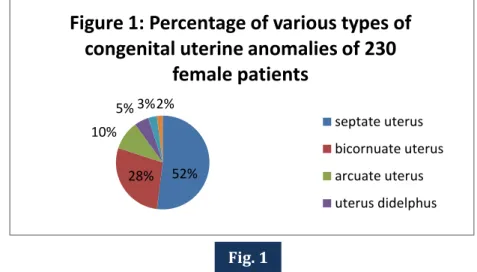
Table 1: Depicts the Number of cases in each type of congenital uterine anomaly in a total of 230 patients diagnosed to have congenital uterine anomaly.
Type of uterine anomaly No. of cases
Septate uterus 119
Bicornuate uterus 65
Arcuate uterus 23
Uterus didelphus 11
Unicornuate uterus 7
Hypoplasia of uterus 5
J of Evolution of Med and Dent Sci/ eISSN- 2278-4802, pISSN- 2278-4748/ Vol. 4/ Issue 26/ Mar 30, 2015 Page 4458 The percentage of varying types of congenital uterine anomaly of these 230 patients is illustrated in figure 1.
Septate uterus is the most common anomaly found in maduranthagam region of Tamil Nadu, as per this study. On further analysis, in the present study, septate uterus is found in 75% (n=61) of patients having recurrent abortions and 69% (n=101) of patients in infertility population. This study is statistically significant at p<0.05.
Figures 2a, 2b, 2c, 3, 4, 5 & 6 shows the ultrasound (USG) and hysterosalpingogram (HSG) pictures of various types of congenital uterine anomalies noted in this present study.
Figure 2a: A transvaginal USG showing septate uterus.
Figure 2b: HSG picture showing septate uterus.
52% 28% 10%
5% 3%2%
Figure 1: Percentage of various types of
congenital uterine anomalies of 230
female patients
septate uterus
bicornuate uterus
arcuate uterus
uterus didelphus
Fig. 1
Fig. 2A Fig. 2B
J of Evolution of Med and Dent Sci/ eISSN- 2278-4802, pISSN- 2278-4748/ Vol. 4/ Issue 26/ Mar 30, 2015 Page 4459
Figure 2c: A transabdominal USG showing septate uterus
Figure 3: A transabdominal USG showing hypoplasia of uterus.
Figure 4: HSG picture showing bicornuate uterus.
Figure 5: HSG picture showing arcuate uterus.
Figure 6: HSG picture showing unicornuate uterus
DISCUSSION: In this present study, septate uterus is the most common major uterine anomaly. Septate uterus anomaly composes of approximately 55% of uterine anomalies.[3, 4, 5] A literature
review showed that the most common major uterine anomaly in patients with recurrent pregnancy loss is subseptate uterus (Homer et al., 2000). The result of our study is similar, showing septate uterus as the most common uterine anomaly among patients with recurrent pregnancy loss, accounting to 75% of the cases.
Embryology: The male and female genitalia are indistinguishable in appearance upto 6 weeks of gestational age. Two sets of paired ducts namely, paramesonephric (Mullerian) duct and mesonephric (Wolffian) duct are present. In the absence of the testis-determining factor of the Y chromosome, the mesonephric ducts begin to degenerate and synchronously, the paramesonephric ducts develop bi directionally along the lateral aspects of the gonads. The female reproductive tract
Fig. 4 Fig. 5
J of Evolution of Med and Dent Sci/ eISSN- 2278-4802, pISSN- 2278-4748/ Vol. 4/ Issue 26/ Mar 30, 2015 Page 4460 develops from the pair of paramesonephric duct and forms the fallopian tube, uterus and upper one third of vagina. The ovaries develop from the germ cells that migrate from the primitive yolk sac and lower two third of vagina develops from the sino-vaginal bulb.
Organogenesis of mullerian ducts, fusion and septal resorption are the three phases which aid in the normal development of the female reproductive tract from the paramesonephric ducts. Defect in organogenesis leads to agenesis, hypoplasia of uterus or unicornuate uterus. Defect in fusion of mullerian ducts leads to bicornuate or didelphys uterus. Failure of septal resorption leads to septate or arcuate uterus. Regression of uterine septum has been proposed to be a result of apoptosis mediated by Bcl2 gene.[6] Absence of this Bcl2 gene results in persistence of septum in uterus.
Classification of Mullerian Duct Anomalies: The classification system proposed by Buttram and Gibbons in 1979 was modified in 1988 by the American Fertility Society, now called as American Society of Reproductive Medicine (ASRM) into seven classes.
Table 2 shows the classification of mullerian duct anomalies according to ASRM:
Classification Clinical Finding Embryological Reason
Class I Segmental agenesis/
Hypoplasia of uterus
Early developmental failure of mullerian ducts.
Class II
Unicornuate uterus with:
a. Absent rudimentary horn. b. Non cavitary rudimentary horn. c. Cavitary non-communicating
rudimentary horn. d. Cavitary communicating
rudimentary horn.
Complete or partial arrested development of one of the mullerian duct
Class III Uterus didelphys Complete non fusion of both
mullerian ducts
Class IV Bicornuate uterus Incomplete fusion of superior segments
of utero vaginal canal
Class V
Septate uterus: a. Complete b.Partial
Complete or partial non resorption of uterovaginal septum
Class VI Arcuate uterus Near complete resorption of
uterovaginal septum
Class VII Diethyl stilbesterol drug exposure related uterine anomaly- hypoplasia of uterus with T- shaped uterine cavity.
Table 2: Showing classification of mullerian duct anomalies according to ASRM and the embryological reason behind the anomalies
J of Evolution of Med and Dent Sci/ eISSN- 2278-4802, pISSN- 2278-4748/ Vol. 4/ Issue 26/ Mar 30, 2015 Page 4461 nourishment because of scanty vascularity of the septum, as the septum disrupts the arrangement of blood vessels in the myometrium.[7]
This view is supported by histological evaluation of the septum, which showed reduced vascularity as compared to rest of the uterus. (Nakada et al., 1989; Dabirashafi et al., 1995)
A study conducted on patients with septate uterus, looked into the site of implantation of embryo and found that, 8 out of 12 pregnancies that miscarried were found on the uterine septum. 4 pregnancies that did not miscarry were found on the lateral wall of uterus.[8] A study has also noted that the distortion of uterine anatomy in sub septate uterus is greater in women with recurrent pregnancy loss. Hence the likelihood of septal implantation, which is more prone for recurrent pregnancy loss, increases with the increasing ratio of septal size to functional cavity.[9]
In another prospective study, septal and non-septal tissue samples were obtained from the posterior uterine wall at the time of Tompkins metroplasty. Increased muscular tissue and less connective tissue were demonstrated in the septum by taking multiple biopsies. It was concluded that decreased connective tissue may result in poor decidualization and implantation, while increased muscular tissue would result in increased contractility of the tissue, predisposing the patient to spontaneous abortion.[8] An overlying endometrium over the uterine septum has been found to be defective,[10] and a scanning electron microscopy showed the septal endometrium to be irregular with decrease in sensitivity to preovulatory hormonal changes.[11] Reduction in endometrial cavity by uterine septum has also been implicated to result in poor obstetric outcomes.[12]
In patients with bicornuate uterus, spontaneous abortion rates are reported to range from 28% to 35% (pooled data, 30%).[12,13,14,15] Premature birth rates range from 14% to 23% (pooled data, 20%); and fetal survival rates range from 57% to 63% (pooled data, 60%).[12,13,15,16,17] Spontaneous abortion rates and preterm delivery are reported to be higher in women with a complete bicornuate uterus than in those with a partial bicornuate uterus.[13]
First trimester pregnancy loss is associated more with the septate and bicornuate uterus and second trimester loss with arcuate, septate and bicornuate uterus. Preterm delivery complication before 37 weeks of gestational age is noted in all types of uterine anomalies. Preterm delivery before 27 weeks is associated with bicornuate uterus.[18]
Other obstetric complications associated with uterine anomalies are malpresentation of fetus, low birth weight babies which is significantly associated with uterus didelphys, bicornuate and unicornuate uterus, increased risk of perinatal mortality in patients with septate and bicornuate uterus, increased risk of intrauterine growth restriction of fetus and increased risk of placental abruption as in the case of patients with arcuate and septate uterus.[18]
Resection of the uterine septum by hysteroscopic metroplasty has shown significant positive pregnancy outcomes. A retrospective cohort study of women undergoing hysteroscopic resection of a uterine septum demonstrated a significant decrease in miscarriage rates from 80% to 17% and an increase in the live birth rates from 18% to 91%.[19]
In general, uterine anomalies present some difficulty in pregnancy retention and overall pregnancy outcome with natural conception and in assisted reproductive techniques. Correctable form of anomalies like septate uterus can be corrected to ensure better pregnancy outcomes.
J of Evolution of Med and Dent Sci/ eISSN- 2278-4802, pISSN- 2278-4748/ Vol. 4/ Issue 26/ Mar 30, 2015 Page 4462 non invasive, cost effective procedure, which has no radiation hazards and is suitable for the first line of investigation. MRI is the gold standard investigation for diagnosing mullerian duct anomalies.
However the role of USG and HSG in screening and initiating the diagnosis has to be considered, especially when non affordable poor people are considered. In our present study, congenital uterine anomaly is found in 15.8% of patients with recurrent pregnancy loss and 7.5% of patients in infertility population, with septate uterus as the most common entity. Since literature has showed better reproductive outcomes with surgical correction of septate uterus, early diagnosis is beneficial for the patients. In non-correctable conditions psycho-social counseling is done to stabilize the patient and the family. A proper knowledge of female reproductive tract anatomy, a knowledge of anomalies associated with them and embryological reasons behind these anomalies will help in proper planning and management of the patient.
ACKNOWLEDGEMENTS: We acknowledge our institutional heads, our Dean Dr. A.R. Chakravarthy, professor of Obstetrics and Gynaecology and the Radiology department of our institution for extending their valuable support in the preparation of this article.
REFERENCES:
1. The American Fertility Society classifications of adnexal adhesions, distal tubal obstruction, tubal occlusions secondary to tubal ligation, tubal pregnancies, Mullerian anomalies and intrauterine adhesions. Fertil Steril 1998; 49:944–55.
2. Standring S (ed). Section 8, Abdomen & pelvis. In: Gray’s Anatomy. The Anatomical Basis of Clinical Practice.40th ed., Elsevier Churchill Livingstone. London, 2008:1314.
3. Homer HA, Li TC, Cooke ID. The septate uterus: a review of management and reproductive outcome. Fertil Steril 2000; 73:1-14.
4. Raga F, Bauset C, Remohi J, Bonilla-Musoles F, Simon C, Pellicer A. Reproductive impact of congenital mulleri ananomalies. Hum Reprod 1997; 12:2277-2281.
5. Fedele L, Bianchi S. Hysteroscopic metroplasty for septate uterus. Obstet Gynecol Clin North Am 1995; 22:473-489.
6. Lee DM, Osathanondh R, Yeh J. Localization of Bcl-2 in the human fetalmullerian tract. FertilSteril 1998; 70:135-140.
7. Candiani GB, Fedele L, Zamberletti D, De Virgiliis D, Carinelli S. Endometrial pattern in malformed uteri. Acta Eur Fertil 1983; 5:311.
8. Dabirashrafi H, Bahadori M, Mohammad K, et al. Septate uterus: new idea on the histologic features of the septum in the abnormal uterus. Am J Obstet Gynecol 1995; 172(1 pt 1):105-107.
9. Salim R, Regan L, Woelfer B, Backos M, Jurkovic D: A comparative study of the morphology of
congenital uterine anomalies in women with and without a history of recurrent first trimester
miscarriage; Hum. Reprod. (2003) 18 (1): 162-166 doi:10.1093/humrep/deg030.
10.Candiani GB, Fedele L, Zamberletti D, De Virgiliis D, Carinelli S. Endometrial patterns in malformed uteri. Acta Eur Fertil 1983; 14:311-318.
11.Fedele L, Bianchi S, Marchini M, Franchi D, Tozzi L, Dorta M. Ultrastructural aspects of endometrium in infertile women with septate uterus. Fertil Steril 1996; 65:750-752.
J of Evolution of Med and Dent Sci/ eISSN- 2278-4802, pISSN- 2278-4748/ Vol. 4/ Issue 26/ Mar 30, 2015 Page 4463 13.Heinonen PK, Saarikoski S, Pystynen P. Reproductive performance of women with uterine
anomalies: an evaluation of 182 cases. Acta Obstet Gynecol Scand 1982; 61:157-162.
14.Propst AM, Hill JA. Anatomic factors associated with recurrent pregnancy loss. Semin Reprod Med 2000; 18:341-350.
15.Buttram VC. Mullerian anomalies and their management. Fertil Steril 1983; 40:159-163.
16.Rock JA, Parmley T, Murphy AA, Jones HW. Malposition of the ovary associated with uterine anomalies. Fertil Steril 1986; 45:561-563.
17.Rock JA, Jones HW. The clinical management of the double uterus. Fertil Steril 1977; 28:798-806.
18.Venetis CA, Papadopoulos SP, Campo R, Gordts S, Tarlatzis BC, Grimbizis GF : Clinical Implications of Congenital Uterine Anomalies: A Meta-analysis of Comparative Studies : Reprod Biomed Online. 2014; 29:665-683.
19.Daly DC, Maier D, Soto-Albors C. Hysteroscopicmetroplasty: Six years’ experience. Obstet Gynecol 1989; 73: 201.
AUTHORS:
1. T. L. Anbumani 2. S. Anthony Ammal 3. A. Thamarai Selvi 4. T. L. Selvakumari
PARTICULARS OF CONTRIBUTORS:
1. Professor& HOD, Department of Anatomy, Karpaga Vinayaga Institute of Medical Sciences.
2. Post Graduate, Department of Anatomy, Karpaga Vinayaga Institute of Medical Sciences.
FINANCIAL OR OTHER
COMPETING INTERESTS: None
3. Post Graduate, Department of Anatomy, Karpaga Vinayaga Institute of Medical Sciences.
4. Professor, Department of Anatomy, Sri Venkateshwara Dental College, Chennai
NAME ADDRESS EMAIL ID OF THE CORRESPONDING AUTHOR: Dr. T. L. Anbumani,
Professor & HOD, Department of Anatomy, Karpaga Vinayaga Institute of Medical Sciences, Maduranthagam.
E-mail: anbumanitl@gmail.com