Identification of an
Xiap
-Like Pseudogene on Mouse
Chromosome 7
Aneta Kotevski1, Wendy D. Cook1, David L. Vaux1*, Bernard A. Callus1,2
1Department of Biochemistry, La Trobe University, Bundoora, Victoria, Australia,2Western Australian Institute of Medical Research and School of Biomolecular, Biomedical and Chemical Sciences, University of Western Australia, Crawley, Western Australia, Australia
Abstract
The most thoroughly characterized mammalian IAP is XIAP/BIRC4, which can inhibit caspases 9, 3 and 7, but may also regulate apoptosis through interactions with other proteins such as Smac/DIABLO, HtrA2/Omi, XAF1, TAK1, cIAP1, and cIAP2. High throughput sequencing of the mouse genome revealed the existence of a gene resembling Xiap/Birc4 on mouse chromosome 7. To confirm the existence of this gene, and to determine its functional significance, we performed Southern and Northern blot analysis. This showed the presence of the Xiap-like gene in both wild-type and Xiapgene knock-out mice, but the corresponding mRNA was not detected in any tissues examined by Northern blot. Analysis of the gene sequence in all three possible reading frames predicts that expression of this gene would not give rise to a full-length protein, but only non-functional truncated polypeptides. Because its nucleotide sequence is 92% identical toXiap, but it has no introns corresponding to those ofXiap, we conclude thatXiap-ps1is a pseudogene generated by retro-transposition of a splicedXiapmessage to chromosome 7.
Citation:Kotevski A, Cook WD, Vaux DL, Callus BA (2009) Identification of anXiap-Like Pseudogene on Mouse Chromosome 7. PLoS ONE 4(11): e8078. doi:10.1371/journal.pone.0008078
Editor:Andreas Bergmann, University of Texas MD Anderson Cancer Center, United States of America
ReceivedOctober 20, 2009;AcceptedNovember 4, 2009;PublishedNovember 30, 2009
Copyright:ß2009 Kotevski et al. This is an open-access article distributed under the terms of the Creative Commons Attribution License, which permits unrestricted use, distribution, and reproduction in any medium, provided the original author and source are credited.
Funding:Funding was received from the National Health and Medical Research Council (433063, 461221) and a Leukemia and Lymphoma Center Grant. The funders had no role in study design, data collection and analysis, decision to publish, or preparation of the manuscript.
Competing Interests:The authors have declared that no competing interests exist.
* E-mail: [email protected]
Introduction
The inhibitor of apoptosis proteins (IAPs) are a family of proteins that bear one or more baculoviral IAP repeat (BIR) domains [1,2]. The most thoroughly characterized mammalian IAP is XIAP/BIRC4, which can inhibit caspases 9, 3 and 7 [2,3], but may also regulate apoptosis through interactions with other proteins such as Smac/DIABLO, HtrA2/Omi, XAF1, TAK1, cIAP1, and cIAP2 [4,5].
Surprisingly, although XIAP is the most potent caspase inhibitor of the IAPs [6], the phenotype of Xiap knockout mice is very mild [7] and humans harbouringXIAP/BIRC4mutations have immune system defects due to abnormal NK cell function, but are otherwise normal [8].
We were alerted (Anthony Uren pers. comm.) to the presence of a
Xiap-like sequence found on mouse chromosome 7 during sequencing of the mouse genome (see: http://apr2006.archive.ensembl.org/ Mus_musculus/domainview?domainentry = IPR001370). To deter-mine whether this gene was functional, and whether redundancy with
Xiap might explain the subtle phenotype of Xiap KO mice, we analysed the sequence of this putativeXiap-like gene, confirmed its existence by Southern blot, and used Northern analysis to determine whether it was expressed.
Materials and Methods
Ethics Statement
All mouse work was done according to the requirements of La Trobe University Animal Ethics Committees with ethics approval
number: AEC 09-01-B. Animals were sacrificed using CO2 asphyxiation and the appropriate organs harvested.
Cell Culture
Primary wild-type andXiap2/2MEFs isolated from C57BL/6 mice were grown in FMA medium (Dulbecco’s Modified Eagle (DME) medium supplemented with 10% (v/v) foetal bovine serum (Gibco, Melbourne VIC), penicillin G (50 U/ml), streptomycin (50mg/ml), L-glutamine (2 mM), 270mM L-asparagine and
50mM 2-mercaptoethanol) in a humidified atmosphere of 10%
CO2 at 37uC. Confluent 10 cm plates were used for preparing
genomic DNA. Cells were harvested, washed and the cell pellets stored at280uC until required.
Tissue Samples
Tissue samples (liver, brain, lung, spleen, heart and intestine) were harvested from wild-type andXiap2/2C57BL/6 mice and were snap frozen in liquid nitrogen and stored at280uC until required.
Genomic DNA Preparation
The frozen samples were incubated overnight with shaking at 55uC in 3 ml genomic lysis buffer (100 mM Tris-HCl pH 8, 5 mM EDTA pH 8, 0.5% (w/v) SDS, 200 mM NaCl, 500mg/ml
Proteinase K). Genomic DNA was precipitated using isopropanol and resuspended in TE buffer (10 mM Tris, 1 mM EDTA). Genomic DNA (12mg) was digested to completion usingBamH1,
Figure 1. Schematic diagram of theXiapgenes.(a) The murineXiapgene spans 42 kb on the X-chromosome and consists of 7 exons.XiapK/O locus has exon 2 removed via homologous recombination. TheXiap-like gene is found on chromosome 7 and lacks any intronic sequences, giving rise to one exon that is 92% identical to spliced WTXiap. A DNA probe designed to detect this pseudogene was produced from a 510 bp region of exon 2 in WTXiapthat is unable to hybridise to theXiapK/O locus.(b) The splicing of the 7 exons ofXiapgives rise to mRNA encoding the XIAP protein. Two codons from the beginning and the end of each exon were aligned toXiap-ps1. The nucleotide sequences ofXiapandXiap-ps1are identical around the exon boundaries with the exceptions of two C.T transitions, one silent at the beginning of exon 4 and another coding at the
end of exon 5.
Southern Blot Analyses
Digested DNA samples were separated by electrophoresis on 1% agarose TAE gels containing 0.2mg/ml ethidium bromide.
Afterwards, the gel was denatured in 1.5 M NaCl/0.5 M NaOH for 45 min and then neutralised in 1 M Tris (pH 7.4)/1.5 M NaCl. Separated DNA was transferred onto Zeta-probe (BioRad) nitrocellulose membrane by overnight capillary transfer and fixed by baking at 80uC for 2 hr. The membrane was pre-hybridised for 30 min at 65uC in Rapid-Hyb buffer (Amersham) followed by hybridisation with32P labelledmXiapDNA probe at 65uC for 2 hr in Rapid-Hyb buffer according to the manufacturer’s specifica-tions. The membrane was washed twice at 65uC for 30 min in 0.3X SSC. The hybridised probe was detected using a Typhoon phosphoimager (Amersham).
The DNA probe was obtained by digestingXiapcDNA to make a 510 bp fragment identical to bases 524 to 1034 of the cDNA of
Xiap, a region within exon 2 and encoding amino acids 105 to 275 of the protein. Alignment of the probe toXiap-ps1 showed 92% identity, indicating that the probe would be able to hybridise to both genes at high stringency.
RNA Extraction and Northern Blot Analyses
To isolate RNA, frozen tissue samples were homogenised in Trizol (Invitrogen) and RNA purified according to the manufacturer’s instructions. Total RNA (10–15mg) was separated on 1.2% agarose formaldehyde gels, transferred to Hybond-N (Amersham) membrane by capillary transfer and fixed by baking at 80uC. Membranes were pre-hybridised for 30 min at 65uC in Rapid-Hyb buffer followed by hybridisation with the32P labelledmXiapDNA probe at 65uC for 2 hr. The final wash was three times for 20 min at 65uC in 0.3X SSC+0.5% SDS, and the hybridised probe was detected using a Typhoon phosphorimager (Amersham).
Results
Identification of aXiap-Like Gene (Xiap-ps1) on Chromosome 7
On mouse chromosome 7 band B3 location 37,599,271– 37,600,785 is a sequence with 92% nucleotide identity to Xiap
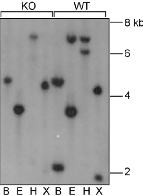
cDNA (Fig. 1a,b). To confirm the existence of theXiap-ps1gene in the C57BL/6 mouse genome we performed Southern blot analysis of genomic DNA from both wild-type (WT) and Xiap deleted mouse embryonic fibroblasts (MEFs), as well as from tissues from C57BL/6 WT andXiapdeleted mice.
As indicated in Fig. 1a, digestion of WT DNA with BamHI,
EcoRI, HindIII and XbaI restriction enzymes is predicted to produce fragments of the mouseXiapgene of 2.1 kb, 6.8 kb, 6 kb and 2 kb, respectively, that would be able to hybridise to anXiap
cDNA probe containing the coding region of exon II. In theXiap
mutant mice, exon II of Xiap has been deleted by homologous recombination, and therefore genomic DNA isolated from these mice does not contain any Xiap sequences that are capable of hybridising with the probe.
Any bands that hybridise at high stringency (65uC; 0.3x SSC) to the
Xiap probe in the DNA from Xiap knockout mice indicate the presence of another gene similar to Xiap. As seen in Fig. 2, hybridisation of the probe to WT samples not only gave rise to fragments of the expected size forXiap, but also additional bands that were also seen in the DNA from theXiap-deleted mice. This confirms the presence of a Xiap-like gene that we have designatedXiap-ps1. These bands of 4.2 kb, 3.4 kb, 6.9 kb and 4.4 kb in samples digested with BamHI, EcoRI, HindIII and XbaI, respectively, are consistent with those predicted from the digestion of theXiap-ps1gene sequence on chromosome 7 in Genbank (http://apr2006.archive.ensembl.
org/Mus_musculus/domainview?domainentry = IPR001370) with the same restriction enzymes (Fig. 1a).
Xiap-ps1Is Not Expressed
To determine whetherXiap-ps1is expressed we performed RNA Northern blot analyses. Tissues from WT andXiapdeleted mice were harvested and total RNA was isolated. The RNA samples were separated on denaturing formaldehyde agarose gels, transferred to membrane and probed with the same32P-labelled probe that was Figure 2. Detection of an Xiap pseudogene by Southern analysis.Genomic DNA was digested to completion withBamHI (B),
EcoRI (E),HindIII (H), orXbaI (X), and probed with a 510 bp fragment from exon 2 ofXiap. Bands of the expected size for theXiapgene were revealed in the WT DNA. In addition, both WT and XiapK/O DNA showed bands of the sizes predicted from the sequence of theXiap-like gene on chromosome 7.
doi:10.1371/journal.pone.0008078.g002
Figure 3. Confirmation of RNA expression by Northern analysis.Total RNA was harvested from C57BL/6 andXiapK/O tissues, separated by electrophoresis, blotted and probed with a 510 bpXiap
fragment as indicated in Figure 1. The only band detected (,6.6 kb) is the reported size for XiapmRNA, and was only found in wild-type tissues and not in theXiapknockout tissues. Ab-actin probe was used to re-probe the same blots to show relative loading of lanes. doi:10.1371/journal.pone.0008078.g003
used in the genomic Southern analyses. As shown in Fig. 3, in all WT tissue samples a single band of approximately 6.6 kb was detected, consistent with previous reports of the size of mouseXiap
mRNA [7,9]. In contrast, in samples fromXiapknockout mice no bands were detected, indicating that the Xiap-ps1 mRNA is not expressed at detectable levels in these tissuesin vivo.
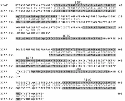
Xiap-ps1Bears Premature Stop Codons
Analysis of theXiap-ps1nucleotide sequence showed that it does not code for a full-length protein due to the presence of premature stop codons. Translation of the gene sequence in all three possible reading frames gives rise to several truncated polypeptide sequences (Fig. 4). This indicates that even if Xiap-ps1 were transcribed, the presence of these premature stop codons would prevent translation of a functional IAP protein.
Discussion
From the Southern analyses (Fig. 2) we have confirmed that a novel Xiap-like gene exists on mouse chromosome 7. As we detected the presence of the Xiap-ps1 gene in genomic DNA samples from Xiap2/2tissues, the bands detected were not the result of the probe hybridising to the Xiap gene on the X chromosome. We failed to detect any evidence by Northern analysis thatXiap-ps1is expressed in tissues in whichXiapis clearly expressed at the mRNA level. Analysis of the nucleotide sequence showed thatXiap-ps1gene is devoid of intronic sequences found in
Xiapand analysis of the various splicing variants showed that
Xiap-ps1 shows similarity to the regions in common to all variants, starting 7 nucleotides upstream of the initiation codon in exon 2 of
Xiap (Fig. 1b). This suggests that Xiap-ps1 has arisen from retrotransposition of a processedXiapmRNA to chromosome 7. We hypothesise that a retroviral infection occurred in mouse germ cells that allowed insertion of reverse-transcribed spliced Xiap
mRNA into chromosome 7. A similar event has been shown to have occurred during evolution of the great apes, leading in that case to a transcribed and translated product, BIRC8 (ILP-2), from a single-exon gene [10].
Examination of the flanking genomic sequences of Xiap-ps1
using the Transfac database revealed several transcriptional elements, the closest to the gene being a retroviral TATA box and a CAP signal for transcription initiation 1692 and 1629 bp upstream of the initiating methionine respectively. The presence of viral elements supports our hypothesis that retrotransposition of theXiapmRNA occurred.
Translation of the sequence for Xiap-ps1indicates that no full length protein could be produced. Although three of the larger peptide sequences were very similar to regions of XIAP, it is likely that even if these peptides were expressed within cells they would not be able to function as inhibitors of apoptosis.
Author Contributions
Conceived and designed the experiments: AK DLV. Performed the experiments: AK. Analyzed the data: AK WDC DLV BAC. Wrote the paper: AK WDC DLV BAC.
References
1. Hinds MG, Norton RS, Vaux DL, Day CL (1999) Solution structure of a baculoviral inhibitor of apoptosis (IAP) repeat. Nature Structural Biology 6: 648–651.
2. Sun C, Cai M, Gunasekera AH, Meadows RP, Wang H, et al. (1999) NMR structure and mutagenesis of the inhibitor-of-apoptosis protein XIAP. Nature 401: 818–821.
3. Sun CH, Cai ML, Meadows RP, Xu N, Gunasekera AH, et al. (2000) NMR structure and mutagenesis of the third Bir domain of the inhibitor of apoptosis protein XIAP. Journal of Biological Chemistry 275: 33777–33781.
4. Ekert PG, Silke J, Hawkins CJ, Verhagen AM, Vaux DL (2001) DIABLO promotes apoptosis by removing MIHA/XIAP from processed caspase 9. Journal of Cell Biology 152: 483–490.
5. Wu G, Chai JJ, Suber TL, Wu JW, Du CY, et al. (2000) Structural basis of IAP recognition by Smac/DIABLO. Nature 408: 1008–1012.
6. Deveraux QL, Roy N, Stennicke HR, Vanarsdale T, Zhou Q, et al. (1998) Iaps block apoptotic events induced by caspase-8 and cytochrome c by direct inhibition of distinct caspases. Embo Journal 17: 2215–2223.
7. Harlin H, Reffey SB, Duckett CS, Lindsten T, Thompson CB (2001) Characterization of XIAP-deficient mice. Molecular & Cellular Biology 21: 3604–3608.
8. Rigaud S, Fondaneche MC, Lambert N, Pasquier B, Mateo V, et al. (2006) XIAP deficiency in humans causes an X-linked lymphoproliferative syndrome. Nature 444: 110–114.
9. Lu X, Lee M, Tran T, Block T (2005) High level expression of apoptosis inhibitor in hepatoma cell line expressing Hepatitis B virus. International Journal of Medical Science 2: 30–35.
10. Richter BWM, Mir SS, Eiben LJ, Lewis J, Reffey SB, et al. (2001) Molecular cloning of ILP-2, a novel member of the inhibitor of apoptosis protein family. Molecular & Cellular Biology 21: 4292–4301.