Ar
ti
cle
0103 - 5053 $6.00+0.00
*e-mail: [email protected]
Studies on Terrein as a New Class of Proteasome Inhibitors
M. Demasi,*,a A. L. Felicio,a A. O. Pacheco,b H. G. Leite,b C. Limaa and L. H. Andrade*,b
aInstituto Butantan, 05503-900 São Paulo-SP, Brazil
bInstituto de Química, Universidade de São Paulo, 05508-900 São Paulo-SP, Brazil
O proteassomo é uma protease intracelular multicatalítica envolvida na regulação do ciclo e sinalização celular, apresentação antigênica e apoptose. Uma vez que inibidores do proteassomo promovem morte celular por apoptose, esses têm sido propostos como novas drogas anti-tumorais. A terreína, um metabólito secundário secretado pelo fungo Aspergillus terreus, foi primeiramente descrita em 1935. Neste trabalho demonstramos que a terreína, isolada através da bioprospecção de inibidores do proteassomo, mostrou efeito inibitório das atividades do tipo quimiotripsina e tripsina da unidade catalítica do proteassomo 20S. Apesar da alta concentração inibitória determinada in vitro, aquela veriicada após incubação de células em cultura na presença de terreína (ibroblasto e tumor pulmonar humano) foi 4 vezes menor, o que sugere que o proteassomo seja um alvo intracelular especíico. A terreína promoveu morte celular por apoptose nas duas linhagens ensaiadas. Embora as concentrações de terreína necessárias para desencadear apoptose nos modelos celulares aqui testados tenham sido altas (ordem de mM) quando comparadas com doses utilizadas de outros inibidores descritos recentemente (ordem de µM e nM), sua estrutura química não está relacionada a nenhum outro inibidor conhecido até o momento. Concluímos que estes resultados apontam para a possibilidade de explorar a terreína como um novo fragmento molecular para o desenvolvimento de inibidores sintéticos do proteassomo.
The proteasome is an intracellular multicatalytic protease involved in the cell cycle regulation, signaling response, antigen presentation and apoptosis. Since proteasome inhibitors promote cell death by apoptosis, they have been proposed as new anti-tumoral drugs. Terrein, a secondary metabolite secreted by the fungus Aspergillus terreus, was irstly described in 1935. In the present work we report that terrein isolated through the screening for inhibitors of the 20S proteasome showed inhibitory effect upon both chymotrypsin- and trypsin-like activities of the multicatalytic core particle, the 20S proteasome. Despite of the high inhibitory concentration determined in vitro, that veriied by incubating cells (ibroblasts and a pulmonary tumor cell line) in the presence of terrein was 4-fold lower indicating the proteasome as a selective intracellular target. Moreover, terrein promoted apoptotic cell death on both ibroblasts and pulmonary tumor cell line tested. Although terrein concentrations (mM range) necessary to elicit apoptosis in the cellular models herein tried were high when compared to those (µM and nM range) of other inhibitors recently described, its chemical structure is not correlated to any other inhibitor reported thus far. Therefore, the present results point out for the possibility of exploring terrein as a new molecular fragment for the development of synthetic proteasome inhibitors.
Keywords: anti-tumoral drugs,proteasome inhibitors, apoptosis, cyclopentenone derivatives, terrein
Introduction
The ubiquitin-26S proteasome system (UBP) mediates major intracellular protein degradation essential for normal cellular functioning. The 20S proteasome represents the UBP catalytic unit that triggers protein breakdown through
its multicatalytic activity.1 The 20S proteasome is composed
Studies on Terrein as a New Class of Proteasome Inhibitors J. Braz. Chem. Soc. 300
trypsin-like and the post-acidic cleavage.2 Substrates of the
ubiquitin-proteasome system include proteins mediating various intracellular functions and defects in the system machinery are linked to many human diseases. Targeting the 20S proteasome catalytic activity has been exploited as new therapeutic approach. For example, blockade of proteasome activity abrogates growth/survival mechanisms, thereby triggering apoptosis. In such context, 20S proteasome was validated as a therapeutic target and led to the FDA approval of the first proteasome inhibitor bortezomib (Velcade®) in 2003.3 Since then, the interest in discovery
and development of other novel proteasome inhibitors was renewed and greatly intensiied.3,4 In the present work we
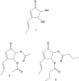
describe a proteasome inhibitor that possesses a chemical structure belonging to no other chemical class of inhibitors among those described thus far (Figure 1, 1). Terrein (1) was irstly puriied from A. terreus in 1935.5 In the last
decade it was described as an inhibitor of melanogenesis6,7
and fat cell differentiation.8,9 Terrein was puriied in our
lab through screening for proteasome inhibitors among samples obtained from culture media after growing a series of fungi species. After complete puriication and structure determination, terrein was identiied as one of the active compounds. Experiments performed in vitro and ex vivo with puriied terrein showed its inhibitory effect upon proteasomal activity and its ability to promote cell death by apoptosis. We also performed structural modiications of terrein that showed the free hydroxyl groups at positions 2 and 3 of the cyclopentenone ring as the structural feature necessary for its interaction with the catalytic particle.
Results and Discussion
Our starting point in the present investigation was the screening for catalytic inhibitors of the 20S proteasome
from natural sources. The screening was performed by testing the ability of crude extracts, obtained from media after growing different fungi species (Aspergillus terreus, Aspergillus niger, Aspergillus foetidus, Rhizopus oryzae and Penicillium decumbens), to modify the catalytic activity of puriied preparations of the 20S proteasome extracted from horse erythrocytes. Those crude extracts at 0.5% concentration (m/v), able to promote at least 90% proteasomal inhibition, were processed till complete purification of the active compound. Results herein reported were performed after accomplishing the multi-step screening procedure followed by structural identiication of the puriied active compound from Aspergillus terreus identiied as being terrein (Figure 1, structure 1), irstly reported in 1935 from same source.5
Firstly we performed biochemical assays to determine terrein IC50 with puriied proteasome preparations. Among the three reported catalytic activities of the 20S proteasome,2
terrein was able to inhibit the chymo and the trypsin-like activities with an IC50 of 0.3 mM. No inhibition of the post-acidic cleavage (caspase-like) was observed.
Next we performed assays to evaluate the proteolytic proteasomal activity, instead of the site speciic or peptidase activity, in the presence of terrein (Table 1). Puriied proteasome preparations were incubated for 15 min in the presence of terrein followed by the addition of casein-FITC. Results obtained showed that terrein was also effective in the inhibition of proteolysis. We veriied 25% and 37.5% inhibition of casein hydrolysis by the 20S proteasome incubated at 0.1 mM and 0.15 mM terrein concentrations, respectively.
Our next goal was to verify whether terrein would be able to be absorbed by cells and to inhibit the intracellular proteasomal activity. For that purpose, ibroblast cells were incubated for 2 h in the presence of increasing terrein concentration followed by the determination of proteasomal Table 1. Effect of terrein upon proteasomal proteolytic activity. Puriied 20S proteasome preparations (10 µg) were incubated in the presence of 20 µg casein-FITC for 1 h at 37 oC in 20 mM Tris buffer, pH 7.5,
after a pre-incubation (10 min) with terrein at indicated concentrations. After incubation, samples were taken for luorescence measurement (wavelengths: excitation 365 nm and emission 525 nm)
Control terrein (mM)
0.10 0.15 0.3
Casein-FITC fragmentation (AU × 103)
16 ± 2 12 ± 1a 10 ± 1a 7.5 ± 0.5 ap ≥ 0.006 (ANOVA followed by Student’s t test).AU: arbitrary units.
Results shown are means ± SD of 3 independent experiments performed in triplicates.
Figure 1. Terrein and its derivatives. Structure 1 is terrein and 2 and 3 are ethyl and butyl ester derivatives, respectively.
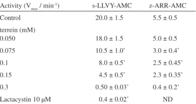
activity in the cell extracts, as described in Materials and Methods. According to the results obtained (Table 2), both chymotrypsin- and trypsin-like like activities were almost completely inhibited in extracts of cells incubated with 0.3 mM terrein similar to the already expected inhibition obtained after incubation of cells with 10 µM lactacystin, a well established proteasome inhibitor.10 However, terrein
concentration (0.3 mM) necessary to produce similar inhibition by lactacystin was much above lactacystin concentration (10 µM). Nevertheless, terrein concentration necessary to produce 50% of proteasome inhibition was 0.075 mM, 4-fold lower than the in vitro IC50 concentration (0.3 mM). This result is indicative that the 20S proteasome is a selective intracellular terrein target.
Afterwards, we evaluated the effect of terrein upon cellular viability by comparing lactacystin and terrein (Figure 2). The human lung tumoral cells and ibroblasts were incubated for 4 h and 24 h in the presence of terrein. Amazingly, results obtained revealed that 4 h incubation (Figure 2A) of ibroblast cells in the presence of 0.15 and 0.25 mM terrein was enough to promote 80% of cell death, the same percentage determined after 24 h incubation with same concentrations of terrein (Figure 2B). Figure 2B clearly shows dose-dependent terrein effect upon cell viability. Differently from lactacystin, a standard proteasome inhibitor, terrein pro-apoptotic effect was much faster: 5 µM lactacystin promoted 20% and 50% of cell death after 4 h and 24 h incubation, respectively whereas 0.15 mM terrein promoted 80% of cell death already after 4 h incubation. So, our conclusion is that though terrein effective dose is quite high (mM range), its effect is fast,
presumably because of its speciic intracellular interaction with the proteasome suggesting a high selectivity.
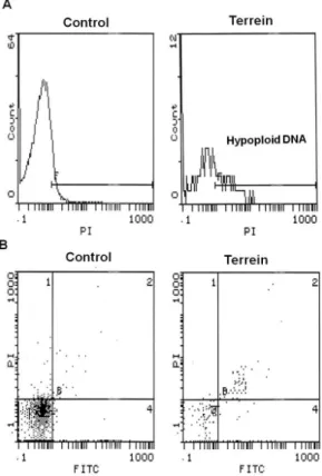
Finally, terrein-induced cell death was characterized by low luorocytometry (FACS) by labeling cells with the fluorescent probe Anexine V-FITC and with propidium iodide. Anexine V-FITC labeling analyzes phosphatidyl serine exposure on the external cell membrane surface, a hallmark in apoptotic cells, whereas propidium iodide labeling to DNA is a useful parameter to analyze both DNA fragmentation, typical of apoptotic cells, and cell permeabilization. These tools are largely utilized to distinguish apoptotic from necrotic cells.11 As depicted in Figure 3A, DNA analysis of human
lung tumoral cells incubated for 2 h in the presence of 0.3 mM terrein showed hypoploid DNA luorescence pattern, typical of apoptotic cells, when compared to Control cells showing a typical DNA luorescence peak. When cells were analyzed after 4 h incubation by double-labeling with Anexin V-FITC and PI, 30% of the cells presented the double-labeling pattern (Anexine V-FITC and PI) characteristic of late apoptotic onset (Figure 3B and Table 3). Our conclusion was that cell death promoted by terrein is likely an apoptotic process, interesting for therapeutic purposes.
Table 2. Hydrolytic activity determination in the extracts of ibroblast cells incubated for 2 h with terrein.Cells were incubated in the presence of terrein at indicated concentrations (terrein), 0.5% DMSO (Control) or 10 µM lactacystin (Lactacystin). After incubation (4 h), cells were lysed and 50 µg 100 µL-1 total protein were taken for the determination
of the hydrolytic activity with the indicated substrates, as described in Materials and Methods
Activity (Vmax / min-1) s-LLVY-AMC z-ARR-AMC
Control 20.0 ± 1.5*1 5.5 ± 0.51*
terrein (mM)
0.050 18.0 ± 1.5*1 5.0 ± 0.51*
0.075 10.5 ± 1.0*1 3.0 ± 0.4*1
0.1 18.0 ± 0.5*1 2.5 ± 0.45*
0.15 14.5 ± 0.5*1 2.3 ± 0.35*
0.3 0.50 ± 0.03* 0.4 ± 0.2*1
Lactacystin 10 µM 10.4 ± 0.02* ND
Results are means ± SD of two independent experiments performed in triplicate. ap ≥ 0.0007 (ANOVA). ND, not determined.
Figure 2. Cellular viability evaluation. Human lung tumoral cells were incubated: (A) 4 h and (B) 24 h in the presence of terrein and lactacystin. Lactacystin concentration was 5 µM and terrein concentrations shown were in the mM range.
a)
Studies on Terrein as a New Class of Proteasome Inhibitors J. Braz. Chem. Soc. 302
In another set of experiments we tested compounds derived from terrein structural modiications. The modiied structures are shown in Figure 1 (2 and 3).
The synthesis of the ethyl and butyl derivatives 2 and 3 were performed with terrein isolated from A. terreus using chemical esteriication with the corresponding acid anhydride. Having in hands these compounds, they were tested in vitro for the proteasomal hydrolytic activity at increasing concentrations up to 1 mM. No inhibition was observed at any concentration tested (results not shown). According to the structure of the derivatives, we deduced that those derivatives were not effective as inhibitors because the free hydroxyl groups are not present at the inal structure. So, our conclusion is that besides of cyclopentenone core, free hydroxyl groups at positions 2 and 3 of the cyclopentenone core (Figure 1, 1) are the key structure feature for the inhibition of proteasomal catalytic activity.
Conclusions
In summary, the present work revealed a new chemical scaffold, the 2,3-dihydroxyl cyclopentenone ring, represented by the natural compound terrein, as a potential proteasome inhibitor. The starting-point of this investigation was the evaluation of mixtures obtained from material secreted from fungi cultured in synthetic medium. Crude material of positive results was processed in a multi-step procedure till the complete puriication of the active compound, as described in Materials and Methods. After puriication and the elucidation of the chemical structure of the puriied compound, we found out that in reality this compound had been already described in the literature.5
However, the activity herein reported has never been reported before. Terrein is a cyclopentenone, a structure not related to any of the proteasome inhibitors described so far.3,4 It seems that terrein cyclopentenone ring and its free
hydroxyl groups are crucial for the proteasome inhibition. Indeed, terrein might be a new lead structure for the design of new proteasome inhibitors.
Materials and Methods
Chemicals
suc-Leu-Leu-Val-Tyr-7-amido-4-methylcoumarin (suc-LLVY-AMC); z-Leu-Leu-Glu-AMC (z-LLE-AMC) and, z-Ala-Arg-Arg-AMC (z-ARR-AMC) were purchased from Calbiochem (San Diego, CA-USA). Casein-FITC was purchased from SIGMA (St. Louis, MO - USA). Bradford reagent was obtained from BioRad (Hercules, CA-USA). Cell culture media and complements were obtained from Invitrogen. Anexin V-FITC and propidium iodide (PI) were purchased from BD Biosciences (San Jose, CA-USA). All other reagents were of analytical grade.
Table 3. Flow citometry of cells incubated for 4 h in the presence of terrein. Fibroblast cells were incubated as described in Materials and Methods for 4 h in the presence of terrein or DMSO (terrein solvent) at indicated concentrations. After incubation, cells were double labeled with PI and Anexine A-FITC and analyzed by low luorocytometry as described in Materials and Methods. Results shown were obtained as per Figure 3B by measuring the double and single labeling
Anexin V- FITC PI Anexin V FITC × PI
Control 10.5% 10.5% 3.0%
0.5% DMSO 10.5% 10.0 % 3.5%
0.3 mM terrein 44% 35% 30%
Microorganisms library
The microorganisms Penicillium decumbens SSP 1944, Aspergillus niger SSP 1078 and Aspergillus terreus SSP 1498 were obtained from the Fungal Culture Collection of the Botanical Institute of São Paulo (São Paulo, Brazil). Aspergillus foetidus CCT 2683 and Rhizopus oryzae CCT 4964 were obtained from the Culture Collection of the André Tosello Foundation. Sterile materials were used to perform the experiments and the microorganisms were handled in a laminar low cabinet.
Culture media
Composition for the culture medium of A. niger, A. foetidus , R. oryzae and P. decumbens was dextrose 20 g L-1,
peptone 10 g L-1and of A. terreus was glucose 20 g L-1, malt
extract 20 g L-1, peptone 1 g L-1. All these components were
purchased from Oxoid.
Fungi culture
The fungi were cultivated in Erlenmeyer flasks (250 mL) containing 100 mL of the appropriate culture medium at 27 °C in an orbital shaker (160 rpm) for 96 h. The cells were harvested by iltration and the iltrate was used in the in vitro assay with puriied preparations of proteasome, as described below.
Isolation of metabolites from EtOAc extract of the culture medium of A. terreus
The fungus A. terreus was cultivated in an Erlenmeyer lask (2 L) containing 1 L of the culture medium (glucose 20 g, malt extract 20 g, peptone 1 g) at 27 °C for 96 h stationarily. After 96 h, the culture broth was iltered off. The iltrate was concentrated to 30% of the initial volume under reduced pressure (50 °C; 72 mbar). Then, the residue was extracted with ethyl acetate (3×300 mL). The organic phases were combined and dried over magnesium sulphate (MgSO4). The solvent was removed in vacuum and brown crystals were obtained. These crystals were puriied by re-crystallization from ethyl acetate to give the terrein (0.4 g). The spectral data of the terrein were in agreement with those reported in the literature.12
Derivatization of terrein
To a Schlenck-flask, terrein (154 mg, 1 mmol), tetrahydrofuran (10 mL), appropriate acid anhydride (2.5 mmol, butyric or acetic anhydride), pyridine (316 mg,
4 mmol) and 4-dimethylaminopyridine (24.4 mg, 0.2 mmol) were added. The resulting solution was stirred for 18 h at room temperature. After this period, the solvent was removed in vacuum. Ethyl acetate (30 mL) was added to the residue and the resulting organic solution was washed with copper sulphate (CuSO4; 2×10 mL). The organic phase was dried over MgSO4. The solvent was removed
in vacuum and the residue puriied by silica gel column chromatography, using a mixture of hexane and ethyl acetate (9:1) as solvent to give the esters (55-65% yield):
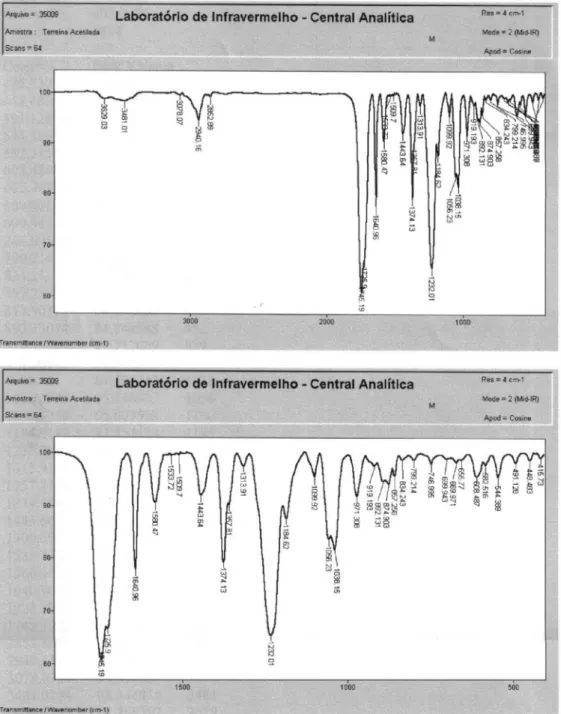
(E)-5-Oxo-3-(Prop-1-enyl)cyclopent-3-ene-1,2-diyl diacetate: ethyl derivative(2)
LRMS [EI], m/z (relative abundance): 238 (2), 210 (1), 196 (4), 178 (24), 153 (16), 136 (82), 138 (20), 121(88), 108 (15), 95 (7), 79 (25), 65 (8), 43 (100). IR νmax/cm-1: 2910,
2852, 1745, 1640, 1580, 1374, 1232, 1056. 1H NMR (200
MHz, CDCl3) δ 6.4 (dq, 1H, J 6Hz, 16.2 Hz), 6.31 (d, 1H, J 16.2Hz), 6.20 (s, 1H), 6.08(d, 1H, J 2.4Hz), 5.22 (d, 1H, J 2.4Hz), 2.16 (s, 3H), 2.15 (s, 3H), 1.94 (d, 3H, J 6Hz).
13C NMR (50 MHz) δ 196.8, 170.2, 170.1, 165.1, 140.2,
128.1, 124.5, 78.3, 74.8, 20.8, 20.5, 19.5.
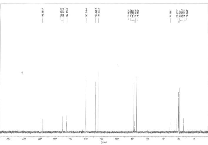
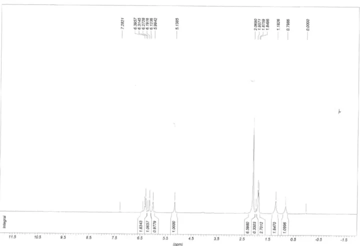
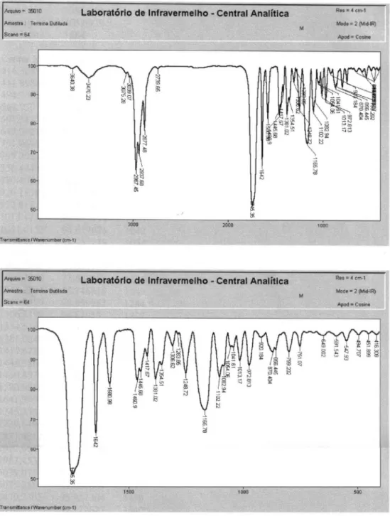
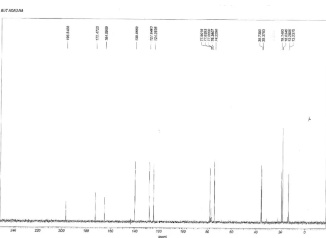
(E)-5-oxo-3-(Prop-1-enyl)cyclopent-3-ene-1,2-diyl dibutyrate: Butyl derivative (3)
LRMS [EI], m/z (relative abundance): 294 (1), 223 (1), 206 (15), 178 (2), 153 (10), 136 (74), 121 (93), 108 (12), 91 (4), 71 (97), 43 (100). IR νmax/cm-1: 2967, 2877,
1745, 1642, 1580, 1445, 1381, 1165. 1H NMR (200 MHz,
CDCl3) δ 6.36 (dq, 1H, J 6Hz, J 15.9Hz), 6.30 (d, 1H, J 15.9Hz), 6.19 (s, 1H), 6.10 (d, 1H, J 2.4Hz), 5.21 (d, 1H, J 2.4Hz), 2.43-2.30 (M, 4H), 0.97 (t, 6H, J 7.5Hz). 13C NMR
(50 MHz) δ 197.2, 178.3, 172.8, 165.2, 140.1, 128.2, 124.5, 78.1, 74.4, 36.0, 35.7, 19.4, 18.3, 18.2, 13.6, 13.5.
Proteasome puriication
Mammal 20S proteasome preparations were obtained from horse erythrocytes by a 3-step chromatographic procedure as described.13 The human 20S proteasome was
purchased from Afinity Bioreagents (Rockford, IL-USA).
Determination of the proteasome catalytic activity by hydrolysis of luorogenic peptides
F l u o r o g e n i c p e p t i d e s ( A M C , 7 a m i d o 4 -methylcoumarin as the luorescent probe) were utilized for the determination of proteasomal activity, as referred elsewhere.2 suc-LLVY-AMC was utilized as a standard
Studies on Terrein as a New Class of Proteasome Inhibitors J. Braz. Chem. Soc. 304
z-ARR-AMC for the trypsin-like activity. 20S proteasome (0.5-3 µg 100 µL-1) was incubated at 37 oC in 20 mM Tris/
HCl buffer, pH 7.5, herein referred to as standard buffer. Incubation was started by the addition of 10-50 µ mM of peptide. Fluorescence emission was recorded at 440 nm (excitation at 365 nm). AMC released from the substrates was calculated from a standard curve of free AMC. In vitro assays consisted of 15 min pre-incubation of puriied proteasome preparations with purified compounds, e.g. terrein, and crude extracts to be tested (screening procedure), followed by the hydrolytic assay as described above. Proteasomal activity was determined in cell extracts by incubating 50 mg aliquots of cellular protein at 37 oC
with same substrates described above at (125 µM inal concentration) and the emission was recorded for 45 min. In parallel, similar samples were previously incubated (15 min) in the presence of 40 µM lactacystin followed by the hydrolytic activity determination. 40 µM lactacystin was chosen because this concentration was able to inhibit up to 90% of both chymotrypsin- and trypsin-like proteasomal activities from control samples (result not shown). The proteasomal activity was attributed to the difference between the hydrolytic activity determined in the absence (total hydrolytic activity) and in the presence of lactacystin (non-proteasomal hydrolytic activity). 50 µg protein samples of cell extracts were used for all experiments based on data obtained from hydrolytic assays performed at increasing protein concentration (25-100 µg) of cellular extract (data not shown). All the assays were performed in triplicate. Cellular extract preparation is described below.
IC
50 determination
The IC50 was determined in vitro by incubating puriied proteasome preparations (1 µg 100 µL-1) at increasing
terrein concentrations (10-500 µM). After 15 min terrein pre-incubation with proteasome preparations, hydrolytic activity was performed as described above. The assays were performed in quadruplicate. The IC50 was calculated by the software Prisma version 3.0. Results are expressed as mean ± SD; p ≤ 0.00012
Proteolysis assay
Proteolysis rate was evaluated by incubating puriied proteasome preparations with FITC-modiied casein for 1 h at 37 oC. The FITC (luorescent probe) casein was
obtained as described.14 After incubation, 1 volume of 10%
trichloroacetic acid was added. Samples were placed on ice for 30 min followed by centrifugation at 15,000 × g. The supernatant, containing casein fragments (hydrolytic
products), was taken for the luorescence measurement (365 nm excitation and 525 nm emission).
Determination of protein concentration
Protein concentration was determined with the Bradford assay (BioRad).
Cell lines and culture
Cell lines utilized were C3H mouse NIH-3T3 ibroblast cells and the human tumoral cell line NCI-H292, both purchased from the American Type Culture Collection (ATCC). Fibroblast cells were cultured in DMEM High Glucose medium supplemented with 10% bovine fetal serum and 4 mM glutamine. The NCI-H292 cells were cultured in RPMI 1640 supplemented with 10% bovine fetal serum, 2 mM glutamine and 1 mM sodium piruvate. Cells were cultured in 10 mL medium into 75 cm2 bottles.
Treatments described were performed when cells were 90% conluent. Cell pellets were obtained by incubation in trypsin/EDTA solution followed by centrifugation and PBS washing. After treatment, cellular pellets were resuspended in 50 mM Tris-HCl pH 7.5, containing 150 mM NaCl, 1 mM EGTA, 10% (v/v) glycerol, 0.5% (v/v) Nonidet P-40 and 1 mM MgCl2. After lysing the cells by passing the cell suspension through insulin-syringes (10 times) followed by 30 min on ice, samples were centrifuged at 15,000 × g for 30 min. Protein concentration in the supernatant was then assessed and aliquots of total protein were taken for the determination of proteasomal activity.
Cellular viability assay
Cells were transferred (10-20 × 103 cells / well) to
96-wells microplates. After treatment cells were washed three times with PBS and incubated in the presence of MTT, 3-(4,5,-Dimethyl thiazolyl-2)-2,5-diphenyl tetrazolium bromide (0.5 mg mL-1 of PBS).15 After 3 h
MTT solution was removed and 100 µL DMSO was added. After solubilization the absorbance at 540 nm was read. The assays were performed in quadruplicate. Lactacystin (Lac) was utilized at 5 µmM (inal concentration) as a positive control of loss of cell viability due to proteasome inhibition.10
Cell death evaluation by low citometry
by a FACSCalibur (BD Biosciences) low cytometer and analyzed by the CellQuest software.16
Acknowledgments
M. D. was supported by grants from FAPESP (04/0763-6; 06/06969-0) and CNPq (471689/07-6); F. A. L. and P. A. O. are FAPESP fellows. L. H. A. thanks FAPESP and CNPq for support.
Supplementary Information
Supplementary data are available free of charge at http://jbcs.sbq.org.br as PDF ile.
References
1. Voges, D.; Zwickl, P.; Baumeister, W.; Annu. Rev. Biochem.
1999, 68, 1015.
2. Gaczynska, M.; Osmulski, P. A.; Methods Enzymol. 2005, 398,
425.
3. Moore, B. S.; Eustáqio, A. S.; McGlinchey, R. P.; Curr. Opin. Chem. Biol. 2008, 130, 7822.
4. Borissenko, L.; Groll, M.; Chem. Rev. 2007, 107, 687. 5. Raistrick, H.; Smith, G.; Biochem. J. 1935, 29, 606.
6. Park, S. H.; Kim, D. S.; Kim, W. G.; Ryoo, I. J.; Lee, D. H.; Huh, C. H.; Youn, S. W.; Yoo, I. D.; Park, K. C.; Cell. Mol. Life Sci. 2004, 61, 2878.
7. Lee, S.; Kim, W. G.; Kim, E.; Ryoo, I. J.; Lee, H. K.; Kim, J. N.; Jung, S. H.; Yoo, I. D.; Bioorg. Med. Chem. Lett. 2005, 15, 471.
8. Kim, D. S.; Cho, H. J.; Lee, H. K.; Lee, W. H.; Park, E. S.; Youn, S. W.; Park, K. C.; J. Dermatol. Sci. 2007, 46, 65. 9. Kim, D. S.; Lee, H. K.; Park, S. H.; Lee, S.; Ryoo, I. J.; Kim,
W. G.; Yoo, I. D.; Na, J. I.; Kwon, S. B.; Park, K. C.; Exp. Dermatol. 2008, 17, 312.
10. Fenteany, G.; Schreiber, S. L.; J. Biol. Chem.1998, 273, 8545 11. Krysko, D. V.; Berghe, T. V.; Parthoens, E.; D’Herde, K.;
Vandenabeele, P.; Methods Enzymol. 2008, 442, 307.
12. Kim, W. G.; Ryoo, I. J.; Park, S. H.; Kim, D. S.; Lee, S.; Park, K. C.; Yoo, I. D.; J. Microbiol. Biotechnol. 2005, 15, 891. 13. Twining, S. S.; Anal. Biochem. 1984, 143, 30.
14. Demasi, M.; Shringarpure, R.; Davies, K. J.; Arch. Biochem. Biophys. 2001, 389, 254.
15. Mosmann, T.; J. Immunol. Methods 1983, 65, 55.
16. Spidlen, J.; Gentleman, R. C. ; Haaland, P. D.; Langille, M.; Meur, N. L.; Ochs, M. F.; Schmitt, C.; Smith, C. A.; Treister, A. S.; Brinkman, R. R.; OMICS 2006, 10, 209.
Received: January 31, 2009 Web Release Date: November 19, 2009
Su
pp
le
m
enta
ry
Inf
or
m
ati
on
J. Braz. Chem. Soc., Vol. 21, No. 2, S1-S5, 2010. Printed in Brazil - ©2010 Sociedade Brasileira de Química 0103 - 5053 $6.00+0.00
*e-mail: [email protected]
Studies on Terrein as a New Class of Proteasome Inhibitors
M. Demasi,*,a A. L. Felicio,a A. O. Pacheco,b H. G. Leite,b C. Limaa and L. H. Andrade*,b
aInstituto Butantan, 05503-900 São Paulo-SP, Brazil
bInstituto de Química, Universidade de São Paulo, 05508-900 São Paulo-SP, Brazil
Figure S1.13C NMR (50 MHz, CDCl
Figure S2.1H NMR (200 MHz, CDCl
Demasi et al. S3 Vol. 21, No. 2, 2010
Figure S4.13C NMR (50 MHz, CDCl
3) Spectrum of the terrein butyl derivative 3.
Figure S5.1H NMR (200 MHz, CDCl
Demasi et al. S5 Vol. 21, No. 2, 2010