The re gulatio n o f apo pto tic
ce ll de ath
1Departamento de Imunologia, Instituto de Ciências Biomédicas,
Universidade de São Paulo, São Paulo, SP, Brasil
2La Jolla Institute for Allergy and Immunology, San Diego, CA, USA
G.P. Amarante-Mendes1
and D.R. Green2
Abstract
Apoptosis is a fundamental biological phenomenon in which the death of a cell is genetically and biochemically regulated. Different mol-ecules are involved in the regulation of the apoptotic process. Death receptors, coupled to distinct members of the caspases as well as other adapter molecules, are involved in the initiation of the stress signals (The Indictment). Members of the Bcl-2 family control at the mito-chondrial level the decision between life and death (The Judgement). The effector caspases are responsible for all morphological and bio-chemical changes related to apoptosis including the eat-me signals perceived by phagocytes and neighboring cells (The Execution). Fi-nally, apoptosis would have little biological significance without the recognition and removal of the dying cells (The Burial).
Co rre spo nde nce
G.P. Amarante-Mendes Departamento de Imunologia ICB, USP
Av. Prof. Lineu Prestes, 1730 05508-900 São Paulo, SP Brasil
Fax: + 55-11-818-7224 E-mail: gpam@ usp.br
Presented at the XIII Annual Meeting of the Federação de Sociedades de Biologia Experimental, Caxambu, MG, Brasil, August 26-29, 1998.
G.P. Amarante-Mendes was supported by FAPESP and CNPq. D.R. Green was supported by NIH and American Cancer Society.
Received April 6, 1999 Accepted July 12, 1999
Ke y wo rds ·Apoptosis ·Cell death ·Caspase ·Mitochondria ·Bcl-2 ·O ncogene
Phase s o f the apo pto tic de ath
Apoptosis has been one of the hottest topics in cellular and molecular biology in the last few years. Since its morphological definition in the early 70s (1), many of the molecular players have been characterized and the biochemical pathway that regulates this death process has been dissected to the limit. Molecules involved in apoptosis range from proteases, kinases, adapter molecules, transcription factors, and so on. Despite the early findings that apoptosis required de novo
protein synthesis, we now agree that most of the players are already present in a non-active form just waiting for a sign to initiate a self-destruction program. Cells appear to sense that their presence is undesirable to the body as a whole; they silently commit suicide and are swiftly cleared by their neigh-bors.
This is what apoptosis is about. The mor-phological characterization that distinguishes apoptosis from necrosis was a pivotal event for the advancement of the field of cell death. However, apoptosis can no longer be viewed as a morphological outcome but rather as an evolutionary conserved physiological pro-gram vital for the development and life of multicellular organisms.
it is totally dependent on the activation of certain members of the caspases which are responsible for the entire morphological and biochemical outcome of apoptosis. How-ever, apoptosis has very little biological meaning without the fourth and last step in this death track - the recognition and elimi-nation of the apoptotic cells without endan-gering any other segment of their micro-environment. We call this step the burial.
For pedagogic reasons and in order to maintain the sequence of information pre-sented at the XIII Annual Meeting of the Federação de Sociedades de Biologia Exper-imental we will describe the apoptotic pro-cess from the point of cell demise to the initiation of the death signaling cascade.
Re co gnizing and re mo ving the de ad - the bio lo gy o f apo pto sis
The first important concept that we should keep in mind is that apoptosis is not a rare event. On the contrary, the vast majority of the cell deaths that occur during the life-time of an organism involve apoptosis. If that is so, why then is apoptosis rarely seen in situ? Recognition, uptake and degradation of apoptotic cells by their neighbor cells are events that occur very rapidly, making it difficult for us to morphologically detect apoptotic cell death in situ. Also important is the fact that this swift elimination somehow prevents the occurrence of an inflammatory
process which would be easily noticed both at the macro- and microscopic levels.
Since the dying cells are removed before they lose their cell membrane integrity, it is easy to imagine that they will no longer release their cellular content into the intra-cellular space and therefore will not initiate any coagulative injury to the adjacent tissue. But this seems to be only part of the reason why the engulfment of apoptotic cells does not elicit an inflammatory reaction. It is very intriguing that whereas the bacterial lipo-polysaccharide (LPS) receptor CD14 trig-gers inflammatory responses upon binding to LPS, the recognition of apoptotic cells via CD14 does not elicit the release of pro-inflammatory cytokines by the same phago-cytes (2). So, is it possible that apoptotic cells initiate an anti-inflammatory signaling cascade in phagocytes? The answer to this question was provided recently by two groups showing that recognition of apoptotic neu-trophils and peripheral blood lymphocytes by monocyte-derived macrophages induces the release of anti-inflammatory cytokines while blocking both zymosan and LPS in-duction of pro-inflammatory cytokines (3,4). Consequently, evidence began to emerge suggesting that the failure to induce an in-flammatory response during apoptotic death might be due to a great extent to an active mechanism mediated by signaling through phagocyte receptors for apoptotic cells. One of those molecules seems to be CD36, which has been shown to participate in the recogni-tion of apoptotic cells (5-7) and its binding by agonistic antibodies mimics the anti-in-flammatory properties of apoptotic cells (3). Other phagocyte molecules involved in recognition and uptake of apoptotic cells may include class A and class B scavenger receptors, CD68, the avß3 integrin,
phago-cyte lectins and the ABC1 transporter (8,9) (Figure 1). Some of the phagocyte receptors do not directly interact with molecules pres-ent on the apoptotic cell membrane but re-quire the presence of bridging molecules.
Apoptotic cell
PSR? ß2-GPI
PS
Gas-6
TSP
avß3
? ?
CD68 CD14
SRA
SRB1 CD36 lectin? ABC1
Phagocyte Figure 1 - M olecules involved in
The best example of this kind of molecule is thrombospondin, which mediates the recog-nition of apoptotic cells through the avß3/
CD36 complex (7). In addition, ß2
-glycopro-tein I and gas-6 are two good candidates to perform the bridging between apoptotic cells and phagocytes (8). Finally, on the side of the apoptotic cells, the most notable eat me signal is the externalization of phospha-tidylserine residues normally found on the inner leaflet of the cell membrane (10). This is one of the first events during the apoptosis process (11) and is totally dependent on the activation of the execution program medi-ated by caspases (12).
Turning o n the apo pto tic pro gram - a pro te o lytic finale
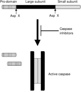
Caspases are a special set of cysteine proteases with an unusual specificity for aspartic acid (reviewed in 13,14). The only other protease known to exhibit the same specificity is granzyme B, a serine protease contained in cytotoxic cell granules which functions to initiate the apoptotic death of target cells. All caspases share a similar structure and are synthesized as a precursor with little or no activity which consists of a pro-domain localized at its amino terminus, a large subunit in the middle of the molecule and a carboxy-terminal small subunit (Fig-ure 2). Activation of caspases proceeds by proteolytic cleavage of the constitutively ex-pressed pro-form at two caspase consensus sites, one that clips off the pro-domain and the other that separates the two subunits. The intriguing nature of the cleavage sites indi-cates that these molecules can undergo auto-activation and also that they can activate each other in an enzymatic cascade similar to the coagulation and the complement cas-cades. Although the large subunit is the one that contains the catalytic domain, it is only active when associated with the small sub-unit. In fact, crystallographic studies revealed that the active caspases are tetramers formed
by the association of two heterodimers and therefore contain two independent catalytic sites (15-17) (Figure 2).
How do we know that caspases are the central executioners in the process of apop-tosis? First, activation of caspases is a very early event which occurs in all forms of apoptosis. Second, prevention of caspase activation by viral or oncogenic proteins, or peptide inhibitors, blocks all the morpho-logical features associated with apoptosis. Third, administration of recombinant cas-pases in cell-free systems results in apopto-tic cytoplasmic and nuclear changes. How-ever, not all members of the caspases are effector molecules in this program. Caspase-6, -7 and -3 are the ones directly implicated with the execution of apoptotic cells. Caspase-8, -10, -9 and -2 are initiator or regulatory caspases, which means that their activities do not directly account for the morphological features of apoptosis. Instead, the primary role of these caspases is to func-tion as signaling molecules, therefore trans-ducing stress signals capable of activating the effector caspases. Other members in-clude caspase-1, -4, -5, -11, -12, and -13 which appear to be primarily involved in inflammatory processes, probably via
pro-Pro-domain Large subunit Small subunit
Asp X
Caspase inhibitors
Active caspase Asp X
teolytic processing of proinflammatory cy-tokines such as IL-1 and IL-18 (18-20).
During apoptosis stimulation, caspases are first activated in the context of multimo-lecular structures called apoptosomes, which can be divided into two subgroups (Figure 3). The first one is related to cell death initiated by cross-linking of so-called death receptors such as CD95 and TNFR1 (see below) while the second is involved in genotoxic stimulation and is composed of three different molecules, i.e., apoptosis ac-tivating factor-1 (APAF-1), cytochrome c and pro-caspase-9. In normal cells, inactive forms of APAF-1 and caspase-9 are present in the cytoplasm whereas cytochrome c par-ticipates in the electron transport chain at the mitochondrial level. Upon stress signals, cy-tochrome c is released from the mitochon-drial intermembrane space into the cyto-plasm where it binds to APAF-1. This inter-action changes the conformation of APAF-1 which subsequently associates with pro-caspase-9, thereby inducing its activation, the activation of the effector caspases (in particular caspase-3) and the initiation of the proteolytic cascade observed in apoptosis
(21).
The fact that a proteolytic activity inside the cell is responsible for its demise led us to think that the main role of caspases was to destroy intracellular proteins in such a way that the cell would not be able to function any longer (22). This view is only partially true. Substrates such as DNA-PKcs,
U1snRNP-70k, lamins, fodrin and the focal adhesion kinase are indeed neutralized by caspase-mediated proteolytic cleavage, thereby contributing to inactivation of both the DNA repair and the RNA splicing ma-chinery, disassembly of the nuclear lamina and alteration in cytoskeleton structure, and cell-cell contact. However, some of the sub-strates are instead activated by caspase cleav-age. For instance, cleavage of the p21-acti-vated kinase PAK2 generates a constitutively active fragment that mediates membrane blebbing and the formation of apoptotic bod-ies (23). Similar activation was also reported for gelsolin (24). Other examples include the cleavage of the Bcl-2 family member Bid (25) and one of the subunits of the DNA fragmentation factor, iCAD/DFF45, so that the other subunit (CAD/DFF40) is released resulting in oligonucleosomal DNA frag-mentation (26).
Judge m e nt in the intrace llular re alm - the m ito cho ndria de cide if ce lls live o r die
So, does this mean that if we block the activity of caspases we will prevent every sign of apoptosis and therefore keep the cells alive? The answer is yes in two situations: a) when apoptosis induction involves an initia-tor caspase, as is the case for apoptosis trig-gered by cross-linking of death-receptors (see below), and b) during developmental cell death when it appears that up-regulation of the expression of effector caspases, such as caspase-3, may lead to the oligomeriza-tion of these molecules thereby activating them and turning on the apoptosis program.
CD95L
CD95
FADD
DED DD
Pró-Caspase-8
Cyt.c
APAF-1
CARD
Pró-Caspase-9
Executor
apoptosome
Initiator
apoptosome Figure 3 - Proposed structure of
t he t w o classes of apopt o-som es. Apoptoo-som es associ-ated w ith the indictment phase of the apoptotic process and therefore related to the death receptors are represented by the CD95L/CD95/FADD/pro-cas-pase-8 apoptosome. In the case of TNFR1- or DR3-initiated apop-totic cascade, the adapter mole-cule TRADD links the cytoplas-mic domain of the death recep-t ors recep-t o FADD/pro-caspase-8 (or -10). The only apoptosome associated w ith the execution
But the answer is no when apoptosis occurs as a consequence of genotoxic stimulation. In this case, pan-caspase inhibitors do block the apoptosis phenotype but are unable to prevent the cell demise which occurs by a non-apoptotic mechanism, apparently coor-dinated by the mitochondria (27-30).
The experimental results obtained in the last few years were fundamental to define mitochondria as stress sensors and as the rheostat that determine whether the cells live or die. In our opinion, perhaps the most important cell death event generated at the mitochondrial level is the release of cyto-chrome c from the mitochondrial intermem-brane space to the cytosol. This brings about at least three major deleterious pathways for the cells. First, as we mentioned above, cyto-chrome c will associate with APAF-1 and initiate the activation of the effector cas-pases leading to apoptosis. Second, cyto-chrome c release from mitochondria dis-rupts the electron transport chain with con-sequent impairment of the production of energy. If cells are not able to compensate for such disturbance, they will certainly die regardless of the activation or not of the caspase cascade. There will be just not enough energy to sustain the basic metabolic reactions that keep cells alive. Third, this same event will increase the generation of reactive oxygen species which are potent cytotoxic agents. Thus, these observations strongly suggest that it is highly unlikely that a cell will remain viable and continue to proliferate when the stress is enough to sig-nal the mitochondria to release their cyto-chrome c. Indeed, we and others have found that such stress signals as cytotoxic drugs, UV- or g-irradiation, enforced expression of Bax or c-myc, glucocorticoids, etc., lead to cytochrome c release and cell death despite the complete inactivation of the apoptosis program by pan-caspase inhibitors (27-30). Therefore, we would like to consider the translocation of cytochrome c from mito-chondria to the cytosol as the moment when
a cell is committed to death. It is important to point out, however, that other mitochondrial events such as the release of other pro-apop-totic factors or the opening of the so-called mitochondrial permeability transition pore may also contribute to this commitment point
(discussed in 31,32). In this regard, it still remains elusive how cytochrome c escapes from the mitochondria.
Like the effector caspase-mediated apop-totic program, the mitochondria-derived com-mitment decision is also molecularly regu-lated. Pro- and anti-apoptotic members of the Bcl-2 family control the mitochondrial rheostat of cell death (33). Recent evidence pointed at two possible survival mechanisms in the mammalian system that operate at the mitochondrial level. One resembles the CED-9/CED-4/CED-3 pathway described in
Caenorhabiditis elegans, and proposes that Bcl-xL binds APAF-1 thus preventing the
activation of caspase-9 and subsequent ap-optosis (34). The other implicates anti-apop-totic members such as Bcl-2 and Bcl-xL in
the maintenance of mitochondrial integrity (reviewed in 35) and the regulation of their membrane permeability to small pro-apop-totic molecules such as cytochrome c (36,37) and apoptosis inducing factor (AIF) (38). It is not completely understood how Bcl-2 members control mitochondrial integrity but, based on the structure of Bcl-xL which
re-sembles pore-forming bacterial toxins and the fact that Bcl-2, Bcl-xL and Bax do form
Bax-in-duced cell death is counteracted by Bcl-2 or Bcl-xL but not by inhibitors of caspases
(30,40). There is, of course, another way the pro-apoptotic members of Bcl-2 family can sensitize a cell for death. They can cause the release of cytochrome c.
In any case, it is important to note that whereas the effector caspases are the central executioners in the apoptosis process, mito-chondria are the arbiter of the life and death decision inside a cell. Also, in many in-stances apoptosis would be more properly considered as a disposal program rather than a killer agenda.
Indictme nt by the e nviro nme nt -signaling thro ugh de ath re ce pto rs
We still know very little about the nature of the pro-death signals that have an impact on the mitochondria. However, as we previ-ously mentioned in this review, initiator cas-pases are able to transduce signals that stimu-late pro-apoptotic changes in the mitochon-dria. One of the initiator caspase-dependent signals involves the proteolytic activation of Bid, a pro-apoptotic member of the Bcl-2 family, by caspase-8. Activated Bid acts on the mitochondria to orchestrate the release of cytochrome c and other cell death events which culminate in apoptosis (25,41).
In comparison to pro-caspase-9 which is activated in the context of the cytochrome c/ APAF-1 apoptosome, caspases-8, -10 and -2 are activated in apoptosomes formed by some members of the tumor necrosis factor tor (TNFR) family, known as death recep-tors, and adapter molecules such as FADD (Fas-associated death domain), TRADD (TNFR-associated death domain), RIP (re-ceptor-interacting protein) and RAIDD (RIP-associated Ich-1/CED-3 homologous protein with death domain). Five death receptors have been described so far in mammals -CD95 (Fas/Apo-1), TNFR1, DR3 (Apo-3/ Wsl/TRAMP/LARD), DR4 and DR5 (Apo-2R/TRAIL-R2/TRICK-2/KILLER), all of
them presenting typical extracellular cys-teine-rich domains that characterize the TNFR members and additional intracellular death domains (DD) (reviewed in 42). Interestingly, every ligand for such molecules belongs to the TNF superfamily and is pro-duced as a trimer. Consequently, these death signals are initiated by trimerization of the receptors, resulting in the association of their DD. This association leads to homotypic interaction with adapter proteins containing DD. FADD binds to CD95 whereas TRADD binds to TNFR1. Besides the DD, FADD has a different domain called DED (death ef-fector domain) which specifically interacts with DED present in the prodomain of cas-pases-8 and -10. The apoptosome CD95/ FADD agglutinates caspase molecules, thereby stimulating their proteolytic activity and initiating the apoptosis cascade (43). TRADD does not contain DED and there-fore cannot bind to caspases. Thus, in order to transduce a death signal, TNFR1/TRADD needs to bring FADD to the complex form-ing the TNFR1/TRADD/FADD apoptosome which afterwards will recruit caspase8 or -10 (44). In addition, TNFR1 may activate a FADD-independent death pathway by en-gaging a different TRADD-binding adapter protein called RIP (45). RIP associates fur-ther with RAIDD (46) which has yet anofur-ther domain responsible for homotypic adhesion. This domain, called CARD (caspase recruit-ment domain) (47), is also present in APAF-1 and in caspases-9 and -2 (48). Whereas the activation of caspase-8 and -10 is dependent on FADD, caspase-2 is activated in a FADD-independent manner, upon formation of the TNFR1/TRADD/RIP/RAIDD apoptosome. However, it is important to note that the predominant death pathway in both cases is the FADD/caspase-8.
seems to be mostly independent of FADD since cells from FADD-deficient mice are fully sensitive to DR4-induced apoptosis, despite the fact that they display a strong resistance to CD95-, TNFR1- and DR3-in-duced cell death (49).
To further complicate the puzzle of mo-lecular interactions involved in death recep-tor-mediated apoptosis, there is another set of membrane proteins designated decoy re-ceptors (DcR). These molecules, represented by DcR1 (TRID/TRAIL-R3/LIT) (50) and DcR2 (TRAIL-R4/TRUNDD) (51), resemble DR4 and DR5 in their extracellular domain but lack a proper intracytoplasmic tail. There-fore, they compete for Apo2L/TRAIL liga-tion without transducing any kind of bio-chemical signal.
Since initiator caspases are directly linked to the death receptor apoptosomes and are required for the proper propagation of the death signal, it is not surprising that pan-caspase inhibitors not only block CD95 or TNFR1-induced apoptosis but also confer resistance to cell death in these circumstances by preventing the mitochondrial event we described as the commitment point.
At least two observations indicate that, even if caspase-8 can bypass the need for mitochondrial events and induce apoptosis by directly activating the effector caspases such as caspase-3, the death receptor-associ-ated caspases seem to induce apoptosis through a mitochondria-dependent mech-anism in most cases. First, in many, but not all cells, CD95-induced apoptosis is blocked by Bcl-2, and this apoptotic pathway is even better prevented by Bcl-xL (52,53). Second,
the addition of mitochondria to cell-free sys-tems results in a full-blown nuclear apopto-sis in conditions where low doses of caspase-8 are not sufficient to induce the typical
nuclear changes. In this system, Bcl-2 is able to block apoptosis induced by low but not high doses of caspase-8 (54).
Co ncluding re marks
Cells may die either by apoptosis or by non-apoptotic mechanisms. If damage is too violent (pathologic), cells have no choice but to undergo the genetically uncontrolled necrotic form of death - they lose membrane integrity, swell and may quickly burst, re-leasing their cellular contents. On the other hand, when damage is more subtle (physi-ologic) it will generate biochemical signals that converge on the mitochondria, special organelles that act as stress sensors having the onus to decide whether the cell may continue to live or should die. In this case, death normally occurs through apoptosis. However, it is important to note that when-ever a cell moves forward to the mitochon-drial commitment point it can no longer be rescued. Not even the complete blockage of the activity of the effector caspases will res-cue this cell from its demise - instead of undergoing the more common apoptosis, the cell will die by a non-apoptotic mechanism. To sum up, the decision between life and death is up to the mitochondria and is regu-lated by members of the Bcl-2 family. In contrast, the execution of the apoptotic pro-gram and the consequent emergence of all morphological changes peculiar to apopto-sis are dependent on the effector caspases.
Ackno wle dgm e nts
Re fe re nce s
1. Kerr JFR, Wyllie AH & Currie AR (1972). Apoptosis: a basic biological phenome-non w ith w ide-ranging implications in tis-sue kinetics. British Journal of Cancer, 26: 239-257.
2. Devitt A, M offatt OD, Raykundalia C, Capra JD, Simmons DL & Gregory CD (1998). Human CD14 mediates recogni-tion and phagocytosis of apoptotic cells.
Nature, 392: 505-509.
3. Voll RE, Herrmann M , Roth EA, Stach C, Kalden JR & Girkontaite I (1997). Immu-nosuppressive effects of apoptotic cells.
Nature, 390: 350-351.
4. Fadok VA, Bratton DL, Konow al A, Freed PW, Westcott JY & Henson PM (1998). M acrophages that have ingested apopto-tic cells in vitro inhibit proinflammatory cytokine production through autocrine/ paracrine m echanism s involving TGF-beta, PGE2, and PAF. Journal of Clinical Investigation, 101: 890-898.
5. Ren Y, Silverstein RL, Allen J & Savill J (1995). CD36 gene transfer confers ca-pacity for phagocytosis of cells undergo-ing apoptosis. Journal of Experimental M edicine, 181: 1857-1862.
6. Savill JS, Dransfield I, Hogg N & Haslett C (1990). Vit ronect in recept or-m ediat ed phagocytosis of cells undergoing apopto-sis. Nature, 343: 170-173.
7. Savill JS, Hogg N, Ren Y & Haslett C (1992). Thrombospondin cooperates w ith CD36 and the vitronectin receptor in mac-rophage recognition of neutrophils under-going apoptosis. Journal of Clinical Inves-tigation, 90: 1513-1518.
8. Fadok VA, Bratton DL, Frasch SC, Warner M L & Henson PM (1998). The role of phosphatidylserine in recognition of apop-totic cells by phagocytes. Cell Death and Differentiation, 5: 551-562.
9. Platt N, da Silva RP & Gordon S (1998). Recognizing death: the phagocytosis of apoptotic cells. Trends in Cell Biology, 8: 365-372.
10. Fadok VA, Voelker DR, Campbell PA, Cohen JJ, Bratton DL & Henson PM (1992). Exposure of phosphatidylserine on the surface of apoptotic lymphocytes trig-gers specific recognition and removal by macrophages. Journal of Immunology, 148: 2207-2216.
11. M art in SJ, Reut elingsperger CPM , M cGahon AJ, Rader J, van Schie RCAA, LaFace DM & Green DR (1995). Early re-distribution of plasma membrane phos-phatidylserine is a general feature of ap-optosis regardless of the initiating
stimu-lus: inhibition by overexpression of Bcl-2 and Abl. Journal of Experimental M edi-cine, 182: 1-12.
12. M art in SJ, Finucane DM , Am arant e-M endes GP, O’Brien GA & Green DR (1996). Phosphatidylserine externalization during CD95-induced apoptosis of cells and cytoplasts requires ICE/CED-3 pro-tease activity. Journal of Biological Chem-istry, 271: 28753-28756.
13. Thornberry NA (1998). Caspases: key me-diators of apoptosis. Chemistry and Biol-ogy, 5: R97-R103.
14. Thornberry NA & Lazebnik Y (1998). Cas-pases: enemies w ithin. Science, 281: 1312-1316.
15. Rotonda J, Nicholson DW, Fazil KM , Gal-lant M , Gareau Y, Labelle M , Peterson EP, Rasper DM , Ruel R, Vaillancourt JP, Thornberry NA & Becker JW (1996). The three-dimensional structure of apopain/ CPP32, a key mediator of apoptosis. Na-ture Structural Biology, 3: 619-625. 16. Walker NP, Talanian RV, Brady KD, Dang
LC, Bump NJ, Ferenz CR, Franklin S, Ghayur T, Hackett M C, Ham m ill LD, Herzog L, Hugunin M , Houy W , M ankovich JA, M cGuiness L, Orlew icz E, Paskind M , Pratt CA, Reis P, Summani A, Terranova M , Welch JP, Xiong L, M öller A, Tracey DE, Kamen R & Wong WW (1994). Crystal structure of the cysteine protease interleukin-1 beta-converting en-zyme: a (p20/p10)2 homodimer. Cell, 78: 343-352.
17. Wilson KP, Black JA, Thomson JA, Kim EE, Griffith JP, Navia M A, M urcko M A, Chambers SP, Aldape RA, Raybuck SA & Livingston DJ (1994). Structure and mech-anism of interleukin-1 beta converting en-zyme. Nature, 370: 270-275.
18. Dinarello CA (1998). Interleuk1 beta, in-terleukin-18, and the interleukin-1 beta converting enzyme. Annals of the New York Academy of Sciences, 856: 1-11. 19. Fant uzzi G, Puren AJ, Harding M W ,
Livingston DJ & Dinarello CA (1998). In-terleukin-18 regulation of interferon gam-ma production and cell proliferation as show n in interleukin-1beta-converting en-zyme (caspase-1)-deficient mice. Blood, 91: 2118-2125.
20. Kim YM , Talanian RV, Li J & Billiar TR (1998). Nitric oxide prevents IL-1beta and IFN-gamma-inducing factor (IL-18) release from macrophages by inhibiting caspase-1 (IL-caspase-1beta-converting enzyme). Journal of Immunology, 161: 4122-4128. 21. Li P, Nijhaw an D, Budihardjo I, Srinivasula
SM , Ahmad M , Alnemri ES & Wang X (1997). Cytochrome c and dATP-depend-ent formation of Apaf-1/caspase-9 com-plex initiates an apoptotic protease cas-cade. Cell, 91: 479-489.
22. M artin SJ & Green DR (1995). Protease activation during apoptosis: death by a thousand cuts? Cell, 82: 349-352. 23. Rudel T & Bokoch GM (1997). M embrane
and morphological changes in apoptotic cells regulated by caspase-mediated acti-vation of PAK2. Science, 276: 1571-1574. 24. Kothakota S, Azuma T, Reinhard C, Klippel A, Tang J, Chu K, M cGarry TJ, Kirschner M W, Koths K, Kw iatkow ski DJ & Williams LT (1997). Caspase-3-generated fragment of gelsolin: effector of m orphological change in apopt osis. Science, 278: 294-298.
25. Li H, Zhu H, Xu CJ & Yuan J (1998). Cleav-age of BID by caspase 8 mediates the mitochondrial damage in the Fas pathw ay of apoptosis. Cell, 94: 491-501.
26. Liu X, Li P, Widlak P, Zou H, Luo X, Garrard WT & Wang X (1998). The 40-kDa subunit of DNA fragmentation factor induces DNA fragmentation and chromatin condensa-tion during apoptosis. Proceedings of the National Academy of Sciences, USA, 95: 8461-8466.
27. Brunet CL, Gunby RH, Benson RSP, Hickman JA, Watson AJM & Brady G (1998). Commitment to cell death meas-ured by loss of clonogenicity is separable from the appearance of apoptotic mark-ers. Cell Death and Differentiation, 5: 107-115.
28. Am arante-M endes GP, Finucane DM , M artin SJ, Cotter TG, Salvesen GS & Green DR (1998). Anti-apoptotic onco-genes prevent caspase-dependent and -independent commitment for cell death.
Cell Death and Differentiation, 5: 298-306. 29. M cCarthy NJ, Whyte M KB, Gilbert CS & Evan GI (1997). Inhibition of Ced-3/ICE-related proteases does not prevent cell death induced by oncogenes, DNA dam-age, or the Bcl-2 homologue Bak. Journal of Cell Biology, 136: 215-227.
30. Xiang J, Chao DT & Korsmeyer SJ (1996). BAX-induced cell death may not require interleukin 1 beta-converting enzyme-like proteases. Proceedings of the National Academy of Sciences, USA, 93: 14559-14563.
32. Green DR & Reed JC (1998). M itochon-dria and apoptosis. Science, 281: 1309-1312.
33. Reed JC (1997). Double identity for pro-teins of the Bcl-2 family. Nature, 387: 773-776.
34. Pan G, O’Rourke K & Dixit VM (1998). Caspase-9, Bcl-XL, and Apaf-1 form a ter-nary complex. Journal of Biological Chem-istry, 273: 5841-5845.
35. M ignotte B & Vayssiere JL (1998). M ito-chondria and apoptosis. European Journal of Biochemistry, 252: 1-15.
36. Yang J, Liu X, Bhalla K, Kim CN, Ibrado AM , Cai J, Peng TI, Jones DP & Wang X (1997). Prevention of apoptosis by Bcl-2: release of cytochrome c from mitochon-dria blocked. Science, 275: 1129-1132. 37. Kluck RM , M artin SJ, Hoffman BM , Zhou
JS, Green DR & New meyer DD (1997). Cytochrome c activation of CPP32-like proteolysis plays a critical role in a Xeno-pus cell-free apoptosis system. EM BO Journal, 16: 4639-4649.
38. Susin SA, Zamzami N, Castedo M , Hirsch T, M archetti P, M acho A, Daugas E, Geuskens M & Kroemer G (1996). Bcl-2 inhibits the mitochondrial release of an apoptogenic protease. Journal of Experi-mental M edicine, 184: 1331-1341. 39. Adams JM & Cory S (1998). The Bcl-2
protein family: arbiters of cell survival. Sci-ence, 281: 1322-1326.
40. Finucane DM , Bossy-Wetzel E, Water-house NJ, Cotter TG & Green DR (1999). Bax-induced caspase activation and apop-tosis via cytochrome c release from mito-chondria is inhibitable by Bcl-xL. Journal of Biological Chemistry, 274: 2225-2233.
41. Luo X, Budihardjo I, Zou H, Slaughter C & Wang X (1998). Bid, a Bcl2 interacting protein, mediates cytochrome c release from mitochondria in response to activa-tion of cell surface death receptors. Cell, 94: 481-490.
42. Ashkenazi A & Dixit VM (1998). Death receptors: signaling and modulation. Sci-ence, 281: 1305-1308.
43. M uzio M , Stockw ell BR, Stennicke HR, Salvesen GS & Dixit VM (1998). An in-duced proximity model for caspase-8 acti-vation. Journal of Biological Chemistry, 273: 2926-2930.
44. Boldin M P, Goncharov TM , Goltsev YV & Wallach D (1996). Involvement of M ACH, a novel M ORT1/FADD-interacting pro-tease, in Fas/APO-1- and TNF receptor-induced cell death. Cell, 85: 803-815. 45. Hsu H, Huang J, Shu HB, Baichw al V &
Goeddel DV (1996). TNF-dependent re-cruitment of the protein kinase RIP to the TNF receptor-1 signaling complex. Immu-nity, 4: 387-396.
46. Duan H & Dixit VM (1997). RAIDD is a new ‘death’ adaptor molecule. Nature, 385: 86-89.
47. Hofmann K, Bucher P & Tschopp J (1997). The CARD domain: a new apoptotic sig-nalling motif. Trends in Biochemical Sci-ences, 22: 155-156.
48. Chou JJ, M atsuo H, Duan H & Wagner G (1998). Solution structure of the RAIDD CARD and model for CARD/CARD inter-action in caspase-2 and caspase-9 recruit-ment. Cell, 94: 171-180.
49. Yeh WC, Pompa JL, M cCurrach M E, Shu HB, Elia AJ, Shahinian A, Ng M , Wakeham A, Khoo W, M itchell K, El Deiry WS, Low e
SW , Goeddel DV & M ak TW (1998). FADD: essential for embryo development and signaling from some, but not all, in-ducers of apoptosis. Science, 279: 1954-1958.
50. Sheridan JP, M arsters SA, Pitti RM , Gur-ney A, Skubat ch M , Baldw in D, Ramakrishnan L, Gray CL, Baker K, Wood W I, Goddard AD, Godow ski P & Ashkenazi A (1997). Control of TRAIL-in-duced apoptosis by a family of signaling and decoy receptors. Science, 277: 818-821.
51. M arst ers SA, Sheridan JP, Pit ti RM , Huang A, Skubatch M , Baldw in D, Yuan J, Gurney A, Goddard AD, Godow ski P & Ashkenazi A (1997). A novel receptor for Apo2L/TRAIL contains a truncated death domain. Current Biology, 7: 1003-1006. 52. M edema JP, Scaffidi C, Krammer PH &
Peter M E (1998). Bcl-xL acts dow nstream of caspase-8 activation by the CD95 death-inducing signaling complex. Jour-nal of Biological Chemistry, 273: 3388-3393.
53. Srinivasan A, Li F, Wong A, Kodandapani L, Smidt Jr R, Krebs JF, Fritz LC, Wu JC & Tom aselli KJ (1998). Bcl-xL functions dow nstream of caspase-8 to inhibit Fas-and tumor necrosis factor receptor 1-in-duced apoptosis of M CF7 breast carcino-ma cells. Journal of Biological Chemistry, 273: 4523-4529.