Ar
ti
cl
e
*e-mail: [email protected]
Analysis of
α
4β
1Integrin Speciic Antagonists Binding Modes: Structural Insights
by Molecular Docking, Molecular Dynamics and Linear Interaction Energy Method
for Free Energy Calculations
João Hermínio Martins da Silva,a,b Laurent Emmanuel Dardenne,c Wilson Savinob and
Ernesto Raul Caffarena*,a
aPrograma de Computação Cientíica - Fundação Oswaldo Cruz, Antiga Residência Oicial,
Av Brasil 4365, 21045-900 Rio de Janeiro-RJ Brazil
bLaboratório de Pesquisa sobre o Timo, Instituto Oswaldo Cruz - Fundação Oswaldo Cruz,
Av Brasil 4365, 21045-900 Rio de Janeiro-RJ Brazil
cGrupo de Modelagem Molecular em Sistemas Biológicos, Laboratório Nacional de Computação
Cientíica, Av Getúlio Vargas 333, 25651-075 Petrópolis-RJ Brazil
A integrina VLA-4 (integrina α4β1) participa da isiopatologia de uma grande variedade de
doenças, incluindo asma, esclerose múltipla, artrite reumatóide e diabetes. A seletividade de ligantes por essa integrina permanece ainda um problema sem resposta, principalmente devido ao fato das estruturas 3D da maioria delas serem ainda desconhecida. Inicialmente, construímos um modelo computacional tridimensional do sítio ativo da integrina α4B1, utilizando a estrutura cristalográica
da integrina αVβ3 como molde. Após, realizamos um estudo por docking molecular usando um
conjunto de sete antagonistas de α4β1 derivados de 4-[N’-(2-metilfenil)ureido]fenilacetil, avaliando os seus modos de ligação. A técnica de dinâmica molecular foi utilizada de modo a aumentar a
eiciência da exploração do peril energético obtido pelo algoritmo de docking. Segundo a nossa
análise, os compostos foram sistematicamente agrupados em dois modos de ligação principais, sendo a conformação estendida o modo prevalente de ligação dos antagonistas no sítio. Cálculos de energia livre também conirmaram essa constatação. Esse estudo aumenta a compreensão dos mecanismos de ligação dessa família de antagonistas e pode fornecer informações úteis ao desenho racional de fármacos especíicos para o VLA-4.
The VLA-4 antigen (α4β1 integrin) is involved in the pathophysiology of a variety of diseases including asthma, multiple sclerosis, rheumatoid arthritis and diabetes. The ligand selectivity toward this integrin remains a dificult problem, mainly due to the fact that 3D structures of most integrins are still unknown. We initially built a 3D computational model of the α4β1 ligand binding site, taking the crystal structure of the integrin αVβ3 as template. Then, we performed a computational study on a set of seven α4β1 antagonists, evaluating the binding modes of 4-[N’-(2-methylphenyl)
ureido]phenylacetyl and derivatives by molecular docking. Molecular dynamics simulations were used to improve the receptor-ligand energy landscape exploration by the docking algorithm. The compounds were systematically arranged in two main binding modes, and in all cases, pointed out that these antagonists preferably bind to the α4β1 integrin active site in an extended conformation that resembles the one in solution. LIE (linear interaction energy) calculations also conirmed this statement given that the most prevailing binding mode is also the energetically most favored one. This study beneits the comprehension of the mechanism of this family of antagonists and may provide useful information for rational drug design.
Introduction
Integrins of the β1 sub-family (formerly named
very late antigens of VLAs) correspond to at least nine
transmembrane heterodimers composed of a given α
subunit noncovalently coupled to the β1-integrin chain
(CD29). These molecules are able to bind one or more extracellular matrix (ECM) ligands, although some of them can also interact with a cell membrane counter-receptor.
Functionally, β1 integrins are related to cell migration,
both in normal and pathological conditions, such as
inlammation and metastasis.1-3
Of particular interest is α4β1 integrin (VLA-4 or CD49d/
CD29), which is expressed by a variety of hemopoietic cell types including monocytes, T and B lymphocytes, basophils, and eosinhophils. This integrin binds the alternatively spliced type III connecting segment (CS-1) of ibronectin, as well as the vascular cell adhesion molecule-1 (VCAM-1). From a therapeutic point of view, it has been shown that blocking VLA-4/VCAM-1 interaction resulted in amelioration of distinct inlammatory diseases, such as asthma, multiple sclerosis, rheumatoid arthritis and type
1 diabetes.4-8
The α4β1 integrin binds to ibronectin in the ECM and to
VCAM-1 on the endothelium, through the minimum binding
epitope Leu-Asp-Val (LDV), discovered by Komoriya et
al.9 This pattern of three residues is homologous to the
Leu-Asp-Ser (LDS) motif found in VCAM-1, which has been
reported as the VCAM-1 binding site of α4β1, suggesting
that VCAM-1 and ibronectin could share an identical or
overlapping α4β1 binding site.5
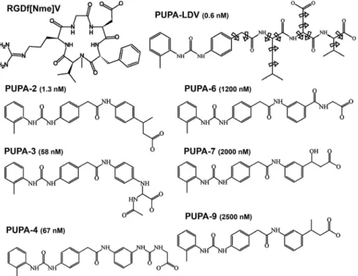
A large number of LDV mimetic moieties have been identiied as potent and often subtype-selective ligands.
Namely, the fibronectin-derived compound 4-[N
’-(2-methylphenyl)ureido]phenylacetyl-Leu-Asp-Val (PUPA-LDV) has been proved to be a potent highly selective
antagonist of the α4β1.10 A set of analogue compounds,
based on the PUPA-LDV leading structure, was obtained from a computational database searching method and their
IC50 values have been measured.10,11
However, detailed structural information about ligand-receptor speciic interactions between this particular family
of ligands and the α4β1 integrin remains relatively poor.
This is likely due to the intrinsic dificulty in obtaining the three-dimensional structure of membrane receptors such as
α4β1 through X-ray crystallography.
We applied a combined approach of homology modeling, ligand-receptor molecular docking and molecular dynamics (MD), in order to determine the
main binding modes of 4-[N’-(2-methylphenyl)ureido]
phenylacetyl-LDV and derivatives within the binding site
of the α4β1 integrin. MD simulations were performed to
identify the most prevalent ligand conformations in solution and establish a connection with its binding modes within the
α4β1 integrin active site. Linear interaction energies (LIE)
calculations were also carried out to identify the preferred binding modes, suggesting that there exists a preference for a speciic way of binding.
Methodology
Comparative Modeling
The alignment between the integrin α4β1 and the
template αVβ3 was obtained using the Align software of
EMBOSS package. Sequences were retrieved from the Entrez Protein database, under NP000876 e P05556 access numbers. The BLOSUM62 matrix was used with a gap and
extension penalties of 11 and 1, respectively.12
A homology model of the β1 subunit α4β1 integrin
has been previously reported by You et al.13 This work
served us as a landmark to model the whole headpiece
of human α4 and β1 subunits with the program Modeller
(version 7.0) according to the comparative protein modeling
methodology.14, 15
It is very unlikely that the conformational transitions
that integrins undergo (i.e., conversions between different
conformations that represent diverse activity levels) would occur spontaneously during MD simulations using
current approaches. Therefore, the structure of the α4β1
integrin headpiece has been modeled using as template
the X-ray structure of the αVβ3 integrin16 in complex with
cyclo(-RGDf[Nme]V-) (from hereafter, RGD-ligand), also known as cilengitide, (PDB entry code: 1L5G) to ensure that the active conformation of the binding site is correctly modeled. This three-dimensional structure, extensively
used in previous works,5,17 is the only available template
so far for building homology model of integrins lacking I-domain in the active state. Moreover, the integrin family is a remarkably well conserved family and this information
is helpful concerning the building of a homology model.18
The human β1 and β3 domains are homologous, sharing
43% identity in the sequence alignment; 33.5% of identity
is found between α4 and αV subunits. The sequence lengths
of the headpiece encompass 237 and 431 residues for
β and α domains, respectively. These percentages of residue identities guarantee an acceptable reliability for the generated structural homology model.
Thirty full-atoms models were built and ranked using the Modeller objective function, which is highly eficient in classifying different models calculated from the same
integrin headpiece was subjected to further reinement through a short minimization protocol using the Gromacs
classical force ield.20 Prior to the minimization, the atomic
coordinates of Mg2+ ions of the Metal Ion Dependent
Adhesion Site (MIDAS) motif were merged into the binding site as present in the crystal structure used as template. In a previous study with the integrin alpha5beta1, the high conservation of the binding site was conirmed between alphaVbeta3 and alpha5beta1. Furthermore, according to
mutagenesis data21, cross linking22 and X-ray structure23
the coordination of MIDAS is conserved and indicates this region as a key element for interacting with ligands similar to RGD.
The stereochemical quality of the resulting model
was checked with the program PROCHECK24,25 and
VERIFY3D.26
Molecular docking
Firstly, we tested the computational approach to αVβ3
integrin in complex with RGD-ligand to set basic rules
and protocols of the docking procedure (i.e., determination
of a suitable set of parameters to reproduce experimental results) and compared the obtained results with the binding mode of RGD-ligand.
The docking of RGD ligand to the αVβ3 integrin
headpiece was carried out using the Autodock program
package version 3.0.527 allowing full lexibility to the
residue side chains.
The Lamarckian-Genetic-Algorithm (LGA), as implemented in the Autodock program, was applied using a protocol of 2048 independent runs with an initial population size of 300 individuals, a maximum number of 500000 evaluations, a mutation rate of 0.02, a crossover rate of 0.80 and an elitism value of 1. For the local search, the Pseudo Solis and Wets algorithm was applied and another non-speciied values were left to their respective default values. Each docking experiment was performed in a grid of
127 points per dimension, with a discretization of 0.275 Å,
encompassing the binding site located in the headpiece of the heterodimer N-terminal.
This set of parameters resulted in an optimum way to reproduce the experimental binding modes of RGD-ligand
in the αVβ3 integrin active site, which was extrapolated
to docking experiments of PUPA ligands within the α4β1
headpiece.
The molecular docking experiments were carried out allowing PUPA-LDV and derivatives (Figure 1) to rotate freely except torsion rotations in amines and peptide bonds.
The crystal structure of the extracellular domains of the
αVβ3 integrin in complex with the RGD-ligand allowed us to
better understand the ligand binding mode. It was observed that the acid oxygen atoms point to the central MIDAS
ion position. Previous reports showed the importance of the MIDAS motif for binding to carboxylic groups of acid
residues.18,28,29 This is the main reason why, out of the 2048
solutions obtained for each ligand, we have only selected those that presented any of their carbonyl oxygen atoms directed towards the central MIDAS ion using a cut-off of 3.5 Å. The exclusion criterion has been previously
applied17 based on structural information observed for
RGD-ligand in the X-ray complex16 and its importance has
been discussed.30 Due to this exclusion procedure, a larger
number of independent runs were necessary to assure a better statistical analysis.
Due to the high number of degrees of freedom of the investigated ligands, the docked conformations differing by less than 3.5 Å in positional root mean square deviation (rmsd) were clustered together and represented by the outcome with the most favorable free energy of binding.
MD simulations
To ind out the preferred conformations of PUPA-LDV and derived compounds isolated in aqueous solution, MD simulations were carried out in explicit solvent.
Calculations were done with the GROMACS package20 and
the Gromos96 classical force ield31 in the Gibbs ensemble
(300 K, 1 bar).32 Atomic partial charges were obtained using
the MMFF94 parameterization.33
Coordinates of the molecular complex were immersed
in a cubic SPC34 water box of volume 125 nm3 and
minimized using periodic boundary conditions to adjust the atomic positions to the force ield. All interatomic
bonds were constrained using the LINCS algorithm.35
Non-bonded interactions were taken into account using the 6-12 Lennard-Jones potential using a cut-off radius of 14 Å and a Coulomb potential with reaction ield with dielectric constant equals to 66 and cut-off radius of 18 Å. MD simulations evolved freely for 5 ns, saving trajectories and velocities every 1 ps to analyze structural features.
Linear interaction energy
Calculations of relatives free energies for different modes of PUPA-LDV and its most active derivatives
were performed using the LIE method36 with the software
package Q37 and the Gromos96 force ield.31 The systems
were solvated with SPC water within a 20 Å sphere and non-bonded interactions across the boundary were excluded. The non-bonded interaction energies were calculated without cut-off restrictions while long-range electrostatics
were treated using a multipole expansion.38 Atoms outside
of the 18.5 Å simulation sphere were harmonically
restrained to their initial positions with a 200 kcal mol-1 Å-2
force constant.
The compounds in the free state were located at the centre of the simulation sphere and restrained with a force
constant of 5 kcal mol-1 Å-2 to guarantee an homogeneous
solvation. All compounds were heated from 1 to 300 K using a stepwise scheme followed by an equilibration period to stabilize ligand surrounding energies before the data collection phase (500 ps). The time step used in the production phase of the simulations was 1 fs, and the temperature was set to 300 K using a weak coupling to
an external bath. SHAKE39 was used to constrain bonds
and angles on solvent molecules. The total simulation time for the production period was 750 ps for the ligands in aqueous solution and 1 ns for the complex. Energies were sampled every 0.5 ps in the production part of the simulations, which were then used in equation (1) to estimate the free energy of binding. The MD run until the collected energies showed stability for a period not shorter than 1000 ps. Stability was addressed by comparing the average values of the irst and second halves of the collection period. Error estimates in the calculated free energies were obtained by adding the errors in the potential energy from both the water and protein simulations, properly scaled through the use of the LIE
α and β factors.
In equation (1), Uel and ULJ are the electrostatic and
Lennard-Jones interaction energies between ligand (l)
and its surroundings (s) in the binding site and in aqueous
solution, respectively; the brackets < >’s denote MD
ensemble averages over trajectories and α, β and γ are
empirical parameters.
∆Gbinding = α(〈ULl-sJ〉bound – 〈ULl-sJ〉free) + β(〈Ul-sel〉bound – 〈Ul-sel〉free) + γ (1)
In detailed studies of ligand binding, Hansson et al.40, 41
have determined that an α value of 0.181 can adequately
reproduce the free energies of binding for a variety of
ligand-protein systems, along with a set of β values
determined by Free Energy Perturbation (FEP), depending on the chemical nature of compounds. According to these
calculations, the β value is 0.5 for charged ligands.
Results and Discussion
Homology Model
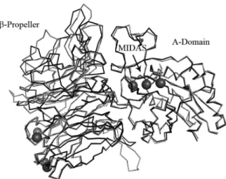
As the complete three-dimensional structure of α4β1
integrin is not available, we used the X-ray crystallographic
model structure of the α4β1 integrin portion containing the ligand binding site (Figure 2).
The β-Propeller and βA domains, responsible for
the ligand binding, constitute the headpiece of the α4β1
integrin. This particular region includes 431 residues of
the α subunit and 237 of the β subunit. Out of these amino
acids only 48 and 37, from α and β subunits respectively,
are in the interface between each other. These values were
calculated using the PISA Server.42 Only three hydrogen
bonds were observed between α and β subunits in the
model. (Tyr253:α-Gly286:Β, Tyr364:α-Ala320:β, and
Glu370:α-Lys289:β). The solvent accessible surface (SAS)
was approximately 29,976 Å2, with only 2,977 Å2 in the
interface region.
The rmsd value between the β-propeller domain of
the template and the α4 domain of the model was 0.75 Å
(considering only the alpha carbons). For the βA domains
(i.e., β1 template and β3 model) we found a rmsd of
0.57 Å. Figure 2 shows the superposition of α4β1 and αVβ3
headpieces.
A particular attention was paid on the modeling of the β
subunit amino acids that form the MIDAS and ADMIDAS regions (Asp150, Lys151, Ser152, Tyr153, Ser154, Glu249, Asp279) due to the crucial role that the divalent ions play in the binding process.
The stereochemical quality of the resulting model was evaluated with the program PROCHECK. Global G-factors
of –0.36 and –0.11 were obtained for β-propeller and βA
domains, respectively.
The majority of the residues of the modeled protein occupied the most favored regions of the Ramachandran plot, while others were located in additional allowed regions. 94.5% (alpha domain) and 96.6% (beta domain) of the residues are in the most favored regions; 4.4% (alpha domain) and 2.4% (beta domain) in additional allowed
regions and only 1.1% (α domain) and 1.0% (β domain)
were in disallowed regions.
The 3D-1D quality was also validated with VERIFY3D and no errors were detected. According to this software,
85.42% of the α subunit residues and 85.71% of β subunit
residues presented an average 3D-1D score greater than 0.2, corroborating the good quality of the modeled structure of the binding site.
Molecular Docking
Firstly, we performed docking essays between
RGD-ligand and αVβ3 as a way to determine the most suitable
docking algorithm parameters to work with. The results obtained with the best parameter set reproduced the crystallographic structure (1L5G) conformation of the ligand in almost 60% of the cases, with at least 0.5 Å of rmsd.
To deine the binding modes for the PUPA’s set of ligands we docked these molecules within the gap of the
integrin headpiece, between the β propeller and βA domains.
After selection (i.e., binding modes where the ligand
directs the carboxyl group of atoms towards the MIDAS ion) and clustering of docked solutions sixty four different families could be found for the PUPA-LDV, with rmsd values less than 3.5 Å between members of the same family. For the remaining compounds, the number of families ranged from 8 to 12 excepting PUPA-2, where only three families fulfilling the selection criterion were found. Despite the large number of families, docking calculations also showed that PUPA-LDV and its derivatives were able to be systematically assembled in two main binding modes, deined by the position of the N terminal cap in the cleft, named hereafter MODE1 and MODE2 (Figure 3). A third (but less deined) binding mode, characterized by exposing the PUPA cap to the solvent, was seldom observed.
A set of common interactions was detected in the binding modes for all investigated ligands. For instance, atoms of the terminal carboxyl group (as well as the side chain oxygen atoms of aspartic acid for PUPA-LDV) were
always coordinating the Mg2+ in the MIDAS region. Ser154,
which also participates in the metal ion coordination, together with Ser247 played a signiicant role in the binding of the compounds setting up hydrogen bond contacts with
Figure 2. Three-dimensional structure of extracellular domains of α4β1
integrin overlapped on the template. The superposition was performed considering all the backbone atoms. α4β1 loops are shown in dark grey,
these atoms. Main chain atoms of Tyr153 and Asn244 also contributed to stabilize the conformation of the ligand.
The main feature of the docked ligands in MODE1 was that they remained completely stretched, inside the crevice
between the propeller and the βA domain on the integrin
head, in such a way that the PUPA cap mainly interacted with the residues Lys158, Lys159, Lys190 and Thr208.
Cation-π interactions were apparently formed between
side chains of Lys159, Lys158, Lys190 and the aromatic rings of the N-terminal cap. Although the force ield has not been parameterized to take into account these special interactions, it is possible to envisage its existence due to the proximity of the intervening groups. Furthermore, the side chain of Thr208 usually hydrogen bonded to the PUPA carbonyl oxygen atom.
In MODE2, the compounds adopted a bent conformation in such a way that PUPA was located in the vicinity of
Val245, Lys246 and Phe247. Cation-π interactions were
also observed between Lys246 and PUPA rings. Peptide bond atoms of Val245 frequently hydrogen bonded the carbonyl or amide atoms of the ligand.
Finally, in MODE3 PUPA cap was situated outside the
gap formed between the β propeller and βA domains. This
particular dizzy mode presented a less deined pattern, when compared to the previous ones, and their free energies of binding were always higher than the ones obtained in previous modes. Also, this speciic mode, which was not observed for all ligands, was less populated and less probable, given that the hydrophobic PUPA rings were completely exposed to water. Interactions between PUPA-LDV (and derivatives) and
the α4β1 integrin binding site are summarized in Table 1.
The existence of interactions was determined based on
distance criteria, namely, cation-π < 4.5 Å, hydrogen bond
(O-O) < 3.5 Å, and electrostatic interaction < 5.0 Å. When analyzing the results for all ligands, we were not able to ind a good correlation between the calculated
free energies of binding and the experimental IC50’s.11 This
result was already expected because even though the current docking functions used to evaluate ligand-receptor binding afinities are suitable for ranking ligands of large databases, they have a poor capability to predict quantitatively, or even qualitatively, useful information about ligand-receptor
afinities.43 It is important to note that the lack of correlation
between the experimental and the calculated ligand-receptor binding afinities are not directly associated to a poor performance of docking programs in reproducing the experimentally observed ligand-receptor binding modes. Actually, the current docking methodologies have a good performance to generate ligand conformations similar to
Figure 3. Binding modes of VLA-4 within the binding site represented as Connolly surface. MODE1 is seen in the upper panel, whereas MODE2 is depicted in the lower panel. The metal ions at the MIDAS region are represented as spheres.
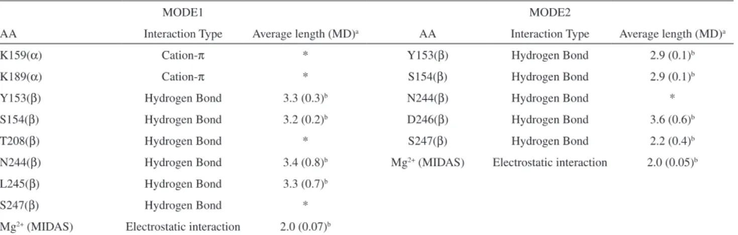
Table 1. Major interactions between PUPA-LDV (and derivatives) and α4β1 integrin binding site. Determination of interactions was based on distance criterion: cation-π < 4.5 Å, hydrogen Bond (O-O) < 3.5 Å, electrostatic interaction < 5.0 Å. Intermolecular interactions were mapped from docking calculation and monitored through MD simulations. Average length column shows the mean interaction distances (Å) held along the MD simulations. The asterisk means that the interaction was not maintained
MODE1 MODE2
AA Interaction Type Average length (MD)a AA Interaction Type Average length (MD)a
K159(α) Cation-π * Y153(β) Hydrogen Bond 2.9 (0.1)b
K189(α) Cation-π * S154(β) Hydrogen Bond 2.9 (0.1)b
Y153(β) Hydrogen Bond 3.3 (0.3)b N244(β) Hydrogen Bond *
S154(β) Hydrogen Bond 3.2 (0.2)b D246(β) Hydrogen Bond 3.6 (0.6)b
T208(β) Hydrogen Bond * S247(β) Hydrogen Bond 2.2 (0.4)b
N244(β) Hydrogen Bond 3.4 (0.8)b Mg2+ (MIDAS) Electrostatic interaction 2.0 (0.05)b
L245(β) Hydrogen Bond 3.3 (0.7)b
S247(β) Hydrogen Bond *
Mg2+ (MIDAS) Electrostatic interaction 2.0 (0.07)b
aAverage length column shows the mean interaction distances (Å) held along the MD simulations; bThe average lenght column shows the mean interaction
crystallographically determined ligand-receptor structures. In most cases, the correct reproduction of the binding mode does not coincide with the binding afinity prediction
performance.43 This lack of correlation is likely due to a poor
representation of important energy contributions, such as protein lexibility, solvation and entropy. However, PUPA-LDV ligand was correctly identiied as the ligand with the best afinity for the receptor. Our results also indicated that the PUPA-LDV binding MODE1 was the preferred one.
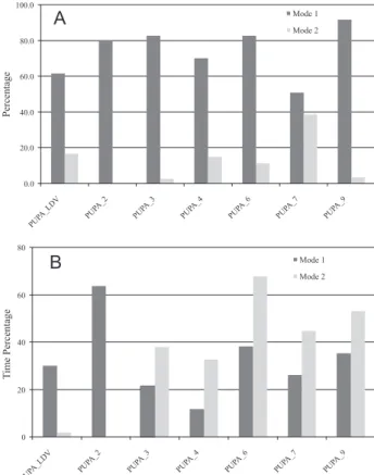
When analyzing the results for the derived compounds, we observed a lack of correlation with experimental ligand binding afinities. However, we observed a clear preference for binding MODE1 for this particular set of ligands (Figure 4A) and also noticed that docking distances
between MIDAS Mg2+ ion and the acid oxygen atoms were
always lower for MODE1 than for MODE2 (Table 2).
MD simulations
The combination of molecular dynamics and docking procedures to investigate how ligands interact with receptors
is well established.44,45 Most of the studies apply molecular
dynamics to improve the position of the ligand within the binding site, after a docking searching. Herein, we used molecular dynamics as an approach attempting to correlate the preferential ligand binding conformations within the
α4β1 integrin active site and their dynamical behavior
when isolated in solution. Molecular dynamics simulations were performed to investigate thermodynamical (LIE calculations) and dynamical aspects (permanence time of docked solutions in water) of the ligands in the binding site and in solution. However, some structural features of the dynamical behavior of ligands within the headpiece of the integrin could be monitored. MD simulations showed that some of docking interactions were unstable, especially
those forming cation-π with the PUPA cap, different from
the hydrogen bonds formed with structural MIDAS amino acids (Ser152, Tyr153, Ser 154, Asn244, Leu245, Asp246 and Ser247). Average distance values and luctuations along 1 ns of simulation are shown in Table 1.
The LIE free energy methodology indicated that the PUPA-LDV, in MODE1, resulted as the best ligand conformation for the whole series of compounds, supporting docking observations. According to the set of parameters used for LIE determination, MODE2 showed
a high energy gap of almost 120 kcal mol-1, (Figure 4B)
with respect to the MODE1 conformation. Looking at the electrostatic energy contribution, we presumed that such interactions might have an important role in the prevalence of that particular binding mode, since this ligand presents 2 negatively charged groups, (Asp side chain and the Val C-terminal). It is important to note that during the dynamics evolution, the C terminal of PUPA-LDV drifted towards the
MIDAS Mg2+ ion, producing a decrease in the electrostatic
energy, facilitated by the high lexibility of this ligand. For the whole set of ligands, the MODE1 showed lower electrostatic energies when compared to MODE2 (Table 3),
Table 2. Distances (Å) between MIDAS Mg2+ and carboxylic oxygen
atoms for both binding modes. The distances were obtained from docking calculation and 1 ns of MD simulations
Ligand MODE1 MODE2
Docking MD Docking MD
PUPA-LDV 2.1 1.99 (0.07)a
2.2 1.97 (0.05)a
PUPA-2 2.2 1.94 (0.04)a ---
---PUPA-3 1.7 1.93 (0.05)a
2.2 1.93 (0.08)a
PUPA-4 1.8 1.97 (0.05)a 3.1 1.94 (0.05)a
PUPA-6 1.6 1.94 (0.05)a
1.8 1.92 (0.05)a
PUPA-7 1.6 1.95 (0.05)a 1.8 1.95 (0.06)a
PUPA-9 1.7 1.94 (0.04)a 2.7 1.94 (0.08)a astandard deviation values.
contributing to a more favorable interaction. The energy difference between the MODE1 and MODE2 showed an
average value of 35 kcal mol-1 for the compounds, except
PUPA-LDV that presented a more accentuated gap between binding modes.
Although, the observed shorter distances between
charged oxygen atoms and Mg2+ for MODE1 obtained
by Docking procedure might explain the prevalence of MODE1 over MODE2, MD simulations showed that once in motion in the integrin active site, the charged groups of compounds maintained similar distance values for both binding modes. Apparently the proximity of the N-terminal CAP with several charged residues as Lys155, Lys159, Lys189, Lys190 and Glu157, in MODE1 appears to be responsible for the lowest electrostatic energies. On the other hand, the MODE2 showed only two charged residues near the PUPA cap: Lys246 and Glu276 with their side chains exposed to the solvent.
Molecules in solution are in constant motion and conformational transitions between different states occur frequently. During the binding process, the guest molecule experiences a conformational entropy loss
(∆S < 0), resulting in a more static conformation within
the active site. This effect of reduction of the ligand conformational entropy during the binding process is
more important for highly lexible ligands (i.e., high
number of conformational degrees of freedom), which generally results in a decrease of the afinity of a lexible
ligand towards a given receptor (i.e., increases the free
energy of binding, ∆G = ∆H - T∆S). For a particular
flexible ligand, a decrease in the conformational entropy can be compensated by strong receptor-ligand interactions. However, it is also natural to expect that the conformational entropy loss effect can be less important if the lexible ligand binds to the receptor active site in a conformation close to that in solution.
Another important point associated with high lexible ligands is the fact that the receptor-ligand docking algorithms present a success rate of inding the correct ligand-receptor binding modes which is very sensitive to the
number of the ligand conformational degrees of freedom.44
The ability of the current docking searching procedures to explore successfully the associated ligand-receptor energy hypersurface decreases with the increasing of the ligand
lexible bonds number.45
Furthermore, we attempted to correlate the preferential
ligand binding conformations within the α4β1 integrin active
site and their dynamical behavior when isolated in solution. In order to verify if the MODE1 and MODE2 docked conformations were feasible in solution, we compared them to the ones obtained from MD simulations trajectories of the molecules in explicit solvent, through the calculation of the rmsd values. Figure 4 shows the percentage of time the conformations obtained from the trajectories present rmsd
≤ 2.0 Å, taking MODE1 and MODE2 docked solutions as
references respectively.
Table 3. Electrostatics and van der Waals energies obtained from LIE calculations. Free energy values are calculated according to LIE equation.
∆Gbind = α(〈U L l-s
J〉 bound – 〈U
L l-s
J〉
free) + β(〈U l l-s
e〉 bound – 〈U
l l-s
e〉
free) + γ where energy units are in kcal mol
–1. Values in parenthesis indicate luctuations
Water Complex
MODE1 MODE2
Eel EvdW Eel EvdW ∆G Eel EvdW ∆G
PUPA-LDV –408.70(18.07) –34.31(0.52) –662.23(7.80) –56.35(2.77) –130.75 –397.79(7.51) –54.26(1.43) –7.59
PUPA-2 –181.46(1.99) –32.48(0.40) –285.81(2.10) –47.15(0.88) –54.83 - -
-PUPA-3 –179.21(2.71) –35.08(0.63) –310.10(6.48) –50.59(1.53) –68.25 –249.59(1.88) –51.49(1.10) –35.54 PUPA-4 –190.83(2.77) –33.15(0.59) –317.28(8.06) –47.65(1.65) –65.85 –248.88(5.10) –51.85(1.79) –31.65
PUPA-6 –190.20(2.88) –32.87(0.65) –350.06(3.74) –43.84(1.60) –81.91 –286.33(6.06) –53.92(2.88) –52.42 PUPA-7 –176.86(3.51) –30.62(0.81) –313.12(7.60) –43.81(1.73) –70.51 –255.76(5.48) –45.39(1.77) –40.87
PUPA-9 –177.47(2.94) –32.46(0.55) –301.46 –50.36(2.52) –65.24 –248.48(8.28) –53.69(1.81) –39.48
The most active ligands UPA-LDV and PUPA-2 exhibited a clear preference for conformations in solution close to the more extended MODE1 docked conformation and MODE2 conformation was not observed in solution for PUPA-2 at all (Figure 5). It is also interesting that as
long as the IC50 values increases, increases the proportion
of MODE2 in solution, as well.
These results provide good evidence that the conformation in solution of compounds is somehow related to their prevalence for binding the receptor. So, the existence of prevailing conformations in solution similar to the observed ones when the inhibitor is within the integrin active site, can be an indication that the energetic costs of ligand conformational relaxation on complex formation are minimized. Likewise, the entropic cost of the binding process is diminished.
Conclusions
Herein, we combined computational methodologies from MD simulations and molecular docking, to predict the binding modes of a set of ligands, derivatives of
the 4-[N’-(2-methylphenyl)ureido]phenylacetyl-LDV
compound within the gap between the α and β subunits
of α4β1 integrin.
For these highly lexible compounds, we used MD simulations results to find the most probable ligand conformations in solution, correlating them with the
potential binding modes within the binding site of α4β1.
Docking experiments of PUPA-LDV and derivatives in the active site of the VLA-4 antigen have identiied two main binding modes, with a well deined binding pattern. These two binding modes cases present interactions
between the antagonists and both subunits of the α4β1
integrin. In MODE1, the ligands remain completely stretched inside the crevice between the propeller and the
βA domain on the integrin head. In MODE2 the ligands
adopt a bent conformation in such a way that the PUPA cap is located in the vicinity of Val245, Lys246 and Phe247
of α subunit.
The amino acids Lys159, Lys189 and LysK190 of the α
subunit, and Tyr153, Ser154, Thr208, Asn244 and Ser247 of
the β subunit associated to the binding MODE1 and Tyr153,
Ser154, Asn244 and Ser247 of the β subunit associated to
the binding MODE2 were identiied as essential residues for the binding process, converting them into interesting targets for site directed mutagenesis experiments.
The analysis of the free energy of binding (LIE), provided by the Autodock scoring function, has correctly identiied PUPA-LDV as the ligand with the best afinity for the receptor. For its derivatives no correlation was
obtained with the reported IC50’s values. However, the
LIE calculations have shown that practically all ligands
have a clear preference for conformations within α4β1
active site associated with binding MODE1, i.e., there
exists a prevailing stretched conformation of PUPA-LDV and derivatives inside the active site cavity. It is very likely that the existence of the stretched conformation in solution helps the molecular recognition, at least for the two most active ones. In this sense, the predominance of an extended conformation in solution is a potential feature that would help in the binding process resulting in an enhanced afinity of the prototype compound for
α4β1 integrin. Additionally, MD simulations of ligands
in solution can be valuable approach to enable prediction
of the binding modes of potentially new α4β1 integrin
antagonists.
Acknowledgements
This work was supported by Fundação Oswaldo Cruz (FIOCRUZ), Laboratório Nacional de Computação Científica (LNCC) and Conselho Nacional de Desenvolvimento Cientíico e Tecnológico (CNPq). The authors also thank inancial support provided by the Centro Argentino-Brasileiro de Biotecnologia (CABBIO).
References
1. Hynes, R. O.; Cell 1992, 69, 11.
2. Ahmed, N.; Riley, C.; Rice, G.; Quinn, M.; Clin. Exp. Metastasis 2005, 22, 391.
3. Giannelli, G.; Astigiano, S.; Antonaci, S.; Morini, M.; Barbieri, O.; Noonan, D. M.; Albini, A.; Clin. Exp. Metastasis 2002, 19, 217.
4. Abraham, W. M.; Sielczak, M. W.; Ahmed, A.; Cortes, A.; Lauredo, I. T.; Kim, J.; Pepinsky, B.; Benjamin, C. D.; Leone, D. R.; Lobb, R. R.; J. Clin. Invest. 1994, 93, 776.
5. Macchiarulo, A.; Costantino, G.; Meniconi, M.; Pleban, K.; Ecker, G.; Bellocchi, D.; Pellicciari, R.; J. Chem. Inf. Comput. Sci. 2004, 44, 1829.
6. Haler, D.A.; J. Clin. Invest. 2004, 113, 788.
7. Yang, X. D.; Karin, N.; Tisch, R.; Steinman, L.; McDevitt, H. O.; Proc. Natl. Acad. Sci. U.S.A 1993, 90, 10494.
8. Seiffge, D.; J. Rheumatol. 1996, 23, 2086.
9. Komoriya, A.; Green, L. J.; Mervic, M.; Yamada, S. S.; Yamada, K. M.; Humphries, M. J.; J. Biol. Chem. 1991, 266, 15075. 10. Lin, K.; Ateeq, H. S.; Hsiung, S. H.; Chong, L. T.; Zimmerman,
11. Singh, J.; Van Vlijmen, H.; Liao, Y.; Lee, W. C.; Cornebise, M.; Harris, M.; Shu, I. H.; Gill, A.; Cuervo, J. H.; Abraham, W. M.; Adams, S. P.; J. Med. Chem. 2002, 45, 2988.
12. Rice, P.; Longden, I.; Bleasby, A.; Trends Genet. 2000, 16, 276. 13. You, T. J.; Maxwell, D. S.; Kogan, T. P.; Chen, Q.; Li, J.; Kassir,
J.; Holland, G. W.; Dixon, R. A.; Biophys. J. 2002, 82, 447. 14. Sali, A.; Blundell, T. L.; J. Mol. Biol. 1993, 234, 779. 15. Sali, A.; Potterton, L.; Yuan, F.; van Vlijmen, H.; Karplus, M.;
Proteins-Struct. Funct. Genet. 1995, 23, 318.
16. Xiong, J. P.; Stehle, T.; Zhang, R.; Joachimiak, A.; Goodman, S. L.; Arnout, S. L.; Science 2002, 296, 151.
17. Marinelli, L.; Gottschalk, K. E.; Meyer, A.; Novellino, E.; Kessler, H.; J. Med. Chem. 2004, 47, 4166.
18. Newham, P.; Humphries, M. J.; Mol. Med. Today 1996, 2, 304. 19. Fiser, A.; Do, R. K.; Sali, A.; Protein Sci. 2000, 9, 1753. 20. Van Der Spoel, D.; Lindahl, E.; Hess, B.; Groenhof, G.; Mark,
A. E.; Berendsen, H. J.; J. Comput. Chem. 2005, 26, 1701. 21. Humphries, M. J.; Symonds, E. J.; Mould, A. P.; Curr. Opin.
Struct. Biol. 2003, 13, 236.
22. Yahalom, D.; Wittelsberger, A.; Mierke, D. F.; Rosenblatt, M.; Alexander, J. M.; Chorev, M.; Biochemistry 2002, 41, 8321. 23. Xiong, J. P.; Stehle, T.; Zhang, R.; Joachimiak, A.; Frech, M.;
Goodman, S. L.; Arnaout, M. A.; Science 2002, 296, 151. 24. Laskowski, R. A.; Moss, D. S.; Thornton, J. M.; J. Mol. Biol.
1993, 231, 1049.
25. Laskowski, R. A.; Rullmannn, J. A.; MacArthur, M. W.; Kaptein, R.; Thornton, J. M.; J. Biomol. NMR 1996, 8, 477.
26. Eisenberg, D.; Luthy, R.; Bowie, J. U.; Methods Enzymol. 1997, 277, 396.
27. Morris, G. M.; Goodsell, D. S.; Huey, R.; Olson, A. J.; J. Comput.-Aided Mol. Des. 1996, 10, 293.
28. Humphries, M. J.; Biochem. Soc. Trans. 2000, 28, 311. 29. Springer, T. A.; Curr. Opin. Struct. Biol. 2002, 12, 802. 30. Arnaout, M. A.; Goodman, S. L.; Xiong, J. P.; Curr. Opin. Cell.
Biol. 2002, 14, 641.
31. Van Gunsteren, W. F.; Billeter, S. R.; Eising, A. A.; Hunenberger, P. H.; Kruger, P.; Mark, A. E.; Scott, W. R. P.; Tironi, I. G.; The GROMOS96 Manual and User Guide; Biomolecular simulation; Groningen, Zurich, 1996.
32. Berendsen, H. J. C.; Postma, J. P. M.; Vangunsteren, W. F.; Dinola, A.; Haak, J. R.; J. Chem. Phys. 1984, 81, 3684. 33. Halgren, T. A.; J. Comput. Chem. 1996, 17, 490.
34. Berendsen, H. J. C.; Postma, J. P. M.; Gunsteren, W. F. V.; Hermans, J.; Intermolecular Forces; Reidel, Dordrecht, 1981. 35. Hess, B.; Bekker, H.; Berendsen, H.; Fraaije, J.; J. Comput.
Chem. 1997, 18, 1463.
36. Aqvist, J.; Medina, C.; Samuelsson, J. E.; Protein Eng. 1994, 7, 385.
37. Marelius, J.; Kolmodin, K.; Feierberg, I.; Aqvist, J.; J. Mol. Graphics Modell. 1998, 16, 213.
38. Lee, F. S.; Warshel, A.; J. Chem. Phys. 1992, 97, 3100. 39. Ryckaert, J. P.; Ciccotti, G.; Berendsen, H. J. C.; J. Comput.
Phys. 1977, 23, 327.
40. Hansson, T.; Marelius, J.; Aqvist, J.; J. Comput.-Aided Mol. Des. 1998, 12, 27.
41. Marelius, J.; Hansson, T.; Aqvist, J.; Int. J. Quantum Chem. 1998, 69, 77.
42. Krissinel, E.; Henrick, K. (2005); Detection of protein assemblies in crystals; Berthold, M. R.; Glen, R., Diederichs, K., Kohlbacher, O., and Fischer, I., eds.; Complife 2005, LNBI 3695, Berlin Heidelberg: Springer-Verlag, 2005.
43. Warren, G. L.; Andrews, C. W.; Capelli, A.; Clarke, B.; LaLonde, J.; Lambert, M. H.; Lindvall, M.; Nevins, N.; Semus, S. F.; Senger, S.; Tedesco, G.; Wall, I. D.; Woolven, J. M.; Peishoff, C. E.; Head, M. S.; J. Med. Chem. 2006, 49 (20), 5912. 44. De Magalhães, C. S.; Barbosa, H. J. C.; Dardenne, L. E.; Lecture
Notes in Computer Science 2004, 3102, 368.
45. De Magalhães, C. S.; Barbosa, H. J. C.; Dardenne, L. E.; Genet. Mol. Biol. 2004, 27, 605.
Received: September 11, 2008