632
BRAZILIAN JOURNAL OFOTORHINOLARYNGOLOGY 74 (4) JULY/AUGUST 2008
http://www.rborl.org.br / e-mail: [email protected]
Rosai-Dorfman disease in the
differential diagnosis of cervical
lymphadenopathy
Summary
Daniele Cristine Gomes Pinto1, Tatiana de Aguiar
Vidigal2, Bruno de Castro3, Bruno Hollanda dos
Santos4, Nicodemos José Alves de Sousa5
1 Otorhinolaryngologist. 2 Otorhinolaryngologist.
3 Otorhinolaryngologist, assistant physician of the Otorhinolaryngology and Head & Neck Surgery Unit, Santa Casa de Belo Horizonte, MG. 4 Otorhinolaryngologist, assistant physician of the Otorhinolaryngology and Head & Neck Surgery Unit, Santa Casa de Belo Horizonte, MG.
5 Otorhinolaryngologist, master’s degree in otorhinolaryngology, UNIFESP. Head of the Otorhinolaryngology and Head & Neck Surgery Unit, Santa Casa de Belo
Hori-zonte, MG. Full professor of Otorhinolaryngology, Faculdade de Ciências Médicas de Minas Gerais. Santa Casa de Misericórdia de Belo Horizonte MG.
Address for correspondence: Daniele Cristine Gomes Pinto - Rua Formosa 245 Santa Tereza Belo Horizonte MG 31015-050. Centro de Estudos Professor Maurílio Soares.
This paper was submitted to the RBORL-SGP (Publishing Manager System) on 30 August 2005. code 961. The article was accepted on 2 November 2007.
R
osai-Dorfman Disease or Sinus Histiocytosis with Massive Lymphadenopathy (SHML) is a rare benign disease of unknown etiology, which presents with cervical lymphadenopathy. It is usually seen in younger patients. The extranodal form affect various regions of the head and neck, and is more common in patients with immune abnormalities. It is a self-limited and seldom life-threatening disease, rendering therapy unnecessary in most cases. For those who require therapy because of persistent or worsening symptoms, treatments modalities include surgery, chemotherapy, radiotherapy and steroids. The authors describe one case of a 43-year-old man with bilateral cervical masses, nasal obstruction, fever, weight loss and decreased vision with 6 months duration. As his social history was positive for tobacco and alcohol use, the initial diagnosis was a possible rhinopharyngeal malignant tumor. Medical investigation established the diagnosis of SHML. After therapy, the 6-month follow-up evidenced the patient´s clinical improvement, although cervical masses persisted. The clinical presentation, histological features, pathogenesis and treatment of this case are discussed.Keywords: cervical lymphadenopathy, rosai-dorfman disease. CASE REPORT
633
BRAZILIAN JOURNAL OFOTORHINOLARYNGOLOGY 74 (4) JULY/AUGUST 2008
http://www.rborl.org.br / e-mail: [email protected]
INTRODUCTION
Sinus histiocytosis with massive lymphadenopathy (SHML) was described in 1969 by Rosai and Dorfman in
a report of four cases;1 the same authors characterized
this disease in greater detail in 1972.2 It is a rare, benign
lymphoproliferative condition presenting with painless cervical lymphadenopathy, fever, leukocytosis with neutrophilia, increased hemosedimentation rate and
polyclonal hypergammaglobulinemia.3,4 Other lymph
node groups may be involved; 30-40% of cases have
extranodal disease.1-3,6 This condition generally affects
children and young adults;5 the mean age at presentation
is 19.7 years.4 It affects equally white and black people,
and is less frequent in Asians. The disease is found worl-dwide and appears to affect males more than females, in
a 1.4:1.0 ratio.6 Its etiology remains uncertain. Possible
causes include altered immune responses and infections by agents such as varicella-zooster and other herpetic viruses, Epstein-Barr and cytomegalovirus, Brucella and
Klebisiela.4 Treatment is controversial. Other modalities
have been attempted, such as surgery, antibiotics, radio-therapy, chemotherapy and steroids, sometime in combi-nation; none has provided consistent results. We present a case that was diagnosed and treated in our clinical unit, and make a brief review of the literature.
CASE REPORT
GCO, a male patient aged 43 years, sought the otorhinolaryngology and head & neck surgery unit pre-senting bilateral neck masses, nasal block, significant weight loss, malaise, evening fever and loss of vision. These symptoms had progressed during the past 6 months. There was no nasal bleeding, odynophagia or dysphonia.
The physical examination showed bilateral neck nodules of different sizes, as shown in figure 2. The nodules were coalescent, adhered to deeper planes and painless. There were no other findings.
Fine needle aspiration biopsy (FNAB) was done of a nodule. Other exams were nasal endoscopy, computed tomography (CT) of the neck, a complete blood count, hepatic function tests, screening for autoimmune diseases, VDRL and HIV serology, and testing for toxoplasmosis. These exams revealed leukocytosis and neutrophilia. Anti-HIV, VDRL and serology for toxoplasmosis were negative. The FNAB was non-conclusive; there were lymphocytes, occasional neutrophils and no neoplastic cells.
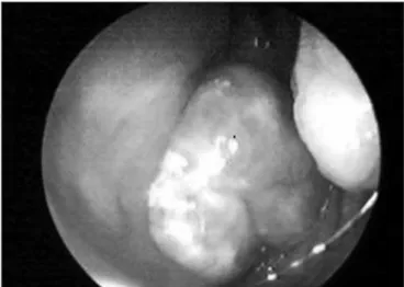
Nasal endoscopy showed a large lobulated tumor in the cavum (Figure 1), which occluded most of the cho-anae. CT of the neck showed an expanding solid lesion in the retropharyngeal and parapharyngeal space, projecting
into the nasopharyngeal lumen. There was also cervical and submandibular lymph node enlargement.
The tumor was biopsied under endoscopic view. Histology showed a diffuse submucosal infiltration by plasma cells containing lymphocytes within their cyto-plasm.
While these exams were done, the patient wor-sened progressively, presenting bilateral axillary and inguinal lymph node enlargement, diffuse facial skin lesions and further loss of vision.
Histological findings suggested extranodal Rosai-Dorfman disease.
Corticosteroid therapy was initiated (prednisone 1mg/kg/day) and adequate clinical control.
The 6-month follow-up showed that the patient improved significantly, but the neck masses regressed only slightly.
Figure 1. Endoscopic image of a tumor in the cavum.
634
BRAZILIAN JOURNAL OFOTORHINOLARYNGOLOGY 74 (4) JULY/AUGUST 2008
http://www.rborl.org.br / e-mail: [email protected]
DISCUSSION
SHML is a rare cause of lymph node enlargement in children and young adults; 80% of cases occur under the age 20 years. Many cases of extranodal involvement have been described since 1969. Apparently, there is no relation between lymph node and extranodal disease;
they may even occur separately.2
The etiology is uncertain, although agents such as the Epstein-Barr or herpes viruses are important in the
pathogenesis.3
The onset of SHML is typically insidiously; the active phase is prolonged, there may be spontaneous
remission and subsequent relapses.3 Factors that
deter-mine recurrences and is frequency are not well unders-tood. Deaths have been reported due to infiltration of
vital organs, such as the liver.4 The neck lymph nodes
are the most frequently involved, followed by inguinal,
axillary and mediastinal lymph nodes.5 The most common
extranodal sites are the skin, the upper respiratory tract and bones. Head and neck involvement - about 22% of
extranodal disease4,5 - include involvement of the nasal
cavity, the paranasal sinuses, the nasopharynx, subman-dibular glands, the parotid, the larynx, the temporal bone, the intratemporal fossa, the pterygoid fossa, the meninges
and the orbit.5 The skin is commonly affected; half of
these patients have another associated extranodal site. Orbit and ocular glove involvement have been reported,
usually as a retroorbitary mass and proptosis.6
The differential diagnosis of extranodal SHML may be a challenge, and is based on the clinical and histological examination. Histology shows typical featu-res, such as diffuse lymphoplasmatic infiltration, Russel bodies, foamy histiocytes and histiocytes with phagocyto-sed lymphocytes within the cytoplasm (emperipolesis). Immunohistochemical features include positive S-100,
alpha-antichymotrypsin and CD1a and CD68 antigens.4,5
Imaging (CT and magnetic resonance imaging) may be used to assess disease extension. If there is cervical lymph node enlargement, FNAB or lymph node biopsies may
be useful for the diagnosis.3
In the case above, FNAB and a cervical biopsy were done to exclude the possibility of neck metastases from local malignancies. Nasal endoscopy was extremely useful in detecting the primary lesion, and CT of the neck demonstrated the disease extension.
The differential diagnosis is made with lymphore-ticular malignancies such as lymphomas, Hodgkin’s dise-ase, malignant histiocytosis and monocytic leukemia, all of which have similar histopathological features. Atypias in cytology and the aggressive clinical course establish the diagnosis in most cases. Other histiocytoses, such as rhinoscleromas, Wegener’s granulomatosis, may also be
included in the differential diagnosis.6 Serology for HIV, toxoplasmosis and syphilis was done since these condi-tions are not rare in our context. Extranodal manifesta-tions in the head and neck are significantly more common in SHML patients with immunological abnormalities.4,5 SHML has been described in HIV-infected patients. An altered immune system and liver, kidney or lower airway involvement are considered as factors that worsen the
prognosis.3
Immunohistochemical exams were not done in our patient; the public health system (Sistema Unico de Saude) did not authorize these tests. The diagnosis, therefore, was confirmed histologically.
The patient next underwent radiological and other image diagnostic tests (a chest X-ray, abdominal ultrasound and CT of the head) to investigate further extranodal involvement. The rhinopharyngeal lesion and diffuse skin lesions on the face were considered extra-nodal involvement of this disease. We did not correlate poor vision with the disease in this patient.
There is no ideal protocol for the treatment of SHML; it is an uncommon, self-limited disease that fre-quently requires no therapy. Treatment is only necessary when lymph node or extranodal tissue enlargement causes significant symptoms, such as airway obstruction or compression of vital organs. Pulsone at al. reviewed 80 cases published between 1969 and 2000; 50% of the-se cathe-ses required no treatment, of which 82% had full
remission.4 The role of surgery is mostly for biopsies
and to relieve obstruction.3 Local recurrence is frequent
following surgical resection. The role of radiotherapy is not well understood; some reports have described full resolution with this treatment, while others have shown
no response.2 Steroids often resolve fever and reduce
lymph node size. Chemotherapy has yielded contro-versial results. A possible efficacy of methotrexate and 6-mercaptopurine requires further investigation. Other reports have suggested using alpha-interferon, although
its side effects have limited its use.4
We treated our patient with corticosteroids, to which he responded well. The patient was sent to an oncology unit for chemotherapy, which was eventually not done. Surgery was not required, given the benign nature of the condition.
FINAL COMMENTS
635
BRAZILIAN JOURNAL OFOTORHINOLARYNGOLOGY 74 (4) JULY/AUGUST 2008 http://www.rborl.org.br / e-mail: [email protected]
REFERENCES
1.Rosai J, Dorfman RF. Sinus histiocytosis with massive lymphadenopathy - A newly recognised benign clinico-pathological entity. Arch Path 1969;87:63-70.
2.Carpenter RJ III, Banks PM, Mc Donald TJ, Sanderson DR. Sinus his-tiocytosis with massive Lynphadenopathy (Rosai-Dorfman disease): Report of a case with respiratory tract involvement. Laryngoscope 1978;88:1963-9.
3.Hazarika P, Nayak DR, Balakrishnan R, Kundaje HG, Rao PL. Rosai-Dorfman disease of the subglottis. J Laryngol Otol 2000;114:970-3.
4.Pulsoni A, Anghel G, Falcucci P, Matera, R Pescarmona, Ribersani M: Treatment of Sinus Histiocytosis Whit Massive Lynfadenopathy (Rosai-Dorfman Disease): Report of a case and literature Review. Am J Hematol 2000;69:61-71.
5.Carbone A, Passannante A, Gloghini A, Devaney KO, Rinaldo A, Fer-lito A: Review of Sinus Histiocytosis Whit Massive Lynfadenopathy (Rosai-Dorfman Disease) of head and neck. Ann Otol Rhinol Laryngol 1999;108:1095-104.