382
Case report
Sao Paulo Med J. 2009; 127(6):382-4
Nonfunctional parathyroid cyst: case report
Cisto de paratireóide não funcionante: relato de caso
Carlos Eduardo Molinari Nardi
I, Ricardo Adriano Nasser Barbosa da Silva
II, Cynthia Maria Massarico Seraim
III,
Rogério Aparecido Dedivitis
IVPostgraduate Course on Health Sciences, Hospital Heliópolis (Hosphel), São Paulo; Head and Neck Surgery Service, Hospital Ana Costa, Santos; and Department of Head and Neck Surgery, Fundação Lusíada – Centro Universitário Lusíadas (Unilus), Santos, São Paulo, Brazil
IMD. Resident in General Surgery, Hospital Ana Costa, Santos, São Paulo, Brazil. IIMD. Resident in Head and Neck Surgery, Hospital Ana Costa, Santos, São Paulo, Brazil.
IIIUndergraduate medical student, Fundação Lusíada – Centro Universitário Lusíada (Unilus), Santos, São Paulo, Brazil.
IVMD, PhD. Full professor, Fundação Lusíada – Centro Universitário Lusíada (Unilus), Santos, São Paulo, and professor, Postgraduate Course on Health Sciences, Hospital Heliópolis
(Hosphel), São Paulo, Brazil.
ABSTRACT
CONTEXT: Parathyroid cysts are rare clinical and pathological entities, with less than 300 cases reported. The inferior parathyroid glands are most commonly involved, with left-side predominance. Parathyroid cysts may be functional or nonfunctional, depending on their association with hypercalcemia.
CASE REPORT: A 25-year-old man presented a palpable asymptomatic left-side neck mass. Ultrasound revealed a cystic structure contiguous with the left thyroid lobe. Serum ionic calcium was normal. The patient underwent left thyroid lobectomy plus isthmectomy with excision of the cyst. The histological indings revealed a parathyroid cyst. Parathyroid cysts typically present as asymptomatic neck masses, and surgical excision appears to be the treatment of choice.
RESUMO
CONTEXTO: Cistos de paratireóide são entidades clínicas e patológicas raras, com menos de 300 casos relatados. As glândulas paratireóides inferiores são mais comumente envolvidas, com predomínio do lado esquerdo. Cistos de paratireóide podem ser funcionais ou não, dependendo de sua associação com hipercalcemia.
RELATO DE CASO: Um homem de 25 anos apresentou-se com massa cervical esquerda palpável assintomática. A ultrassonograia revelou uma estrutura cística contígua com o lobo tireoidiano esquerdo. O cálcio iônico sérico estava normal. O paciente foi submetido a lobectomia esquerda com istmectomia e excisão do cisto. Os achados histopatológicos revelaram cisto de paratireóide. Cistos de paratireóide tipicamente se apresentam como massas cervicais assintomáticas e a ressecção cirúrgica parece ser o tratamento de escolha.
KEY WORDS: Parathyroid diseases. Cysts.
Parathyroid glands. Calcium. Parathyroid hormone.
PALAVRAS-CHAVE: Doenças das paratireóides. Cistos.
Glândulas paratireóides. Cálcio.
Nonfunctional parathyroid cyst: case report
Sao Paulo Med J. 2009; 127(6):382-4
383
INTRODUCTION
Parathyroid cysts are rare clinical and pathological entities, with less than 300 cases reported in the world literature. It has been reported that they occur in 0.5% of parathyroid disease cases and represent 1% of all cystic lesions of the neck.1 In 1880, Sandstrom reported the irst
parathyroid cyst.2 hese cysts commonly occur in the fourth and ifth
decades of life,3 with a female to male ratio of 2.5:1.1 he inferior
para-thyroid glands are most commonly involved, with left-sided predomi-nance. Parathyroid cysts may be functional or nonfunctional, depend-ing on their association with hypercalcemia. he clinical manifestation may consist of a solitary thyroid nodule or a neck mass.3 he treatment
options include ultrasound-guided aspiration. However, carcinoma aris-ing in the parathyroid cysts has been reported. hus, surgical removal of all cysts should be strongly considered.4 he purpose of this study was
to describe a case of a patient with a parathyroid cyst.
CASE REPORT
he patient was a 25-year-old man with a six-month history of asymptomatic left-side neck mass. At the physical examination, two painless masses of sizes 2.5 x 1.0 mm and 1.0 x 1.0 mm were palpable. Ultrasound revealed a 28 x 19 x 8 mm cystic structure contiguous with the left thyroid lobe and a thyroid nodule. Fine-needle aspiration biop-sy was performed, resulting in indings of squamous epithelial cells that suggested a branchial cyst. hyroid nodule biopsy was suggestive of hy-perplasia or adenoma. hyroid function tests, including triiodothyro-nine, thyroxin and thyroid-stimulating hormone were normal.
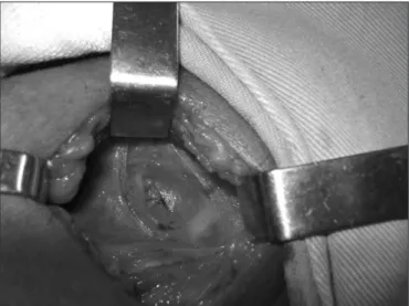
One year later, ultrasonography was performed again, and it re-vealed cystic growth, now measuring 35 x 26 x 16 mm. Surgery was in-dicated. he serum ionic calcium concentration was 1.25 mg/dl (1.16-1.32 mmol/l), parathyroid hormone was 35 pg/ml (11-67 pg/ml), thy-roid-stimulating hormone was 1.21 µΙU/ml (0.4-4.0 µΙU/ml) and free T4 was 0.95 ng/dl (0.8-1.9 ng/ml). he patient underwent neck ex-ploration and left thyroid lobectomy plus isthmectomy, with excision of the parathyroid cyst (Figure 1). he histological indings revealed a
parathyroid cyst (Figure 2). he parathyroid hormone level after surgery was 11.5 pg/ml, while the ionic calcium was 1.17 mg/dl.
DISCUSSION
Parathyroid cysts typically present as asymptomatic neck masses,2 as
in the patient in the current study, although cases of compromised air-ways and recurrent laryngeal nerve palsy secondary to large cysts have been reported.2Table 1 shows the published papers relating to
parathy-roid cysts, according to the databases. he most common presentation is a cystic lesion during a neck incision to treat a thyroid nodule.1
Para-thyroid cysts may be functional or nonfunctional depending on their as-sociation with hypercalcemia.3
At physical examination, parathyroid cysts tend to be soft, mobile, nontender masses, usually located in the lower part of the neck but can arise at any site between the jaw and the mediastinum.5 he
diagnos-tic workup should include a thorough head and neck examination, ine-needle aspiration, ultrasound and thyroid scan. Ultrasound may reveal a nonspeciic cystic structure.3 On radioiodine thyroid scans, parathyroid
cysts appear as areas of absent uptake.1 Computed axial tomography scan
and magnetic resonance imaging may be indicated when there is a soli-tary neck mass that is not diagnosed by other methods.3 Percutaneous
needle aspiration of parathyroid cysts reveals crystal-clear luid, which is highly suggestive of the diagnosis. Elevated parathyroid hormone levels in the cystic luid conirm the diagnosis,2 but do not indicate that the cyst is
Table 1. Published papers relating to parathyroid cysts, according to
database
Database Search strategy Results
PubMed “Parathyroid Diseases” as key word AND “Cyst” as word
165 12 original articles 1 historical article 3 reviews 143 case reports 3 letters to the editor 3 comments on previous articles
Lilacs “Parathyroid Diseases” as key word
4 1 original article 3 case reports Cochrane
Library
“Parathyroid” AND “Cyst” as words
0 0
Figure 2. Typical parathyroid tissue seen in the cyst wound. Hematoxylin
and eosin, 400 X.
Sao Paulo Med J. 2009; 127(6):382-4 Nardi CEM, Silva RANB, Seraim CMM, Dedivitis RA
384
functioning1. he diferential diagnosis should include thyroglossal duct
cyst, branchial cleft cyst, thyroid adenoma, and parathyroid carcinoma. he treatment for parathyroid cyst includes aspiration, injection of sclerosing agents and surgical excision.3 Fine-needle aspiration under
ul-trasound guidance with cystic luid assay for parathyroid hormone lev-els represents the approach of choice for both diagnosis and the initial treatment, since this is safe, easy and repeatable.1 his method is more
efective on nonfunctioning cysts.3 Intracystic tetracycline injection may
also be used on patients with recurrence; however, this has been asso-ciated with neck pain, neurotoxicity and recurrent nerve palsy due to leakage of the sclerosing agents through the disrupted thin cyst wall.1
Surgical excision appears to be the treatment of choice for functioning parathyroid cysts3 and in cases of repeated recurrence.1 Complications
caused by removal of parathyroid cysts include hypocalcemia, hemor-rhage, hypercalcemic crises, tetany and recurrent laryngeal nerve palsy.3
REFERENCES
1. Ippolito G, Palazzo FF, Sebag F, Sierra M, De Micco C, Henry JF. A single-institution 25-year review of true parathyroid cysts. Langenbecks Arch Surg. 2006;391(1):13-8.
2. Ihm PS, Dray T, Sofferman RA, Nathan M, Hardin NJ. Parathyroid cysts: diagnosis and mana-gement. Laryngoscope. 2001;111(9):1576-8.
3. Fortson JK, Patel VG, Henderson VJ. Parathyroid cysts: a case report and review of the litera-ture. Laryngoscope. 2001;111(10):1726-8.
4. Coelho DH, Boey HP. Benign parathyroid cyst causing vocal fold paralysis: a case report and review of the literature. Head Neck. 2006;28(6):564-6.
5. Jha BC, Nagarkar NM, Kochhar S, Mohan H, Dass A. Parathyroid cyst: a rare cause of an anterior neck mass. J Laryngol Otol. 1999;113(1):73-5.
Sources of funding: None
Conlict of interest: None
Date of irst submission: January 6, 2009
Last received: July 23, 2009
Accepted: November 19, 2009
Address for correspondence: Rogério Aparecido Dedivitis
Rua Olinto Rodrigues Dantas, 343 — Conj. 92 Santos (SP) — Brasil