Strain Specific Phage Treatment for
Staphylococcus aureus
Infection Is
Influenced by Host Immunity and Site of
Infection
Nathan B. Pincus, Jensen D. Reckhow☯, Danial Saleem☯, Momodou L. Jammeh, Sandip K. Datta, Ian A. Myles*
Bacterial Pathogenesis Unit, Laboratory of Clinical Infectious Diseases, National Institute of Allergy and Infectious Diseases, National Institutes of Health, Bethesda, Maryland, United States of America
☯These authors contributed equally to this work.
*mylesi@niaid.nih.gov
Abstract
The response to multi-drug resistant bacterial infections must be a global priority. While mounting resistance threatens to create what the World Health Organization has termed a “post-antibiotic era”, the recent discovery that antibiotic use may adversely impact the microbiome adds further urgency to the need for new developmental approaches for anti-pathogen treatments. Methicillin-resistantStaphylococcus aureus(MRSA), in particular, has declared itself a serious threat within the United States and abroad. A potential solution to the problem of antibiotic resistance may not entail looking to the future for completely novel treatments, but instead looking into our history of bacteriophage therapy. This study aimed to test the efficacy, safety, and commercial viability of the use of phages to treat
Staphylococcus aureusinfections using the commercially available phage SATA-8505. We found that SATA-8505 effectively controlsS.aureusgrowth and reduces bacterial viability both in vitro and in a skin infection mouse model. However, this killing effect was not ob-served when phage was cultured in the presence of human whole blood. SATA-8505 did not induce inflammatory responses in peripheral blood mononuclear cultures. However, phage did induce IFN gamma production in primary human keratinocyte cultures and in-duced inflammatory responses in our mouse models, particularly in a mouse model of chronic granulomatous disease. Our findings support the potential efficacy of phage thera-py, although regulatory and market factors may limit its wider investigation and use.
Introduction
Resistance to antibiotics is one of the most significant challenges in healthcare today. While the incidence of antibiotic resistance toStaphylococcus aureushas increased over the nearly nine decades since Alexander Fleming observed the treatment potential ofPenicillium notatum, the
OPEN ACCESS
Citation:Pincus NB, Reckhow JD, Saleem D, Jammeh ML, Datta SK, Myles IA (2015) Strain Specific Phage Treatment forStaphylococcus aureus
Infection Is Influenced by Host Immunity and Site of Infection. PLoS ONE 10(4): e0124280. doi:10.1371/ journal.pone.0124280
Academic Editor:Ulrich Nübel, Leibniz-Institute DSMZ, GERMANY
Received:January 5, 2015
Accepted:March 12, 2015
Published:April 24, 2015
Copyright:This is an open access article, free of all copyright, and may be freely reproduced, distributed, transmitted, modified, built upon, or otherwise used by anyone for any lawful purpose. The work is made available under theCreative Commons CC0public domain dedication.
Data Availability Statement:All relevant data are within the paper.
Funding:This work was supported by the Intramural Research Program of The National Institutes of Health and The National Institute of Allergy and Infectious Disease. The funders had no role in study design, data collection and analysis, decision to publish, or preparation of the manuscript.
discovery and development of new antibiotics has slowed to a trickle [1–3]. Without develop-ment of new treatdevelop-ment modalities, there is a very real threat of approaching what the World Health Organization (WHO) termed a“post-antibiotic era”, where the treatments we have re-lied on are simply no longer effective and we are left unarmed in the unending silent war against disease [2,3]. Methicillin-resistantStaphylococcus aureus(MRSA) has been declared a serious threat by the Centers for Disease Control and Prevention (CDC), causing 80,461 seri-ous infections and 11,285 deaths in the US in 2011 alone [4]. Additionally, the emergence of vancomycin resistant or intermediateStaphylococcus aureus(VRSA or VISA) threatens to cause infections that have few treatment options [1]. The rising problem of antibiotic resistance in bacterial infections, andS.aureusinfections in particular, creates a pressing need for re-search into alternative methods of treatment.
A solution to the problem of antibiotic resistance may not entail discovering completely novel treatments, but instead looking back into history, to bacteriophage therapy. Phage thera-py harnesses the killing power of bacteria’s natural predators, lytic bacteriophages, to fight dis-ease [1,5,6]. The concept of phage therapy predates the use of conventional antibiotics, initially put forth by Felix d’Herelle, co-discoverer of phages, in the early 1920’s [5–7]. Although this was followed by an initial boom of interest and research, phage therapy faded from use follow-ing the discovery of antibiotics [1,5–7]. However, both phage therapy and research have con-tinued in Eastern Europe and former Soviet states, reporting success in treating a variety of bacterial infections, including those byS.aureus[1,6]. The rising problem of antibiotic resis-tance and the need for alternative treatments have led to a resurgence of interest in phage ther-apy. However, many studies to date lack adequate controls and are poorly designed; additional research is needed to substantiate the safety and efficacy of these treatments before they can be integrated into standard clinical care [1,6,7]. A current pilot clinical trial in Belgium is examin-ing the use of topical application ofS.aureusandP.aeruginosaphages to treat burn wound in-fections [1,8]. In addition, staphylococcal phages have other potential healthcare uses, such as application to catheter material to control biofilm formation [9] or in hand wash solutions to lower CFUs ofS.aureuson the skin of healthcare workers [10].
This study aimed to investigate the efficacy, safety, and commercial viability of the use of phages to treatStaphylococcus aureusinfections.S.aureusbacteriophage SATA-8505 (ATCC PTA-9476) was examined for its ability to prevent and treat infections with clinically relevant MRSA strain USA300in vitroin human cells andin vivoin mice. SATA-8505 was selected as a model phage to assess efficacy for treatment of USA300 infection because of its commercial availability and patented claims of activity against USA300in vitrowith classifications 435/ 235.1 (USA), C12P1/06 (International), C12N7/00, C12N2795/00021 (Cooperative), and C12N7/00 (European) (Patent no: US 7,745,194 B2)[11].
Materials and Methods
Materials
Chemical reagents were purchased from Sigma Chemical Company, St. Louis, MO. Blood aglates were from Thermo Scientific, Dubuque, IA. Tryptic Soy Broth and Brain Heart Infusion media/agar were from General Laboratory Products, Yorkville, IL. Fetal Bovine Serum was from Thermo Scientific, Dubuque, IA.
All experiments were done in compliance with the guidelines of the NIAID Institutional Ani-mal Care and Use Committee.
Bacterial and phage strains
USA300 LAC strain of MRSA was a gift from F. DeLeo (Rocky Mountain Labs, NIAID). SATA-8505 bacteriophage, which has been patented for its reported activity against USA300in
vitro(Patent no: US 7,745,194 B2)[11], was obtained from ATCC (ATCC PTA-9476).S.aureus
USA100 strains 71080 (NR-46418, VRS8) and 626 (NR-4668) were provided by the Network on Antimicrobial Resistance inStaphylococcus aureus(NARSA) for distribution by BEI Re-sources, NIAID, NIH.
Phage propagation and selection for efficacy
SATA-8505 was propagated by inoculating 10 mL BHI (Thermo Fisher Scientific) with 100μL
of SATA-8505 stock and 100μL of an overnight culture (ONC) of USA300 and incubating at
37°C overnight with shaking. The resulting lysate was centrifuged at 6000g for 12 minutes, the supernatant put through a 0.22μm filter, and stored at 4°C until use. Plaque forming units
(PFUs) of phage were determined using the standard double agar overlay method [12]. Serial dilutions of SATA-8505 stock (100μL) and 100μL of USA300 ONC were mixed into 4 mL
mol-ten BHI top agar (0.5% agar) and poured over BHI plates. Plates were incubated overnight at 37°C and the resulting plaques counted. In order to select for SATA-8505 virions against USA300, plaques in the top agar were resuspended in 5mL TSB, centrifuged at 6000g for 15 minutes, the supernatant put through a 0.22μm filter, and the resulting phage stock was used
in subsequent propagations. Experiments using alternate strains ofS.aureuswere conducted in an identical manner.
Phage viability assay
A known concentration of SATA-8505 was divided into 2mL aliquots and stored in the follow-ing conditions: room temperature, -80°C, lyophilized then room temperature, lyophilized then 4°C. Phage counts were determined via the top agar method at indicated time points. In addi-tion, the viability of phage stock stored at 4°C was routinely monitored via the double agar overlay method.
Phage killing kinetics of Staphylococcus in media and whole blood
Approximately 106CFUs of mid-exponential growth phase USA300 were added to 5 mL ali-quots of BHI or TSB with SATA-8505 at varying MOI. Samples were incubated with shaking at 37°C, with bacterial concentration over time determined by plating on BHI (BD Biosciences) or blood (Thermo Fisher Scientific) agar. For the whole blood assay, 10mL blood from normal donors and/or CGD patients was obtained from the NIH Clinical Center Department of Trans-fusion Medicine and was added to blood culture bottles containing 30mL of TSB (Thermo Fisher Scientific) with SATA-8505 at varying MOI. In experiments using blood, control blood culture vials were injected with an equal volume of Hanks Buffered Salt Solution (HBSS) (Thermo Fisher Scientific). All human samples were obtained under permission of the Institu-tional Review Board (IRB) for the NaInstitu-tional Institute of Allergy and Infectious Disease (NIAID) and the National Institutes of Health (NIH) Clinical Center. All participants provided their written consent to the research protocol and IRB consent was obtained prior toCytokine response to phage exposure
Keratinocyte cultures were derived as previously described [13]. After cells reached confluence in a 6-well plate (BD Falcon, Bedford, MA), phage was added in escalating doses up to 1010 PFU/culture. Supernatants were harvested at 24 hours and analyzed using multiplex
(BIO-RAD, Hercules, CA). Peripheral blood mono-nuclear cells (PBMC) were isolated using standard Ficol gradient centrifugation (GE Healthcare Life Sciences, Pittsburgh, PA). Cells were suspended at 2x10⁶/mL in culture media of DMEM (Gibco Invitrogen, Carlsbad, CA), 10% FBS, Hepes buffer (Thermo Scientific), non-essential amino acids (Gibco), penicillin/ streptomycin (Gibco), sodium pyruvate (Gibco), and 2-mercapto-ethanol (Gibco). Superna-tants were harvested after 72 hours and analyzed using multiplex (BIO-RAD, Hercules, CA).
Skin infection model
Mice were inoculated subcutaneously with 107CFUS.aureus, and skin lesions assessed as pre-viously described [13–15]. The route of phage delivery has a clear effect on the outcomes of phage therapy, with systemic delivery the most efficacious, even in the case of local infections [7]. When treating with phage, intraperitoneal injections were performed immediately prior to MRSA inoculation with either 107or 109PFU of SATA-8505 for MOI of 1 or 100. PFU/lesion concentrations were determined by homogenizing a 3mm diameter punch biopsy of the skin lesion in 0.5mL of HBSS, then adding the entire volume to in the double agar overlay method as above. Diluent treatment involved injection with supernatants from overnight USA300 cul-tures that had been pelleted and filter sterilized in a manner identical to the phage culcul-tures.
Statistics
Means were compared using two-tailed unpaired t test, or ANOVA with Bonferroni adjust-ment for comparison of multiple samples, with Prism software (GraphPad, San Diego, CA). ns = not significant,= p<0.05,= p<0.01,= p<0.001,= p<0.0001.
Results
SATA-8505 Improves MRSA Skin Infection at Low Multiplicity of
Infection (MOI)
Phages againstS.aureushave been shown to be effective at killing a multitude ofS.aureus
strainsin vitro[10,17,18], and, unlike antibiotics, have the capacity to rapidly evolve during an infection to overcome developing resistance [10]. Furthermore, phages have shown promise in protecting againstS.aureussepsis [19–21] and have been beneficial in treating staphylococcal skin and soft tissue infections in mice [20,22]. Therefore, after demonstrating thein vitro effec-tiveness of SATA-8505 at clearing USA300, we evaluated the potential of this specific phage as an antibiotic agent against USA300in vivo. To do this, we used wild type and immunodeficient CGD mice, which are susceptible toS.aureusdue to their dysfunctional neutrophil response. The mice were inoculated with 107bacteria subcutaneously immediately after intraperitoneal injection of phage. Skin lesion size was quantitatively measured for 6 days after inoculation. Mice treated with phage at a MOI of 1 (1 phage virion to 1 CFU bacteria) showed smaller skin lesion sizes in both wild type and CGD mice as compared to diluent treated controls (Fig 2A). USA300 secretes a variety of virulence factors [23] and thus our phage cultures could stimulate
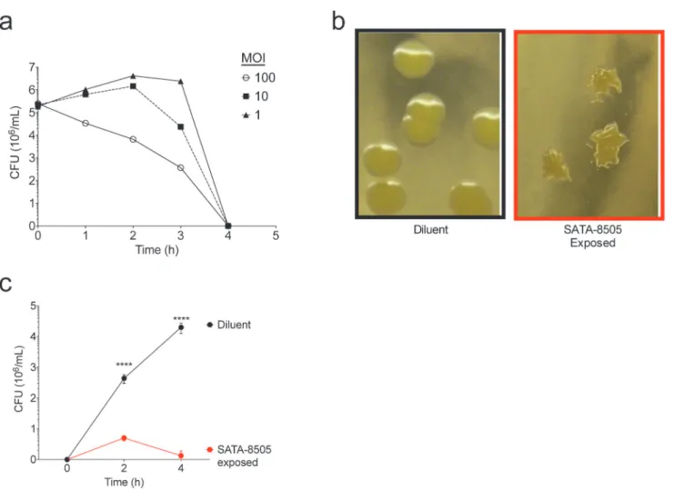
Fig 1. SATA 8505 Effectively Kills USA 300 and Reduces its Viability in vitro.(a) Average colony forming unit (CFU) counts for USA300 cultured with SATA-8505 for up to four hours at ratios ofS.aureus:Phage of 1:1, 1:10, or 1:100 (MOI 1, 10, 100 respectively). (b) Images of surviving colony morphology of USA300 grown in TSB after exposure to BHI (diluent) or SATA-8505. (c) Regrowth of surviving colonies pictured in panel b,S.aureusgrown in TSB after prior exposure to BHI (diluent) or SATA-8505 run in triplicate culture. Data shown are representative of 3 or more independent experiments and displayed as mean + s.e.m.****= p<0.0001.
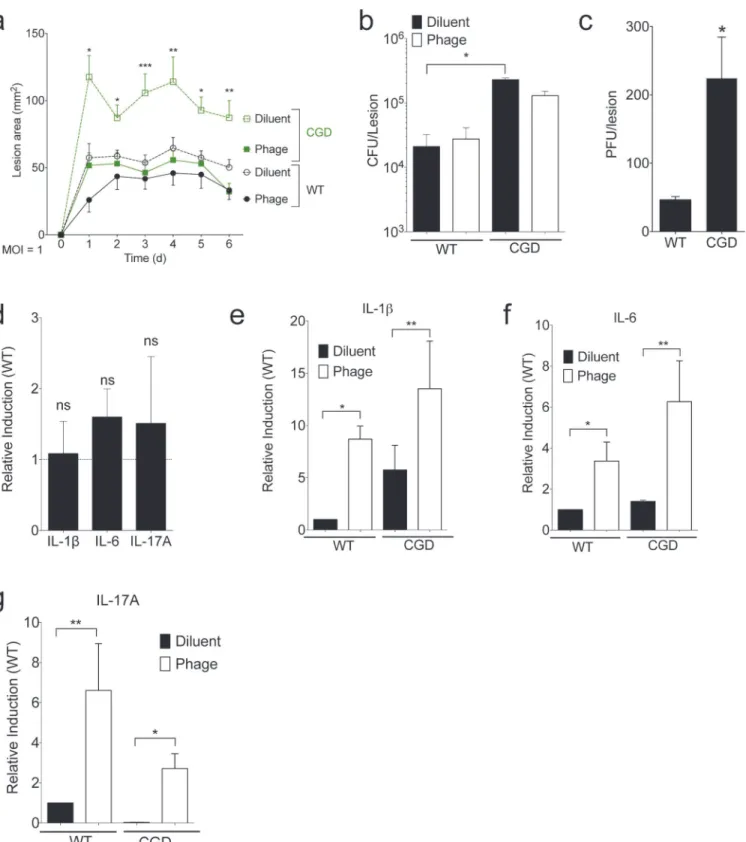
Fig 2. SATA-8505 Improves MRSA Skin Infection at Low MOI.Mice were injected intraperitoneally (I.P.) with 107plaque-forming units (PFU) of
SATA-8505 immediately prior to subcutaneous injections of 107CFU of USA300. (a) Lesion size progression over following six days. On day 6, skin biopsies were
homogenized for culture, total bacterial (b) and phage burden (c) were calculated for individual mice. (d) mRNA transcript levels for IL-1ß, IL-6, and IL-17A in individual CGD mice relative to wild type controls, standardized to GAPDH. (e-g) mRNA transcript levels for IL-1ß, IL-6, and IL-17A in CGD and wild type mice with and without SATA-8505 treatment relative to wild type controls injected with diluent, standardized to GAPDH. Data shown are representative of 2–3 independent experiments using 5 or more mice per group, and displayed as mean + s.e.m. Differences were calculated by ANOVA with Bonferroni correction and depict differences from diluent treated wild type unless otherwise noted. ns = not significant,*= p<0.05,**= p<0.01.
responses due to the presence of theseS.aureusproducts; to control for this effect, all dilutions and diluent treatments were performed using supernatant from an overnight USA300 culture that had been pelleted and filter sterilized in a manner identical to the phage cultures.
CGD mice had larger lesion sizes than wild type mice in a skin infection model ofS.aureus, a novel and somewhat unexpected finding given the lack of cutaneousS.aureusinfections in patients with CGD, who typically getS.aureusinfections in deep-seated tissues such as the liver [24]. While CGD mice had a greater CFU burden than did wild type, phage treatment failed to significantly alter bacteria counts in either strain (Fig 2B). CGD mice also had higher plaque-forming units (PFU) of SATA-8505 in their lesions (Fig 2C). Despite having slightly larger lesion sizes and bacterial burden during infection (Fig 2A and 2B), CGD mice did not have significant differences in their mRNA levels for IL-1ß, IL-6, or IL-17A (Fig 2D), consistent with their immunodeficiency reflecting a defect in neutrophil function rather than cytokine production [25,26]. While phage treatment reduced lesion size (Fig 2A), it increased lesional inflammatory cytokines in both wild type and CGD mice (Fig 2E–2G). The isolated reduction of lesion size without effects on bacterial burden and cytokine responses suggests that phage therapy at this dose may be inhibiting the ability of bacteria to inflict toxin-mediated dermone-crosis without dramatically affecting viability [27].
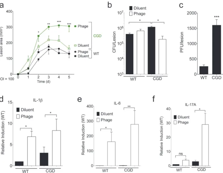
Given these results, we hypothesized that increasing the dosage of the bacteriophage would result in greater bacterial clearance. Inoculation of mice with staphylococcal bacteriophage K at doses as high as 2 x 10¹¹PFU was shown to have no effects on physical condition or survival over one month and no effects on organ pathology after 14 days [19]. Accordingly, we in-creased the dose of phage to an MOI 100 while maintaining a 107CFU inoculation. CGD mice again had larger lesions and higher bacterial counts than wild type, and the higher MOI phage treatment significantly increased lesion size and reduced bacterial burden in CGD mice (Fig 3A and 3B). CGD mice had significantly higher amounts of virus extracted from their skin le-sions than wild type mice (Fig 3C). High dose phage treatment led to increased lesional tran-script levels of IL-1ß, IL-6, and IL-17A, particularly in the CGD mice (Fig 3D–3F). Of note, interferon gamma (IFN gamma) was not induced at either MOI treatment (not shown). In these studies at a high phage MOI, phage effects on lesional bacterial burden were only appar-ent in the setting of CGD and its ineffective neutrophil response. Furthermore, phage therapy induced a vigorous inflammatory response that may have contributed to the increased lesion size despite better bacterial control. Taken together, these studies suggest a complex balance be-tween direct bactericidal activity and induction of an inflammatory response that may contrib-ute to the ultimate efficacy of phage therapy.
by USA300-derived products in our diluent controls. Since primary human KC cultures can contain dendritic cell contamination, we also exposed the HaCaT cell line to phage and found no significant increase in IFN gamma (Fig 4G). This discrepancy may reflect dendritic cell con-tamination, or may be a reflection of the differences in HaCaT cell lines from primary cells [28,29].
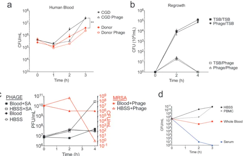
SATA-8505 Does Not Impact USA300 Growth in Human Blood
To determine the effect of phage on bacterial growth in human blood, we inoculated blood from a healthy donor and a patient with CGD with 106CFU of USA300 and 108PFU of phage, monitoring bacterial growth in clinical blood culture bottles for three hours. As expected, CGD patients had greater bacterial growth in their blood than healthy volunteers. However, unlike the bactericidal effects seen in TSB media (Fig 1), phage did not significantly affect bacterialFig 3. SATA-8505 Fails to Improve MRSA Skin Infection at High MOI.Lesion size (a), CFU (b), PFU (c), and transcript data (d-f) for mice injected with 109 plaque-forming units (PFU) of SATA-8505 immediately prior to subcutaneous injections of 107CFU of USA300 processed in an identical manner as MOI of 1
experiments. Data shown are representative of 2–3 independent experiments using 5 or more mice per group, and displayed as mean + s.e.m. Differences were calculated by ANOVA with Bonferroni correction and depict differences from diluent treated wild type unless otherwise noted. ns = not significant,
*= p<0.05,**= p<0.01,***= p<0.001.
growth in blood (Fig 5A) or affect bacterial colony morphology (not shown). Bacteria exposed to phage in blood and then re-cultured in TSB grew just as well as bacteria unexposed to phage, and the bacteria were killed effectively upon a second exposure to phage in TSB (Fig 5B). These results indicated that the failure of phage to clear MRSA from the blood culture was not due to selection of a phage-resistant bacterium, and that the surviving colonies did not harbor phage the way they appeared to when re-cultured after being exposed to phage while grown in TSB (Fig 1C).
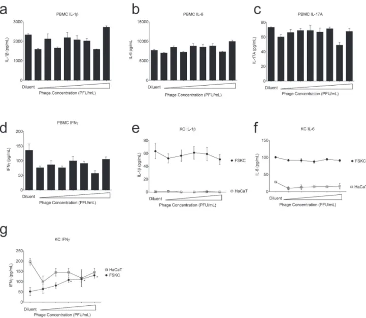
Fig 4. SATA-8505 Induces Interferon Gamma in Primary Human Keratinocytes.Human peripheral mononuclear cells (PBMC) were cultured in triplicate at 26/mL with SATA-8505 at 1 PFU/mL to 108PFU/mL at ten-fold increments or diluent derived from the supernatant of an overnight culture of SA that had
been pelleted and filter sterilized in a manner similar to the phage-containing media. At 72 hours supernatants were harvested and analyzed for IL-1ß (a), IL-6 (b), IL-17A (c), and IFN gamma (d). Human keratinocytes from primary foreskins (foreskin keratinocytes; FSKC) or the HaCaT cell line were cultured to confluence on 6-well plates and incubated in triplicate with SATA-8505 at 104PFU/mL to 108PFU/mL at ten-fold increments or TSB diluent. At 24 hours supernatants were harvested and analyzed for IL-1ß (e), IL-6 (f), and IFN gamma (g). Phage for all experiments was diluted in TSB from overnight culture of USA300 that was centrifuged at 5000rpm for 12 minutes and filter-sterilized through a 0.44 micrometer filter. Data shown are representative of 3 independent experiments using 3 different healthy volunteers (a-d) or a pool of 5 or more foreskin samples (e-g) and displayed as mean + s.e.m.*= p<0.05.
We next hypothesized that the failure of phage to clear a blood culture containing MRSA was due to direct clearance of the phage by the blood cells. However, we found no significant change in phage concentration when grown in blood, irrespective of the presence ofS.aureus
(Fig 5C; left y-axis). Again, phage did not significantly reduce MRSA colonies when grown in human blood, despite clearance of MRSA from culture media without blood (Fig 5C; right y-axis). Therefore, it appeared that the presence of human blood significantly reduced both MRSA and phage proliferation. We next separated whole blood into its cellular and serum components and again cultured USA300 in each of these for three hours. We found that at equal volumes, whole blood provided a bacteriostatic growth environment, PBMCs had no im-pact on growth, and serum had a bactericidal effect (Fig 5D). Given the mortality of MRSA bacteremia [30–32], it is clear thatS.aureusdoes proliferatein vivo, however our results sug-gest that perhaps this growth occurs in seeded tissues rather than in the blood compartment di-rectly. Our findings indicate that SATA-8505 phage treatment of USA300 bacteremia may not be effective at directly clearing the blood. This may reflect alterations in blood-borne USA300
Fig 5. SATA-8505 Does Not Impact USA300 Growth in Human Blood.(a) Hourly quantification of starting culture of 106CFU USA300 in three parts TSB and one part whole blood from healthy donor or a patient with CGD, grown with or without 108PFU SATA-8505 in duplicate. (b) Regrowth of surviving
colonies from whole blood culture run in triplicate. One colony ofS.aureuspreviously grown in 3:1 TSB:Whole blood without phage was subsequently grown in TSB with SATA-8505 (TSB/Phage) or without phage (TSB/TSB); one colony of the survivingS.aureuspreviously grown in the presence of SATA-8505 was subsequently grown in TSB with SATA-8505 (Phage/Phage) or without phage (Phage/TSB). (c) Average quantification of starting culture of 1010CFU of USA300 grown in either 3:1 TSB:Whole blood or 3:1 TSB:HBSS, with or without 107PFU SATA-8505. (d) Average quantification of starting culture of 107
CFU of USA300 grown in three parts TSB with either one part whole blood, HBSS, serum, or peripheral mononuclear cells (PBMC) in equivalent volume HBSS. Data shown are representative of 2–3 independent experiments using 3 or more different healthy volunteers and 2 patients with CGD. Data is displayed as mean + s.e.m.**= p<0.01.
that affect phage uptake and propogation, or may indicate the presence of inhibitory serum fac-tors, such as albumin that may bind cations required for phage adsorption [33,34]. However, our results in the skin infection model as well as prior work showing protection from IV chal-lenge suggest that phage therapy can control MRSAin vivo. Further investigation is needed to elucidate how SATA-8505 and other phages control MRSA pathologyin vivoand to uncover within which compartments these benefits occur.
SATA-8505 Exhibits Commercial Viability but Significant Strain
Limitations
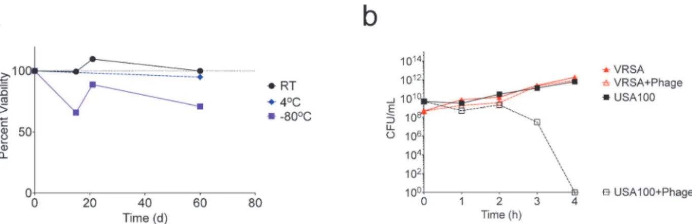
For a therapeutic phage to be clinically useful and a viable alternative to conventional antimi-crobials, it must be easily stored and shelf stable, as pharmacies could not realistically be ex-pected to culture batches de novo. To evaluate phage viability, we stored phage in frozen, refrigerated, and room temperature conditions and quantified PFU’s intermittently for 60 days. Phage was most stable at room temperature, and nearly 100% viable after 60 days (Fig 6A). Commercial labs have successfully lyophilized phage, suggesting lyophilization may be an option to further extend shelf life.
Another real-world challenge to therapeutic use of phages is their bacterial strain specificity. While strain-specificity may be useful for avoiding the unintended impacts on the microbiome seen with standard antibiotic treatment [35–40], it also limits the practical application of phage therapy, as the time needed to identify the causative strain would prohibit its use as a first-line treatment for acutely infected patients. Many strains of bacteria acquire resistance to antibiotics but remain otherwise similarly pathogenic, such as vancomycin resistantStaphylococcus aureus
(VRSA). A VRSA strain on the USA300 background was not readily available, so we repeated the broth inoculation described using the MRSA USA100 strain 626 and a VRSA strain on the USA100 background (VRS8). While SATA-8505 cleared MRSA 626 from the broth culture, it did not have an impact on the growth of VRSA (Fig 6B). Three rounds of serial passage of phage through the VRSA culture to allow selection of potential VRSA-specific mutants did not enhance the killing ability (not shown). Although both MRSA 626 and VRS8 are pulsed-field type USA100 with matching spa type (Ridom t002) and multi-locus sequence type (ST 5), they are not isogenic strains. Thus, the difference in phage susceptibility may reflect vanA-mediated alterations in peptidoglycan structure in the VRSA strain [41], but may also reflect other differ-ences between these two clinical isolates that will need further elucidation [42]. This difference
Fig 6. SATA-8505 Exhibits Commercial Viability but Significant Strain Limitations.(a) SATA-8505 quantified over up to sixty days after storage at either room temperature (RT), 4 degrees Celsius (4°C), or frozen at -80°C. (b) Average hourly quantification of 108
–109CFU of USA100S.aureusor a vancomycin
resistant variant of USA100 (VRSA) cultured with up to 1011PFU of SATA-8505. Data shown are a representative of 2 independent experiments.
in susceptibility, however, does highlight that relatively subtle alterations can render bacteria resistant to phage and may provide a platform for future mechanistic insights.
Discussion
Our results confirm that SATA-8505 reduced viability of USA300, and found that it effectively controlled USA300 infection in human cellsin vitroand in a mouse model of skin infection. This effect wasS.aureusstrain-specific and not observed in whole blood, where USA300 failed to proliferate and thus was not a viable phage target. SATA-8505 remained viable when stored at room temperature for two months. These results highlight the potential viability of phage therapy if limitations based on site of infection and strain of infecting bacteria can be over-come. Our results thus demonstrate both the promising attributes of phage therapy against USA300, and factors that may limit its real-world potential.
SATA-8505 is effective at killing USA300 and USA100 bacteria in standard growth condi-tions, can improve outcomes in MRSA skin infection when dosed appropriately, does not in-duce inflammatory responses in human PBMCs, and can be stored long-term without the need for refrigeration. However, we also found that SATA-8505 may induce inflammation in both mouse and human skin, could worsen MRSA skin infection or induce inflammatory damage in the immunocompromised if the dosing is overly aggressive, and has reduced clearance in mice with CGD. Additionally, SATA-8505 does not appear to kill USA300 when grown in the pres-ence of human blood, and is incapable of killing our selected strain of VRSA even when on an otherwise susceptible USA100 background.
Our results also highlight the potential importance of appropriate dosing if phage therapy is used as a future therapeutic option. Although not an insurmountable limitation, most phage strains are incapable of infecting mammalian tissue and are thus rapidly cleared from the host [43,44]. Thus the timing of phage treatment must also be examined. Our choice to treat mice with phage immediately before infection was done to assure phage titers would be present at time ofS.aureusexposure. Future work will be needed to see the time course of protection in this system as this timing may greatly alter the clinical utility phage as either aS.aureus thera-peutic or prophylactic (for example, in patients on dialysis or patients that are post-operative). However, just because bacteriophage is not infectious to humans does not mean it does not harbor immunogenic potential, which may itself have detrimental effects on health. Like other biologic pharmaceuticals, higher doses may increase treatment effectiveness, yet do so at the risk of increasing treatment-induced complications [44]. More research is required to deter-mine an optimal dosage that maximizes bacterial clearance while minimizing collateral damage to the host. Our results indicate that dosing may be of particular concern with immune com-promised patients, as the higher dose of bacteriophage worsened infectious outcomes in our immunodeficient mice. Given that certain immune compromised individuals are more likely to suffer from MRSA infections, and would thus be the most likely candidates for phage thera-py [4], researchers should be cautious of extrapolating dosing from early trials that are typically limited to healthy volunteers.
infection when formulating phage treatment, given that bacterial growth patterns may be al-tered by changes in the in vivo compartment in ways that may preclude therapeutic benefit.
There are many potential limitations to phage therapy becoming part of routine infection control. Our findings highlight the potential efficacy of phage therapy but also outline its scien-tific and therapeutic complexities such as dosing, host immune status, and site of infection. However, other notable limitations are of the regulatory and financial nature. For example, al-though cocktail treatment appear the best scientific model for the future, under current regula-tions each bacteriophage included in a proposed cocktail would have to undergo individual testing and show safety and effectiveness as an isolated therapy [49,50]. As discussed above, the strain-specificity of bacteriophage would likely render such testing unsuccessful; while each phage could be assessed for safety separately, each phage could only be expected to work in the fraction of cases that are caused by its target strain. Though the effectiveness of each individual phage may be low, the effectiveness of multiple phages may be additive [49]. This additive ben-efit will not be captured unless this treatment is allowed to undergo testing as a combination therapy. There are polyvalent Staphylococcal phages identified that could mitigate the need for combination therapy and SATA-8505 possesses lytic activity against 10–38% of the 60 strains of SA tested for the patent application [11]. However, our results suggest that even relatively minor genetic changes could greatly reduce treatment efficacy as seen in the susceptibility dif-ferences between two USA100 strains, MRSA 626 and VRS8. Therefore, phage therapy may benefit from being regulated in manners more similar to vaccines than chemical pharmaceuti-cals, where multivalent vaccines are not expected to show efficacy of each component in isola-tion and polyvalent phages could be combined for greater coverage through redundancy of strain targets.
When phage is applied to food products for the purposes of controlling bacterial growth, it is deemed“generally regarded as safe”by the Food and Drug Administration and thus requires no pre-market proof of safety, as exemplified by the use of phage in cheese to protect against the growth ofListeria monocytogenes[49]. While such loose regulatory ideals are inappropriate for pharmaceuticals, it is not inconceivable that regulations on phage therapy could be
amended to reflect the current scientific understanding. Other notable regulations proposed for safe clinical use of phages use include the availability of the full phage genome sequence, verification of the lack of toxin producing elements, lack of lysogenic potential, evaluation of transducing abilities, production under Good Manufacturing Practices, and (if possible) propa-gation in a non-pathogenic strain of bacteria [51]. The sequence of SATA-8505 is available and reveals the phage to be from the Myoviridae family, but the remaining details would need to be verified before therapeutic consideration. Accordingly, future work using Myoviridae phage therapies should consider the potential to induce inflammation in human keratinocytes.
Even with regulatory reforms, there would still be concerns related to financial incentives to produce phage therapy. Bacteriophage can be easily produced using basic materials, and could be readily cultured outside of a pharmaceutical laboratory. A second supply stream would threaten the financial benefits of legal production, and may be difficult to control in parts of the world where drug production is not vigilantly regulated. Such a phenomenon is a concern with all pharmaceuticals, but is of particular interest with phage because of this ease of replica-tion. Furthermore, products of nature are not open to being patented, which was upheld as re-cently as 2013 in the United States Supreme Court caseAssociation for Molecular Pathology v.
Myriad Genetics. The seemingly reasonable basis for such patent restrictions, along with the
However, despite all of these limitations, another conclusion that must be drawn from the literature is that the response to multi-drug resistant bacterial infections must be a top priority. Beyond the threat of a“post-antibiotic era”, the newly elucidated potential for antibiotic-in-duced harm to the normal microbiome [35] has further highlighted the need for new ap-proaches when developing targeted anti-pathogen treatments. Urgent facilitation of research into human dosing, efficacy, and best regulatory practices for phage therapy could assure a more timely response if any high-threat, multidrug resistant pathogen should become an emer-gent pandemic [52]. Ultimately, in spite of significant logistical challenges faced by phage ther-apy, this type of research highlights an important direction for science. As both antibiotic resistance and immune compromised populations continue to increase, the need for alternative therapies, and alternative therapeutic mechanisms, is urgent. As natural predators of bacteria, bacteriophages remain a promising option for controlling bacterial infections. However, future research, and perhaps refinement of the legal process of drug development, are needed before this may become a reality.
Acknowledgments
We would like to thank the NIAID building 33 and 14BS animal care and breeder technicians for their assistance as well as Mr. and Mrs. Topolino (NIAID) for their cooperation and sacri-fice during the course of this project.
Author Contributions
Conceived and designed the experiments: NBP SKD IAM. Performed the experiments: NBP JDR DS MLJ IAM. Analyzed the data: NBP IAM SKD. Contributed reagents/materials/analysis tools: SKD IAM. Wrote the paper: NBP JDR DS SKD IAM.
References
1. Klem J, Dömötör D, Schneider G, Kovács T, Tóth A, Rákhely G (2013) Bacteriophage therapy against Staphylococci. Acta Microbiologica et Immunologica Hungarica 60: 411–422. doi:10.1556/AMicr.60. 2013.4.3PMID:24292085
2. (2014) Antimicrobial resistance: global report on surveillance. World Health Organization.
3. Alanis AJ (2005) Resistance to antibiotics: are we in the post-antibiotic era? Archives of Medical Re-search 36: 697–705. PMID:16216651
4. Myles IA, Datta SK (2012) Staphylococcus aureus: an introduction. Semin Immunopathol 34: 181– 184. doi:10.1007/s00281-011-0301-9PMID:22282052
5. Matsuzaki S, Rashel M, Uchiyama J, Sakurai S, Ujihara T, Kuroda M, et al. (2005) Bacteriophage thera-py: a revitalized therapy against bacterial infectious diseases. Journal of Infection and Chemotherapy 11: 211–219. PMID:16258815
6. Sulakvelidze A, Alavidze Z, Morris JG (2001) Bacteriophage Therapy. Antimicrobial Agents and Che-motherapy 45: 649–659. PMID:11181338
7. Ryan EM, Gorman SP, Donnelly RF, Gilmore BF (2011) Recent advances in bacteriophage therapy: how delivery routes, formulation, concentration and timing influence the success of phage therapy. Journal of Pharmacy and Pharmacology 63: 1253–1264. doi:10.1111/j.2042-7158.2011.01324.x PMID:21899540
8. Merabishvili M, Pirnay J-P, Verbeken G, Chanishvili N, Tediashvili M, Lashkhi N, et al. (2009) Quality-controlled small-scale production of a well-defined bacteriophage cocktail for use in human clinical tri-als. PLoS One 4: e4944. doi:10.1371/journal.pone.0004944PMID:19300511
9. Lungren MP, Christensen D, Kankotia R, Falk I, Paxton BE, Kim CY (2013) Bacteriophage K for reduc-tion ofStaphylococcus aureus biofilm on central venous catheter material. Bacteriophage 3: e26825-26821-e26825-26825
11. Pasternack GR, Sulakvelidze A (2010)Staphylococcus aureus: bacteriophage and uses thereof. USA: Intralytix, Inc.
12. Adams MH (1959) Bacteriophages. New York, N.Y.: Interscience Publishers.
13. Myles IA, Fontecilla NM, Valdez PA, Vithayathil PJ, Naik S, Belkaid Y, et al. (2013) Signaling via the IL-20 receptor inhibits cutaneous production of IL-1beta and IL-17A to promote infection with methicillin-resistant Staphylococcus aureus. Nat Immunol 14: 804–811. doi:10.1038/ni.2637PMID:23793061
14. Myles I, Pincus NB, Fontecilla NM, Datta SK (2013) Effects of parental omega-3 fatty acid intake on off-spring microbiome and immunity. PLOS One 9(1): e87181.
15. Myles IA, Fontecilla NM, Janelsins BM, Vithayathil PJ, Segre JA, Datta SK (2013) Parental dietary fat intake alters offspring microbiome and immunity. J Immunol 191: 3200–3209. doi:10.4049/jimmunol. 1301057PMID:23935191
16. Tenover FC, Goering RV (2009) Methicillin-resistantStaphylococcus aureusstrain USA:300: origin and epidemiology. Journal of Antimicrobial Chemotherapy 64: 441–446. doi:10.1093/jac/dkp241 PMID:19608582
17. Vandersteegen K, Mattheus W, Ceyssens P-J, Bilocq F, De Vos D, Pirnay J-P, et al. (2011) Microbio-logical and molecular assessment of bacteriophage ISP for the control ofStaphylococcus aureus. PLoS One 6: e24418. doi:10.1371/journal.pone.0024418PMID:21931710
18. Kraushaar B, Thanh MD, Hammerl JA, Reetz J, Fetsch A, Hertwig S (2012) Isolation and characteriza-tion of phages with lytic activity against methicillin-resistantStaphylococcus aureusstrains belonging to cloncal complex 398. Archives of Virology 158: 2341–2350.
19. Matsuzaki S, Yasuda M, Nishikawa H, Kuroda M, Ujihara T, Shuin T, et al. (2003) Experimental protec-tion of mice against lethalStaphylococcus aureusinfection by novel bacteriophageΦMR11. Journal of
Infectious Diseases 187: 613–624. PMID:12599078
20. Capparelli R, Parlato M, Borriello G, Salvatore P, Iannelli D (2007) Experimental phage therapy against
Staphylococcus aureusin mice. Antimicrobial Agents and Chemotherapy 51: 2765–2773. PMID: 17517843
21. Takemura-Uchiyama I, Uchiyama J, Osanai M, Morimoto N, Asagiri T, Ujihara T, et al. (2014) Experi-mental phage therapy against lethal lung-derived septicemia caused byStaphylococcus aureusin mice. Microbes and Infection.
22. Chhibber S, Kaur T, Kuar S (2013) Co-therapy using lytic bacteriophage and linezolid: effective treat-ment in eliminating methicillin resistantStaphylococcus aureus(MRSA) from diabetic foot Infections. PLoS One 8: e56022. doi:10.1371/journal.pone.0056022PMID:23418497
23. Schlievert PM, Strandberg KL, Lin YC, Peterson ML, Leung DY (2010) Secreted virulence factor com-parison between methicillin-resistant and methicillin-sensitive Staphylococcus aureus, and its rele-vance to atopic dermatitis. J Allergy Clin Immunol 125: 39–49. doi:10.1016/j.jaci.2009.10.039PMID: 20109735
24. Holland SM (2013) Chronic granulomatous disease. Hematol Oncol Clin North Am 27: 89–99, viii. doi: 10.1016/j.hoc.2012.11.002PMID:23351990
25. Rieber N, Hector A, Kuijpers T, Roos D, Hartl D (2012) Current concepts of hyperinflammation in chron-ic granulomatous disease. Clin Dev Immunol 2012: 252460. doi:10.1155/2012/252460PMID: 21808651
26. Song E, Jaishankar GB, Saleh H, Jithpratuck W, Sahni R, Krishnaswamy G (2011) Chronic granuloma-tous disease: a review of the infectious and inflammatory complications. Clin Mol Allergy 9: 10. doi:10. 1186/1476-7961-9-10PMID:21624140
27. Kennedy AD, Bubeck Wardenburg J, Gardner DJ, Long D, Whitney AR, Braughton KR, et al. (2010) Targeting of alpha-hemolysin by active or passive immunization decreases severity of USA300 skin in-fection in a mouse model. J Infect Dis 202: 1050–1058. doi:10.1086/656043PMID:20726702
28. Seo MD, Kang TJ, Lee CH, Lee AY, Noh M (2012) HaCaT Keratinocytes and Primary Epidermal Kerati-nocytes Have Different Transcriptional Profiles of Cornified Envelope-Associated Genes to T Helper Cell Cytokines. Biomol Ther (Seoul) 20: 171–176. doi:10.4062/biomolther.2012.20.2.171PMID: 24116291
29. Sprenger A, Weber S, Zarai M, Engelke R, Nascimento JM, Gretzmeier C, et al. (2013) Consistency of the proteome in primary human keratinocytes with respect to gender, age, and skin localization. Mol Cell Proteomics 12: 2509–2521. doi:10.1074/mcp.M112.025478PMID:23722187
30. Pastagia M, Kleinman LC, Lacerda de la Cruz EG, Jenkins SG (2012) Predicting risk for death from MRSA bacteremia. Emerg Infect Dis 18: 1072–1080. doi:10.3201/eid1807.101371PMID:22709685
32. Hanberger H, Walther S, Leone M, Barie PS, Rello J, Lipman J, et al. (2011) Increased mortality associ-ated with methicillin-resistant Staphylococcus aureus (MRSA) infection in the intensive care unit: re-sults from the EPIC II study. Int J Antimicrob Agents 38: 331–335. doi:10.1016/j.ijantimicag.2011.05. 013PMID:21798720
33. Rountree PM (1955) The role of divalent cations in the multiplication of staphylococcal bacteriophages. J Gen Microbiol 12: 275–287. PMID:14367753
34. Young I, Wang I, Roof WD (2000) Phages will out: strategies of host cell lysis. Trends Microbiol 8: 120– 128. PMID:10707065
35. Blaser M, Bork P, Fraser C, Knight R, Wang J (2013) The microbiome explored: recent insights and fu-ture challenges. Nat Rev Microbiol 11: 213–217. doi:10.1038/nrmicro2973PMID:23377500
36. Jakobsson HE, Jernberg C, Andersson AF, Sjolund-Karlsson M, Jansson JK, Engstrand L (2010) Short-term antibiotic treatment has differing long-term impacts on the human throat and gut micro-biome. PLoS One 5: e9836. doi:10.1371/journal.pone.0009836PMID:20352091
37. Cho I, Yamanishi S, Cox L, Methe BA, Zavadil J, Li K, et al. (2012) Antibiotics in early life alter the mu-rine colonic microbiome and adiposity. Nature 488: 621–626. doi:10.1038/nature11400PMID: 22914093
38. Cho I, Blaser MJ (2012) The human microbiome: at the interface of health and disease. Nat Rev Genet 13: 260–270. doi:10.1038/nrg3182PMID:22411464
39. Pflughoeft KJ, Versalovic J (2012) Human microbiome in health and disease. Annu Rev Pathol 7: 99– 122. doi:10.1146/annurev-pathol-011811-132421PMID:21910623
40. Jernberg C, Lofmark S, Edlund C, Jansson JK (2010) Long-term impacts of antibiotic exposure on the human intestinal microbiota. Microbiology 156: 3216–3223. doi:10.1099/mic.0.040618-0PMID: 20705661
41. Perichon B, Courvalin P (2009) VanA-type vancomycin-resistant Staphylococcus aureus. Antimicrob Agents Chemother 53: 4580–4587. doi:10.1128/AAC.00346-09PMID:19506057
42. Kos VN, Desjardins CA, Griggs A, Cerqueira G, Van Tonder A, Holden MT, et al. (2012) Comparative genomics of vancomycin-resistant Staphylococcus aureus strains and their positions within the clade most commonly associated with Methicillin-resistant S. aureus hospital-acquired infection in the United States. MBio 3.
43. Merril CR, Biswas B, Carlton R, Jensen NC, Creed GJ, Zullo S, et al. (1996) Long-circulating bacterio-phage as antibacterial agents. Proc Natl Acad Sci U S A 93: 3188–3192. PMID:8622911
44. Merril CR, Scholl D, Adhya SL (2003) The prospect for bacteriophage therapy in Western medicine. Nat Rev Drug Discov 2: 489–497. PMID:12776223
45. Fu W, Forster T, Mayer O, Curtin JJ, Lehman SM, Donlan RM (2010) Bacteriophage cocktail for the pre-vention of biofilm formation by Pseudomonas aeruginosa on catheters in an in vitro model system. Anti-microb Agents Chemother 54: 397–404. doi:10.1128/AAC.00669-09PMID:19822702
46. Tanji Y, Shimada T, Fukudomi H, Miyanaga K, Nakai Y, Unno H (2005) Therapeutic use of phage cock-tail for controlling Escherichia coli O157:H7 in gastrointestinal tract of mice. J Biosci Bioeng 100: 280– 287. PMID:16243277
47. Carvalho CM, Gannon BW, Halfhide DE, Santos SB, Hayes CM, Roe JM, et al. (2010) The in vivo effi-cacy of two administration routes of a phage cocktail to reduce numbers of Campylobacter coli and Campylobacter jejuni in chickens. BMC Microbiol 10: 232. doi:10.1186/1471-2180-10-232PMID: 20809975
48. Tanji Y, Shimada T, Yoichi M, Miyanaga K, Hori K, Unno H (2004) Toward rational control of Escheri-chia coli O157:H7 by a phage cocktail. Appl Microbiol Biotechnol 64: 270–274. PMID:13680205
49. Chan BK, Abedon ST, Loc-Carrillo C (2013) Phage cocktails and the future of phage therapy. Future Microbiol 8: 769–783. doi:10.2217/fmb.13.47PMID:23701332
50. Thiel K (2004) Old dogma, new tricks—21st Century phage therapy. Nat Biotechnol 22: 31–36. PMID: 14704699
51. Pirnay JP, Blasdel BG, Bretaudeau L, Buckling A, Chanishvili N, Clark JR, et al. (2015) Quality and Safety Requirements for Sustainable Phage Therapy Products. Pharm Res.