Phase Variable O Antigen Biosynthetic Genes Control
Expression of the Major Protective Antigen and
Bacteriophage Receptor in
Vibrio cholerae
O1
Kimberley D. Seed1, Shah M. Faruque2, John J. Mekalanos3, Stephen B. Calderwood4, Firdausi Qadri5, Andrew Camilli1*
1Howard Hughes Medical Institute and Department of Molecular Biology and Microbiology, Tufts University School of Medicine, Boston, Massachusetts, United States of America,2Molecular Genetics Laboratory, International Centre for Diarrhoeal Disease Research, Bangladesh, Dhaka, Bangladesh,3Department of Microbiology and Molecular Genetics, Harvard Medical School, Boston, Massachusetts, United States of America,4Division of Infectious Diseases, Massachusetts General Hospital, and Harvard Medical School, Boston, Massachusetts, United States of America,5Centre for Vaccine Sciences, International Centre for Diarrhoeal Disease Research, Bangladesh, Dhaka, Bangladesh
Abstract
TheVibrio choleraelipopolysaccharide O1 antigen is a major target of bacteriophages and the human immune system and is of critical importance for vaccine design. We used an O1-specific lytic bacteriophage as a tool to probe the capacity ofV. cholerae to alter its O1 antigen and identified a novel mechanism by which this organism can modulate O antigen expression and exhibit intra-strain heterogeneity. We identified two phase variable genes required for O1 antigen biosynthesis, manA and wbeL. manA resides outside of the previously recognized O1 antigen biosynthetic locus, and encodes for a phosphomannose isomerase critical for the initial step in O1 antigen biosynthesis. We determined thatmanA
andwbeLphase variants are attenuated for virulence, providing functional evidence to further support the critical role of the O1 antigen for infectivity. We provide the first report of phase variation modulating O1 antigen expression inV. cholerae, and show that the maintenance of these phase variable loci is an important means by which this facultative pathogen can generate the diverse subpopulations of cells needed for infecting the host intestinal tract and for escaping predation by an O1-specific phage.
Citation:Seed KD, Faruque SM, Mekalanos JJ, Calderwood SB, Qadri F, et al. (2012) Phase Variable O Antigen Biosynthetic Genes Control Expression of the Major Protective Antigen and Bacteriophage Receptor inVibrio choleraeO1. PLoS Pathog 8(9): e1002917. doi:10.1371/journal.ppat.1002917
Editor:Karla J. F. Satchell, Northwestern University, Feinberg School of Medicine, United States of America
ReceivedMarch 18, 2012;AcceptedAugust 5, 2012;PublishedSeptember 13, 2012
Copyright:ß2012 Seed et al. This is an open-access article distributed under the terms of the Creative Commons Attribution License, which permits unrestricted use, distribution, and reproduction in any medium, provided the original author and source are credited.
Funding:This work was supported by US National Institutes of Health grants AI055058 (AC), AI058935 (SBC) and GM068851 (JJM), and by the International Centre for Diarrheal Disease Research Bangladesh (SMF FQ). AC is a Howard Hughes Medical Institute investigator. The funders had no role in the study design, data collection and analysis, decision to publish, or preparation of the manuscript.
Competing Interests:The authors have declared that no competing interests exist.
* E-mail: [email protected]
Introduction
Lipopolysaccharide (LPS) is a prominent constituent of the outer membrane of Gram-negative bacteria. The LPS molecule is divided into three components; lipid A, core oligosaccharide and O-specific polysaccharide (or O antigen). The structure of the O antigen typically defines the serogroup of an organism, and over 200 serogroups of Vibrio cholerae are currently recognized [1]. Interestingly, the O1 serogroup has been and continues to be the dominant cause of both endemic and epidemic cholera throughout the world, though the reasons for this are unknown. The incidence of cholera worldwide is steadily increasing, and the cumulative number of reported cases in 2010 was nearly double what it was in 2009 [2]. When considering the level of gross under-reporting, the actual global disease burden is estimated to be 3–5 million cases and more than 100,000 deaths [3,4]. The observed increase in reported cases in 2010 is largely due to a recent outbreak of an O1 strain that started in Haiti: Even more concerning is the observation that 53% of the global total of the number of reported deaths from cholera in 2010 occurred in Haiti in a period of only 70 days [2]. These observations highlight the fragile nature
of impoverished and tragedy-struck nations to the rapid onset of cholera epidemics.
(VC0259–VC0260, VC0263) [11] (Fig. 1A). A putative pathway for the biosynthesis of perosamine has been proposed by Stroeher
et al. [12] (Fig. 1B). In this pathway, which is based solely on homology comparisons, the first step is the conversion of fructose-6-phosphate (F6P) to mannose-fructose-6-phosphate (M6P) by ManC (a predicted type II phosphomannose isomerase [PMI]). M6P is then converted to mannose-1-phosphate (M1P) by ManB, and then to GDP-mannose by ManC. GDP-mannose is converted to GDP-4-keto-6 deoxymannose by WbeD and then GDP-perosamine by WbeE. PMIs (E.C. 5.3.1.8) catalyze the reversible isomerization of M6P to F6P and are divided into three families on the basis of amino acid sequence [17]. Type I PMIs are monofunctional enzymes and include proteins from humans to bacteria including
E. coli[18] andSalmonella entericaserovar Typhimurium [19]. Type II enzymes are bacterial bifunctional enzymes possessing both PMI and guanosine diphospho-D-mannose pyrophosphorylase (GMP) activity (for the conversion of M1P into GDP-mannose) in distinct catalytic domains [20]. PMIs play critical roles in mannose catabolism and in the supply of GDP-mannose, which is necessary for the mannosylation of various structures including LPS.
The distal location of the O antigen extending outward from the bacterial surface positions it at the interface between the bacterium and its environment. As such, the O antigen is important for protection from various environmental stresses including antibiot-ics and the host immune response [21,22]. The O antigen is also consequently the target of both the immune system and bacteriophages, which can independently apply powerful selective forces. As such, cell surface structures, including the O antigen, are frequently observed to exhibit high levels of variation [23]. Examples of phase variable surface structures are abundant in bacterial pathogens and include Haemophilus influenzae lipooligo-saccharide (LOS) [24–26], Neiserria meningitidis LOS [27], Helico-bacter pyloriLPS [28,29] andCampylobacter jejuniLOS [30,31]. The loci responsible for phase variable expression of these structures, often referred to as contingency loci, are thought to offer a preemptive strategy to increase diversity necessary for bacterial adaptation in unpredictable environments [32]. Phase variation
can be mediated by DNA polymerase slipped-strand mispairing across simple sequence repeats, and when located in coding sequences, can lead to a frameshift mutation resulting in the production of truncated, often nonfunctional, peptide. Homopol-ymeric nucleotide tracts are one subset of simple sequence repeats commonly observed to undergo frequent expansion and contrac-tion resulting in reversible heritable phenotypic variacontrac-tion [23,32]. One variation of the V. cholerae O1 antigen that has been demonstrated and which defines the two serotypes, Ogawa and Inaba, is the presence or absence, respectively, of a terminal methyl group [16]. The two serotypes can undergo serotype conversion during epidemics or in endemic areas [33–38]. Spontaneous mutations in the predicted methylase wbeT
(VC0258) are linked to this switching phenotype [15] and may be involved in immune evasion as cross-serotype protection is limited. In contrast to the immune pressure being somewhat specific for a given serotype, bacteriophages that target the O1 antigen for use as a receptor may not be serotype-specific. In this regard, it has recently been reported that the absorption of several different O1-specific phages toV. choleraecan be modulated by its cyclic AMP (cAMP)-cAMP receptor protein regulatory system, suggesting that regulatory pathways may exist that alter O1 antigen abundance or surface organization [39]. We recently described ICP1, an O1-specific, but serotype nonspecificV. cholerae
phage that is prevalent in cholera patient stool samples in the cholera endemic region of Bangladesh [40]. ICP1 is likely related to the previously described phage JSF4 [41]. We sought to investigate the mechanisms employed by pathogenicV. choleraeO1 to resist ICP1 infection and discovered two phase variation mechanisms by whichV. choleraeO1 displays intra-strain O antigen heterogeneity. This heterogeneity is mediated by two contingency loci involved in tetronate biosynthesis (wbeL) and a previously unrecognized PMI (manA) critical for perosamine biosynthesis.
Results/Discussion
Identification of contingency loci controllingV. cholerae
O1 antigen biosynthesis
Plaques resulting from the infection of a wild typeV. choleraeO1 strain with the O1-specific phage ICP1 were routinely observed to have colonies growing in the center indicating the presence of phage resistant isolates. Four independent phage resistant isolates were subjected to whole genome resequencing and the majority of the isolates (three of four) had single nucleotide deletions that mapped to homonucleotide (poly-A) tracts within two genes. Two mutants were found to have a deletion in the poly-A (A8) tract
starting at nucleotide position 108 inwbeL, a gene that is predicted to be required for tetronate synthesis [14]. The full-length WbeL protein is 471 amino acids and a single nucleotide deletion within the poly-A tract results in the production of a truncated peptide of 42 amino acids due to a premature stop codon 12 nucleotides downstream of the poly-A tract (Fig. S2).wbeL is unique to V. choleraeO1 strains, and the poly-A tract is 100% conserved in all 37
V. choleraeO1 strains available for analysis through the National Center for Biotechnology Information (NCBI) DNA sequence database.
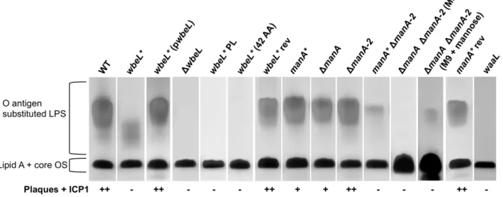
Purified LPS from awbeL*(A7) phase variant shows a distinct
lower molecular weight pattern on a silver stained SDS-PAGE gel (Fig. 2), although the strain exhibits a normal slide agglutination phenotype with anti-Ogawa typing serum (Table 1). The wbeL*
strain is completely resistant to infection with ICP1, and accordingly purified LPS from this strain shows no inhibition of ICP1 (Table 1). To test the possibility that thewbeL* A7frame shift
exerts a polar effect on downstream genes, which also contribute
Author Summary
to tetronate biosynthesis (Fig. 1B), we performed complementation analysis and found that wild type LPS production and ICP1 sensitivity are restored when thewbeL* mutant is complemented with wbeL in trans (Fig. 2). This indicates that the observed phenotypes are a consequence of the loss of WbeL expression and not due to polar effects of thewbeL*mutation. To further confirm this, an in-frame deletion in wbeL was constructed and comple-mentation analyses were performed. TheDwbeLstrain is devoid of O1 antigen (Fig. 2) and accordingly exhibited no agglutination with anti-Ogawa typing serum (Table 1), and these phenotypes can be complemented withwbeL in trans(data not shown). Since the phenotype of the in-frame deletion mutant is not consistent with the originalwbeL*mutation, we hypothesized that thewbeL*
allele maintains some O1 antigen biosynthetic function in the cell. Consistent with this, when theDwbeLstrain is complemented with the wbeL* allele in trans, agglutination in the presence of anti-Ogawa typing serum is restored while the strain maintains complete resistance to ICP1, just like the original wbeL* strain (Table 1). The DwbeL mutant expressing wbeL* in trans also produces a small amount of lower molecular weight LPS like the original wbeL* strain (data not shown). To further address the biosynthetic function of thewbeL*allele, we explored two possible explanations for the phenotype of thewbeL* phase variant. First, that the presence of the truncated 42 amino acid peptide produced by thewbeL*allele is necessary and sufficient to allowV. choleraeO1 to elaborate the lower molecular weight LPS observed (Fig. 2). To test this hypothesis we constructed a deletion in the remainder of the coding sequence downstream of the premature stop codon in
wbeL*. Purified LPS from awbeL* strain expressing only the 42 amino acid peptide lacks O1 antigen substituted LPS (Fig. 2), which rules out this hypothesis. The second hypothesis that we
tested to account for the biosynthetic role of thewbeL*allele is that it allows some functional WbeL protein to be made because the
wbeL*allele is subject to nonstandard decoding (ribosomal frame shifting or transcriptional slippage at the A7 tract). This in turn
would allow for a small amount of tetronate modification to occur, resulting in a lower molecular weight but still compositionally (and antigenically) normal O antigen (which is consistent with the observation that the wbeL* phase variant agglutinates in the presence of anti-Ogawa typing serum (Table 1). To test this hypothesis, we made silent point mutations within the A7tract in
wbeL*(A7toTAAGAAA) designed to prevent non-standard decoding
as well as prevent further slipped-strand mispairing during replication and characterized the ability of this strain (referred to aswbeL*PL for phase-locked) to produce O1 antigen substituted LPS. The point mutations inwbeL* PL abolished the ability of this strain to make O antigen substituted LPS, which supports the conclusion that thewbeL*allele maintains biosynthetic function by allowing some functional WbeL protein to be produced through nonstandard decoding, and that this is dependent on the A7tract.
The mechanism responsible is not currently known, nor is it known if there are other sequence motifs within thewbeL* allele that facilitate the+1 frameshifting needed to restore the reading frame. It is important to emphasize that the observed lower molecular weight pattern of purified LPS fromwbeL*is not due to reversion of A7to A8in the genome of a substantial subset of the
population, because if that were the case we would observe a small amount of wild type length O1 antigen and not the unique species observed for the wbeL* strain. These results further suggest that tetronate acylation of the perosamine backbone is necessary for incorporation of the O1 antigen into the LPS molecule, and this may be required for recognition and subsequent transport of the
Figure 1. Genetic organization of the O1 antigen biosynthetic locus and the proposed pathway for the synthesis of perosamine in
V. choleraeO1.A) Pictorial representation of the genes in the O1 antigen biosynthetic (wbe) locus. The annotated ORFs and their orientation are indicated by block arrows (drawn to scale). The genes currently described as being required for O1 antigen biosynthesis are located between the gmhD andrjgflanking genes. Work presented here however extends the O1 antigen biosynthetic cluster to include VC0269 (manA). The locus can be divided up into five major groups according to putative function that are indicated by the same color arrow. B) Proposed biosynthetic pathway for perosamine inV. choleraeO1. The pathway includes ManA for the conversion of F6P to M6P and eliminates ManC (indicated in grey) as the enzyme responsible for the initial step of perosamine biosynthesis.
doi:10.1371/journal.ppat.1002917.g001
undecaprenyl-linked O antigen polymer to the periplasm by the ABC transporter and/or for efficient ligation of the O antigen polymer to the lipid A-core by WaaL ligase [42]. Importantly, these data demonstrate that the lower molecular weight O1 antigen produced by thewbeL*mutant somehow endowsV. cholerae
with full resistance to ICP1.
The second mutation identified in ICP1-resistant colonies from the center of plaques also mapped to a poly-A tract, and this localized to VC0269, which we designate manA for reasons explained below.manAhas two poly-A (A9) tracts and, as illustrated
in Fig. 1A, is located approximately 4 kbp downstream of the right junction gene (rjg= VC0264), which was thought to delineate the end of the O1 antigen biosynthetic locus in V. cholerae. ManA shows homology to type I PMIs that catalyze the reversible isomerization of F6P to M6P, which is the first step in perosamine biosynthesis (Fig. 1B). LikewbeL,manAis also specific toV. cholerae
O1 strains: All 37V. choleraeO1 strains available for bioinformatic analysis havemanAand it is highly conserved between these strains with the exception of two strains which have themanA* (A8) allele
resulting from a single nucleotide deletion in the first poly-A tract (these latter strains are designated 2740–80 and HC-61A1). The full length ManA protein is 399 amino acids, and a single nucleotide deletion within the first poly-A tract is predicted to result in a truncated peptide 81 amino acids long, while a single nucleotide deletion within the downstream poly-A tract (designat-edmanA*) produces a truncated peptide 207 amino acids long (Fig. S3). LPS purified from manA* after overnight growth looks identical to the parental strain (Fig. 2). However, purified LPS from themanA* strain does not bind and inhibit ICP1 as efficiently as a equal amount of LPS purified from the parental strain (Table 1), which is consistent with this mutant being partially phage resistant (Fig. 2 and Table 1). These results leave open the possibility that whilemanA*produces wild type length O1 antigen, the overall abundance of O1 antigen substituted LPS is less than wild type, and this translates into fewer available receptors for ICP1. Phage infection is a complex process, which can require sequential receptor binding steps. For example T4 infection is initiated by the reversible attachment of at least three of the six long tail fibers to the outer core of the LPS, but does not result in DNA injection unless the six short tail fibers successfully engage their receptors on the inner core of the LPS [43–45]. Throughout the course of this study isolates were obtained with a frameshift in either poly-A tract inmanAand these isolates were phenotypically indistinguishable from one another with regard to phage sensitivity and agglutination with anti-Ogawa typing serum (data not shown). Similarly, a strain harboring an in-frame deletion of manA was phenotypically indistinguishable from the manA phase variants (Fig. 2 and Table 1), indicating that both frame-shift mutations function asmanAnulls.
manAandmanA-2, but notmanC, are important for perosamine biosynthesis
manAdoes not appear to be required for producing full length O antigen as indicated by SDS-PAGE and silver staining of LPS (Fig. 2), and yet the manA* strain is partially resistant to ICP1, which specifically requires the O1 antigen for infection. These observations led us to speculate that there is likely another gene that can contribute to the conversion of the F6P to M6P in V. choleraeO1. We noted the presence of another annotated type I PMI in theV. choleraeO1 genome, VC1827, hereafter designated
manA-2.manA-2is located immediately downstream of a mannose permease encoded by VC1826 [46], but is not linked to the O1 antigen biosynthetic cluster.manAandmanA-2are 65% identical at the nucleotide level over 70% of their sequence, and at the protein
level, they are 59% identical over 97% of their sequence.manA-2
has a higher GC content thanmanA(46.1% compared to 42.1%, respectively) and its GC content is much closer to the overall GC content of the entire V. cholerae N16961 genome (,47% [47]) suggesting manA may have been recently horizontally acquired. Unlike manA, manA-2 is found in non-O1 V. cholerae. In other Gram-negatives, includingE. coliandS. Typhimurium, themanA
gene generally maps as an independent gene not associated with the LPS gene cluster due to its role in mannose metabolism [48,49], although in these organisms the PMI activity of the unlinked manA is required for O antigen synthesis [50,51]. In organisms that do not metabolize mannose, the manA gene is generally absent [49]. Some bacteria have a bifunctional type II PMI-GMP (typically referred to as ManC) encoded in the LPS biosynthetic cluster. Monofunctional and bifunctional forms of ManC are not clearly distinguishable on the basis of size or even sequence similarity [49]. Indeed ManC (VC0241) inV. choleraeO1 has the bioinformatic designation of a type II PMI, however our results below suggest this enzyme lacks PMI activity.
The presence of two putative type I PMIs inV. choleraeO1 led us to investigate the phenotypes of single and double mutants with regards to O antigen biosynthesis. A mutant lackingmanA-2was indistinguishable from wild type with regard to phage sensitivity and LPS pattern on a gel (Fig. 2 and Table 1), indicating that
manA-2 is not important for O antigen biosynthesis under the conditions tested. However, in the absence of manA, manA-2
becomes important since themanA* DmanA-2 double mutant is completely phage resistant and produces very little fully length LPS (Fig. 2). Another potential source of M6P for perosamine biosynthesis is through the conversion of exogenously acquired mannose to M6P by a hexokinase, which led us to determine if trace mannose present in the growth media (Luria-Bertani [LB] broth) contributed to the small amount of O1 antigen still visible in themanA*DmanA-2double mutant. Indeed, this small amount of O1 antigen substituted LPS is absent when the double mutant is grown in M9-glucose, but is present when the double mutant is grown in M9-glucose plus mannose (Fig. 2), demonstrating that exogenous mannose is responsible for the small amount of O1 antigen substituted LPS in the double mutant. Similar results have been observed inE. coliandS.TyphimuriummanAmutants, which are unable to synthesize O antigen without the inclusion of mannose in the growth media [50,51]. Also consistent with these results, we observed that our doublemanA* DmanA-2mutant is unable to grow in M9-mannose owing to the critical nature of type I PMIs in the conversion of M6P to F6P as a substrate for glycolysis. These results indicate thatmanCinV. choleraeO1, which was hypothesized to catalyze the first reaction in biosynthesis of perosamine (Fig. 1B) [12], is not active as a bifunctional PMI-GMP, and is likely only important for the later steps in perosamine biosynthesis for converting M1P to GDP-mannose. As type II PMIs possess two catalytically distinct domains for each PMI and GMP activity [20], attempts were made to construct mutantmanC
alleles that should be defective for PMI activity alone (data not shown) but all constructs resulted in complete depletion of the O1 antigen, suggesting that these mutations had an inadvertent negative impact on the GMP activity of the protein. The reason behindV. choleraeO1 possessing two type I PMIs is thus not clear, although it suggests that they each have their primary roles:manA
in O antigen biosynthesis andmanA-2 in mannose metabolism, though it is unclear what factors define those functional roles. A search for other bacteria harboring multiple annotated type I PMIs reveals a limited number of organisms including strains of
Vibrio vulnificus(Accession No. NC_005140),Vibrio parahaemolyticus
Figure 2. LPS profiles ofwbeL* and manA* phase variants and related strains by SDS-PAGE and silver staining. Strains for LPS purification were grown overnight in LB (or M9-glucose+/2mannose as indicated) at 37uC with shaking. The bacterial strains are indicated above the lanes and the positions of the fully substituted LPS with attached O antigen and lipid A core oligosaccharide (OS) precursor are indicated. Approximately 5mg of purified LPS was loaded into each lane, with the exception of the lanes containing LPS prepared from M9 cultures in which
,30mg of LPS was loaded. A strain lacking O1 antigen transferase activity,waaL, which has no O1 antigen, was used as a control for assessing the
electrophoretic migration of the lipid A-core oligosaccharide. The ability of ICP1 to infect these strains as evidenced by plaque formation with bacteria grown to OD600,0.5 is indicated as follows: (++) clear plaques, (+) turbid plaques, (2) no plaques.
doi:10.1371/journal.ppat.1002917.g002
Table 1.Summary of phenotypes of single and double mutants ofV. choleraeO1 and their reverted or trans-complemented derivatives as analyzed by silver stained SDS-PAGE, phage sensitivity, and agglutination by anti-Ogawa typing serum.
Strain
O antigen substituted LPSa
ICP1 sensitivityb
Log10inhibition of purified LPSc
Agglutination with serum
WT ++ ++ 2.3 +
waaL None 2 n.d. 2
wbeL* + 2 0 +
wbeL* pwbeL ++ ++ n.d. +
DwbeL None 2 0 2
DwbeLpwbeL ++ ++ n.d. +
DwbeLpwbeL* + 2 n.d. +
wbeL*(42 AA) None 2 n.d. 2
wbeL*PL None 2 n.d. 2
wbeL*rev ++ ++ n.d. +
manA* ++ + 1.6 +
DmanA ++ + 1.6 +
DmanA-2 ++ ++ n.d. +
manA*DmanA-2 + 2 0 +
DmanADmanA-2(in M9+glucose) None 2 0 2
DmanADmanA-2(in M9+glucose+mannose) + 2 n.d. +
manArev ++ ++ n.d. ++
aThe pattern of purified LPS from strains grown overnight in LB (unless otherwise indicated) was visualized by SDS-PAGE and silver staining and the phenotype of each
strain was categorized as follows: (++) wild type appearance of O antigen substituted LPS, (+) altered levels or appearance of O antigen substituted LPS, (None) no O antigen substituted LPS was observed.
bThe ability of ICP1 to infect these strains as evidenced by plaque formation with bacteria grown to OD
600,0.5 is indicated as follows: (++) clear plaques, (+) turbid plaques, (2) no plaques.
cThe ability of purified LPS to inhibit plaque formation by ICP1 was determined using 10mg of purified LPS and is indicated as the average log
10inhibition of two
independent assays. n.d. not done. doi:10.1371/journal.ppat.1002917.t001
our knowledge, the functional roles that these enzymes have in these other organisms are not known.
As mentioned previously, the O1 antigen biosynthetic cluster was originally identified through the heterologous expression of the V. cholerae O1 antigen in E. coli [10], and all O1 antigen biosynthetic genes studied thus far have been between the gmhD andrjgflanking genes.manAwas likely not identified as part of this biosynthetic pathway because its function was complemented by theE. coli manAgene, permitting expression of the O1 antigen in this host. Additional genes required for O1 antigen biosynthesis were subsequently identified following the initial report by Manninget al.[11], and were similarly likely missed because the phenotype was masked in E. coli. Blokesch and Schoolnik [52] provided some additional support that further extends the O1wbe
region downstream of rjg. They observed that when serogroup conversion ofV. choleraeO1 to O139 occurred through uptake of O139 donor DNA during natural transformation, the crossovers were often localized within or downstream of VC0271 at the right junction, and the location of the left junction was within or upstream ofgmhD. These results coupled with our observation that
manAparticipates in O1 antigen biosynthesis suggests that thewbe
region extends approximately 8 kbp downstream ofrjg(Fig. 1B). There are six genes currently annotated in addition tomanAin this region (Fig. 1A), however it remains to be seen if these other genes do in fact participate in O1 antigen biosynthesis.
V. choleraeO1wbeLandmanA phase variants are defective for colonization of the small intestine
The ability ofwbeL*and manA*phase variants to colonize the small intestine was assessed in competition assays in the infant mouse model. The wbeL* strain is attenuated over 1000-fold (Fig. 3). We did not anticipate such a high level of attenuation given that this strain still elaborates LPS (although it is a lower molecular weight form, Fig. 2). Interestingly, the altered LPS produced by thewbeL*strain does provide some advantage over not having any O1 antigen, as is apparent by the significantly lower competitive index (CI) for theDwbeLstrain (p,0.05, Mann-Whitney U test). To rule out secondary mutations, we chose to use revertant strainsin vivoto avoid potential complications concerning plasmid loss and non-wild type gene expression levels during infection. The colonization defect observed for thewbeL*strain is absent when the poly-A (A7) tract is reverted to wild type length
(A8) (Fig. 3), demonstrating that the virulence defect is due to the
wbeL* allele. The manA* phase variant, which produces an apparently full length LPS but which is partially resistant to ICP1, is over ten-fold attenuated for colonization, and this defect is absent when the A8tract is reverted to wild type length (A9) (Fig. 3).
We anticipated that amanA*DmanA-2double mutant would be significantly more attenuated owing to a major reduction in O1 antigen produced by the strain (Fig. 2). However, although we did observe a further decrease in the CI formanA*DmanA-2compared to manA*, it was not significant (p = 0.42) (Fig. 3). To try and explain this, we hypothesized that the selective pressure exerted on the manA* DmanA-2 double mutant in the small intestine is sufficient to select for spontaneous revertants which would potentiate the observed moderate drop in CI. To test this, we patchedmanA*DmanA-2isolates recovered from mouse intestines onto M9-mannose agar plates. Consistent with our hypothesis, roughly half of isolates recovered from mice after 24 h of infection had regained the ability to grow on mannose, and subsequent sequencing revealed that these strains had reverted to the wild type, in-frame poly-A tract inmanA. Conversely, we were unable to detect spontaneous revertants of thewbeL*ormanA*strains among the isolates recovered from mouse intestines infected with those
strains, suggesting that the A7wbeL*allele is less prone to reversion
by slipped-strand mispairing, and that the A8allele inmanA*is not
under enough selective pressure to revert as long as ManA-2 is functional. Our inability to detectwbeL*revertants is likely due to the experimental limitation that there are so fewwbeL* isolates recovered from infected mice that we were only able to test ,100 CFU.
ThemanArevertants recovered from intestines of mice infected with the manA* DmanA-2 strain were effectively equivalent to
DmanA-2, which we previously observed has no effect on LPS biosynthesis or virulence (Fig. 2 and Fig. 3). Confirming this, we also competed the double mutant with in-frame deletions in both
manAandmanA-2and observed the anticipated significant increase in attenuation (.2000-fold) compared to amanA* phase variant alone (p,0.05 Mann-Whitney U test) (Fig. 3). Furthermore, the doubleDmanA DmanA-2mutant is significantly more attenuated when grown in M9-glucose prior to infection than when it is grown in LB which contains trace mannose (p,0.05 Mann-Whitney U test). Consistent with this, a comparable CI was observed for the two strains that are devoid of all O1 antigen prior to infection (DmanADmanA-2grown without exogenous mannose, andDwbeL) (Fig. 3).In vitrocontrol competitions revealed that the observed defects were specific to thein vivoenvironment, with the exception of theDwbeLmutant, which has a,10-fold defectin vitro compared to the wild type (data not shown). The nature of this defect is not known, but it could be due to the build-up of O antigen and/or LPS intermediate products in this strain which, unlike the wbeL* phase variant, is unable to elaborate any O1 antigen on the surface. Consistent with this, mutants harboring transposon insertions in wbeL, which also showed a decreased colonization phenotype in infant mice, also exhibited a decreased CI inin vitrocontrol competitions [53].
V. choleraeO1 phase variants are sensitive to the antimicrobial peptide polymyxin B
observed polymyxin sensitivity, but in a growth phase-dependent manner. There was intermediate sensitivity of this strain to polymyxin B when the inoculum used for the killing assay was grown up to early exponential phase (OD600= 0.15) (Fig. 4A). In
contrast, when manA* was grown up to mid-exponential phase prior to treatment with the peptide, wild type levels of survival were observed (Fig. 4A). The phenotype observed at early exponential phase with the manA* phase variant could be fully complemented by expressing manA in trans (Fig. 4A), again indicating the observed phenotypes are due to the loss of ManA. We had also observed that phage sensitivity of themanA* phase variant was growth phase-dependent, and these results parallel the observations in the antimicrobial peptide assay, that is at OD600= 0.15, manA* is completely resistant to ICP1 and at
OD600$0.2, turbid plaques result from ICP1 infection (data not
shown). To address this puzzling observation, we purified LPS from manA*and wild type at early and mid-exponential growth phase (OD600= 0.15 and OD600= 0.5, respectively). In contrast to
the seemingly wild type appearance of purified LPS frommanA*
after overnight growth (Fig. 2) and at mid-exponential phase, the LPS pattern ofmanA*isolated at early exponential growth phase showed very little O1 antigen substituted LPS (Fig. 4B). Since analysis of the doublemanA*DmanA-2mutant indicated that manA-2 is important for O1 antigen synthesis only in the absence of
manA, we interpret these results to suggest the compensatory activity of ManA-2 is incomplete during early exponential phase growth in LB broth. The reason for this is not known, but may relate to differences in expression, activity or localization of ManA-2. In any event, a complete functional redundancy between ManA and ManA-2 would have been at odds with the observation that all V. choleraeO1 strains have a phase variable manAgene: Specifically, the evolution of a contingency locus would be futile if the encoded protein exhibited complete functional redundancy with a non-phase variable gene.
In general, the results of the polymyxin B killing assays (Fig. 4A) parallel the observedin vivo colonization defects (Fig. 3). Strains that produce less O1 antigen substituted LPS (such aswbeL*and
DmanADmanA-2) are highly susceptible to polymyxin B and are more severely attenuatedin vivothanmanA*, which accordingly is less susceptible to polymyxin B. These data suggest that the phase variants are defective forin vivocolonization because they are more susceptible the antimicrobial peptides present in the intestinal tract.
Phase variation ofwbeLoccurs as the dominant mechanism of O1 antigen variation in the face of predation by phage in a simulated natural environment
V. choleraeO1 persists in the environment as a member of the aquatic ecosystem where it is thought to associate with and use the chitinous exoskeletons of zooplankton as a nutrient source [60]. The levels ofV. choleraeO1 phages in the environment, including potentially ICP1, have been shown to inversely correlate with disease burden suggesting that phage predation in the natural environment may contribute to the collapse of a given cholera epidemic [41]. We investigated the potential for phage resistance to develop in a simulated natural environment comprised of chitin and pond water. A thousand CFU from independent cultures of wild type V. cholerae O1 were inoculated into pond water with chitin, and ICP1 was added at an MOI of 0.01. After 24 hours at 30uC, we observed that in all 11 pond microcosms to which ICP1 was added, the phage titer increased at least one million-fold (data not shown). Bacterial levels in the uninfected control had increased 10,000-fold, while the bacterial levels in the infected microcosms varied substantially from below the detection limit to levels nearly comparable to that observed in the uninfected control (Table 2). All isolates recovered from environments to which phage was added were resistant to ICP1 infection. Furthermore, the majority of isolates (63 out of 80 isolates, 79%) from independent
Figure 3.V. choleraeO1 phase variants are attenuatedin vivo.Competition indices (CI) were determined between the strains indicated and the parental strain carrying alacZ deletion in infant mice 24 hours post infection. Each symbol represents the CI for an individual mouse and horizontal lines indicate the median CI for each strain.
doi:10.1371/journal.ppat.1002917.g003
microcosms that had become resistant to ICP1 had the wbeL*
frameshift allele (Table 2). We only observed heterogeneity in the resistance mechanism for isolates from one microcosm (Table 2, number 10), suggesting that in most cases phage predation resulted in the clonal expansion of a single resistant mutant. We did not observe any mutations in the poly-A tracts ofmanA, likely owing to the incomplete ICP1 resistance afforded to V. choleraewith those alleles. Control experiments with manA* and wbeL* variants as input strains in the absence of phage confirmed that neither mutant exhibit a decreased ability to grow in the simulated pond microcosm (data not shown). Isolates from all microcosms to which phage were added were also replica plated onto LB agar containing 350mg/mL polymyxin B (a concentration that permits growth of the wild type parent but notwbeL* ormanA*), and all isolates were unable to grow. This indicates that the mutations that occurred less frequently and that did not map towbeLalso likely affected LPS biosynthesis. These results show that in a simulated natural environment phage predation can occur with consequent selection for bacteria with altered O1 antigen, and that the dominant mechanism by which mutational escape is achieved is through mutations in the poly-A tract inwbeL.
We also investigated the diversity of phage resistant mutants that appeared in the center of plaques on LB plates by similarly sequencing the poly-A tracts in wbeL and manA in 83 phage resistant isolates. In contrast to the experiment designed to mimic the natural aquatic environment, during selection in LB soft agar overlays in which diffusion of phage and bacteria are relatively limited, the mode of phage resistance is much more varied although a substantial portion can be attributed to phase variation inwbeLandmanA(,20%); we found that six out of 83 isolates were wbeL* phase variants and 11 out of 83 weremanA* phase variants (six of the 11 had a deletion that mapped to the first poly-A tract, and the other five to the second poly-A tract). The reason for this difference in frequency ofwbeL* andmanA* occurrence compared
to the pond microcosm is not known. However, as was mentioned previously (and will be addressed below) themanA*mutation was observed in sequenced V. cholerae O1 isolates; therefore it is apparent there are relevant circumstances in whichmanA*phase variants are selected for.
O1 antigen heterogeneity exists between clinical isolates ofV. choleraeO1
We wanted to determine if O antigen heterogeneity exists in the population of V. cholerae excreted along with ICP1 phage from patients during natural infection. To do this we obtained three ICP1-positive stool samples collected from three patients admitted to the International Centre for Diarrhoeal Disease Research, Bangladesh (ICDDR,B) during a cholera epidemic in 2001. Several representative isolates from all three stool samples were analyzed and were found to beV. cholerae O1 Inaba and were sensitive to ICP1. We tested the ability of these stool isolates to become phage resistant by collecting phage resistant mutants from the centers of plaques resulting from ICP1 infection and readily isolatedmanA*andwbeL*phase variants (data not shown), showing that these clinical isolates can phase vary at these contingency loci. Next we screened many thousands of colonies from each of these archived stool samples for the presence ofmanA*andwbeL*phase variants, which we hypothesized might have arisen as a result of ICP1 phage pressure during the patient infection. Since we showed above thatmanA*andwbeL*phase variants are attenuated in the infant mouse model, we expected that their frequency in the human stool samples would be low or perhaps even undetectable due to decreased fitness during infection of the human small intestine. As we had done previously, we took advantage of the observation that both phase variants exhibit increased sensitivity to polymyxin B compared to the parental strain. We determined that both thewbeL* and manA*phase variants obtainedin vitrofrom these clinical isolate strain backgrounds failed to grow on LB agar
Figure 4.V. cholerae O1 phase variants are more sensitive to the antimicrobial peptide polymyxin B. A) Strains were grown to OD600= 0.15 (hatched columns) or OD600= 0.5 (solid black columns) and exposed to 50mg/ml polymyxin B for 2 hours. After treatment, serial dilutions of each culture were plated for enumeration to determine the percent survival compared to a mock-treated control. Data represents the means and standard deviations of two independent experiments performed in technical duplicate. B)manA*phase variant exhibits O1 antigen production dependent on growth phase. Approximately 2mg of purified LPS was loaded into each lane.
plates containing 800mg/ml polymyxin B after replica plating, while the parental strain maintained its ability to grow. We screened approximately 5,000 colonies from each stool sample by replica plating and did not observe any isolates with increased polymyxin B sensitivity.
In the above analysis we were limited by the number of available archived stool samples, since routine practice is to purify a single colony from a stool sample and store that for further examination. While such single colony isolate collections cannot be used to answer the question of whether O antigen heterogeneity exists within the population of V. cholerae excreted from a single patient, it does allow us to determine if O antigen heterogeneity exists between isolates recovered from different patients. We evaluated phage sensitivity of approximately 50 isolates recovered from cholera patients at the ICDDR,B between 2001 and 2005. Three of these isolates displayed resistance to ICP1 and were characterized further. One of the clinical isolates has a mutation in the second poly-A tract inmanA. The other two resistant isolates did not have mutations that mapped to the poly-A tracts in either
manAorwbeL, and examination of purified LPS from these strains showed that they produce very little full length O-antigen substituted LPS (data not shown), however the nature of the observed defects is not known.
From these results it appears that, despite the hypothesized ICP1 phage pressure during infection and despite pressure from the host immune system to reduce or alter the O1 antigen (O1 antigen is the dominant antigen [61,62]), the intra-patient and inter-patient O1 antigen variability is quite low. In agreement with this, previous studies have detected LPS mutants in mice following coinfection of phage andV. choleraeO1, but at very low frequencies. Zahidet al., [63] estimated that the frequency of such mutants was on the order of 1028
, and in accordance with our results, failed to detect phage-resistance heterogeneity amongV. choleraeO1 directly from human stools. Additionally, despite relatively high levels of O1-specific phage in the stool samples,V. cholerae O1 isolated from the same stool samples remain completely susceptible to phage lysis [63–65].
Previous studies of V. cholerae O antigen negative strains or strains with altered LPS structures were also found to be defective in colonization of infant mice [53,59,63,66–69]. These results support the assertion that O1 antigen-deficientV. choleraewould be selected against during human infection due to the high fitness cost associated with mutational escape. When put into the context of our findings, it is fitting that mutational escape is frequently conferred through phase variability, as a key feature defining this mode of variability is its reversible nature [23,32,70]. Inherently each cell will retain its ability to switch between expression states (the switching rates of phase variable genes are typically between 1022
to 1025
[32]), and therefore the phenotype of a clonal population of bacteria capable of phase variation will vary as a function of selection. Predation of V. cholerae O1 in the environment by O1 antigen-dependent lytic phage may rapidly select for the subpopulation with altered O1 antigen mediated by
manA* and wbeL* frame-shifted contingency loci, and even though those subpopulations are less suited for life in the intestinal tract, positive selection (for example when this mixed population is ingested by a human) results in enrichment of the subpopulation with full O1 antigen expression. We were able to experimentally confirm the reversibility of frameshift mutations occurring at the poly-A tract inmanA, however, we were unable to do so forwbeLlikely due to experimental limitations (discussed above). It is possible that the observed mutational escape mediated by the poly-A tract in wbeL is a function of hypermutation and not phase variation (if it is not reversible). However, we have clearly demonstrated the utility of these loci in mediating alterations in the expression of the key V. cholerae
antigen and phage receptor.
Concluding comments
Pathogenic V. cholerae O1 has evolved to live in very diverse environments including fresh water, salt water and the human small intestine. The O1 polysaccharide antigen is the dominant cholera antigen and can induce protective immune responses in
Table 2.Phage predation in a pond microcosm leads to the selection ofwbeL*phage resistant mutants.
Microcosma Phage addedb Total CFU 24 h p.i.c % WTd ICP1 resistant isolates
%wbeL* %manA* % Other
1 2 26107 100 0 0 0
2 + 46106 0 100 0 0
3 + 46105 0 100 0 0
4 + 16106 0 100 0 0
5 + 46103 0 0 0 100
6 + 46105 0 100 0 0
7 + 66105 0 100 0 0
8 + ,d.l. 2 2 2 2
9 + 16107 0 0 0 100
10 + 66105 0 87.5 0 12.5
11 + 66105 0 100 0 0
12 + 46105 0 100 0 0
aIndependent cultures of wild typeV. choleraeO1 were used to inoculate 103CFU per microcosm containing pond water and chitin as a sole nutrient source. bICP1 was simultaneously added at an MOI = 0.01 where indicated.
c
,d.l. is below the limit of detection for this assay (1 CFU).
dEight isolates from each microcosm were tested for resistance to ICP1. If no alteration to sensitivity was observed, the isolates were categorized as WT. If resistance was
observed than the poly-A tracts inwbeLandmanAwere sequenced. If resistance was observed and a frameshift in neitherwbeLnormanAwas observed, than the isolates were categorized as other.
doi:10.1371/journal.ppat.1002917.t002
humans and animals [61,71–73], and thus is a critical immunogen guiding cholera vaccine development. The extent to which V. cholerae can vary expression of the O1 antigen is not currently appreciated. We demonstrate for the first time that the O1 antigen is subject to phase variation and show that this is mediated by three homonucleotide tracts in two genes (wbeLandmanA), which are critical for O1 antigen biosynthesis. The ubiquitous presence of these phase variable homonucleotide tracts in allV. choleraeO1 strains points to the significant role they play in modulating expression of this surface exposed antigen. Moreover, by identifyingmanAas critical for O1 antigen biosynthesis, we have extended the genome boundaries previously believed to contain all the necessary genes for O1 antigen biosynthesis inV. cholerae. Phase variation mediated by homonucleotide tracts has not been previously well-documented inV. cholerae. To our knowledge, the only prior report of phase variation in V. cholerae was that by Carrollet al., [74] in which expression of the membrane bound virulence regulator, TcpH, was observed to be subject to phase variation mediated by a poly-G (G9) tract. However, with the
growing list of currently availableV. choleraeO1 genome sequences, it is clear that this tract is not well-conserved (only three of the available 37 sequenced strains have this tract [data not shown]), and thus this likely does not represent a wide-spread mechanism employed by V. cholerae O1 to alter virulence expression. Examination of the currently available V. cholerae O1 genome sequences may facilitate further exploration of phase variation in this organism; it is interesting to note that there are only twelve homonucleotide tracts of nine or greater nucleotides in length located within coding regions in the V. cholerae O1 N16961 genome, and several of these are located within known virulence factors (data not shown), however the significance this remains to be examined.
The biological role of phase variation in mucosal pathogens is frequently anticipated to facilitate immune evasion in the host [75]. However, in the case of the facultative pathogenV. cholerae, our data point to the primary role for O1 antigen phase variation as a strategy for dealing with the strong opposing selective pressures of phage predation in the environment and the strict requirement of O1 antigen for colonization of the intestinal tract. Phase variation of these genes thus allows for a subset of the population ofV. choleraebeing disseminated from a patient or being ingested in contaminated water, to be resistant to O1-dependent phages or to be virulent, respectively, thus contributing to the overall fitness of this pathogen. We hypothesize that the host immune response represents yet a second strong selective pressure against the O1 antigen, though the effects of this on circulating strains of V. cholerae within immune populations has not been studied.
The ubiquitous presence and overall success of ICP1-related phages is likely, at least in part, due to their use of a critical virulence factor as a receptor [40]. Our observation that mutational escape facilitated bywbeL and manApredominatesex vivostrongly suggests that ICP1 is particularly adept at predation of
V. choleraeO1 within the human host where the requirement for colonization and virulence necessitates the maintenance of the O1 antigen. This may suggest a mechanism whereby this phage and the human host act synergistically to limit V. cholerae during infection, and perhaps how phage contribute to the overall decline of a given cholera epidemic as has been hypothesized [41,64]. It remains to be seen if there are additional mechanisms employed byV. choleraeO1 to evade phage predation, specifically within the human intestinal tract, and how this arms race between ICP1 and its bacterial host shapes the evolution of the circulatingV. cholerae
O1 strains within the endemic region of Bangladesh.
Materials and Methods
Growth conditions
Strains were grown on Luria-Bertani (LB) agar or in LB broth at 37uC with 100mg/ml streptomycin (Sm). When indicated M9 minimal media (supplemented with trace metals, vitamins (Gibco MEM Vitamins, Invitrogen), 0.1% casamino acids) with 0.4% glucose and/or 0.4% mannose was used. Strains containing the pMMB67EH vector were grown in the presence of 100mg/ml Sm and 50mg/ml ampicillin (Amp). Expression from the Ptac
promoter was induced by the addition of 1 mM isopropyl-b-D -thiogalactopyranoside (IPTG). Phage susceptibility was deter-mined by the soft agar overlay method as described previously [40] and/or by measuring growth of a bacterial isolate in the presence of ICP1 (to an approximate MOI = 1) in LB plus Sm broth culture using a Bio-Tek microplate reader.
Isolation of spontaneousmanAandwbeLphase variants and whole genome resequencing
A wild typeV. cholerae O1 strain (E7946) was used in standard plaque assays with phage ICP1 as previously described [40]. Following overnight incubation, colonies were routinely observed in the center of plaques indicating the presence of phage resistant isolates. Four independent colonies were chosen for further analysis including phage resistance assays and whole genome sequencing using an Illumina genome analyzer II (Tufts University Core facility) as previously described [40]. Assembled genomes were aligned to theV. choleraeO1 N16961 [47] and E7946 (unpublished data) reference genomes. Two of the independently isolated phage resistant strains had a single nucleotide deletion in the poly-A tract ofwbeL(designatedwbeL*), while one phage resistant derivative had a single nucleotide deletion in the second poly-A tract of manA
(designated manA*). The other derivative not chosen for further study had a nonsynonymous substitution inmanB.
Bacterial strain construction
PCRs for sequencing and cloning were carried out using EasyA polymerase (Agilent). Primer sequences are available upon request. In-frame unmarked deletions were constructed using splicing by overlap extension (SOE) PCR [76] and introduced using pCVD442-lac [77]. Deletion alleles constructed in this study are missing the entire open reading frame, except for the start and stop codons (with the exception of thewbeLdeletion allele which also preserved a single codon immediately upstream of the stop codon). Expression plasmids were constructed by cloning the desired open reading frame(s) (including the predicted ribosome binding site) into the multiple cloning site of pMMB67EH. Expression vectors were transferred into V. cholerae by conjugation with E. coli
SM10lpir and selection of SmRAmpRcolonies. Strains utilized in this study are shown in Table 3.
LPS analysis
Slide agglutination tests were performed using V. cholerae O1 Ogawa polyclonal rabbit antiserum (Difco). LPS was extracted from overnight cultures unless otherwise indicated, as described previously [72]. Briefly, cultures were centrifuged and washed twice in TM buffer (50 mM Tris [pH 7.5], 10 mM MgCl2)
centrifuged as before. Purified LPS was separated on a 4–12% NuPage Bis-Tris gel (Invitrogen) and visualized by silver-staining (SilverQuest, Invitrogen). The concentration ofV. choleraeLPS was determined by comparison to a standard curve ofE. coliO26:B6 LPS (Sigma) using a Fujifilm FLA-900 scanner as previously described [72]. The ability of purified LPS to neutralize plaque formation was determined as previously described [40].
Infant mouse colonization assays
In vivocompetition experiments were done using 4–5 day old CD-1 mice. The dams and their litters were housed with food and waterad libitumand monitored in accordance with the rules of the Department of Laboratory Animal Medicine at Tufts Medical Center. The inoculum was prepared as a 1:1 mixture of the strain of interest (lacZ+) and the appropriate control strain (DlacZ). Mice were infected intragastrically with approximately,105CFU and sacrificed 24 hours post-infection. Small intestines were homoge-nized in 1 ml LB+16% glycerol, diluted in LB broth, and plated on LB agar plates containing 100mg/ml Sm and 40mg/ml 5-bromo-4-chloro-3-indolyl-b-D-galactopyranoside (X-gal). The competitive index was calculated as the ratio of the mutant compared to the control strain normalized to the input ratio.In vitrocontrols were included in each of these experiments in which the same inoculum was diluted 1:100 into at least five independent LB cultures and the output ratios of mutant to the control strain were determined on Sm X-gal agar plates as above.
Polymyxin B killing assays
Polymyxin B killing assays were done as previously described with minor modifications [57]. Briefly, overnight cultures were
subcultured 1:100 into LB and grown at 37uC to the desired OD (OD600= 0.15 and OD600= 0.5). 5ml polymyxin B (Invitrogen) at
500mg/ml was added to 45ml of the above culture in a well of a 96-well polypropylene microtiter plate to obtain a final test concentration of 50mg/ml polymyxin B. After three hours of incubation at 37uC with shaking, serial dilutions of each culture were plated on LB Sm plates. The percent survival was calculated as (CFU(polymyxin treatment)/CFU(untreated))6100. The average
per-cent survival was determined from two biological replicates, each having been done in technical duplicate.
Evaluation of phage resistance during growth on chitin Overnight cultures of wild type V. choleraeE7946 were serially diluted in filter sterilized pond water to approximately 105CFU/ ml. 10ml of diluted culture (103CFU) was used to inoculate 1 ml chitin solution (1% chitin from crab shells [Sigma] in filter sterilized pond water). To assess the impact of phage on the appearance of phase variants under these conditions, approxi-mately 10 PFU of ICP1 was immediately added following inoculation of bacteria (MOI = 0.01). The mixture was allowed to incubate for 24 hours at 30uC statically at which time the mixture was vortexed and plated for CFU. ICP1 was enumerated by adding chloroform to a 100ml aliquot of the above solution, diluted and plated for PFU with E7946 using the soft agar overlay method as described above.
Evaluation of the presence of phase variants in cholera stool samples
Three cholera stool samples collected at the ICDDR,B in 2001 and stored in the presence of glycerol were assayed for the
Table 3.Strains used in this study.
V. choleraestrains Relevant genotype/description Reference or Source
WT (E7946) Spontaneous SmR derivative of E7946, El Tor Ogawa [79]
wbeL* Spontaneous phage resistant isolate, single nucleotide deletion in poly-A tract
(A8RA7) inwbeL(VC0249)
This study
DwbeL In-frame deletion ofwbeL This study
wbeL* (pwbeL) wbeL* (pMMB67EH::wbeL) This study
DwbeL(pwbeL) DwbeL(pMMB67EH::wbeL) This study
DwbeL(pwbeL*) DwbeL(pMMB67EH::wbeL*) This study
wbeL* (42 AA) wbeL* with a deletion in the remainder of the coding sequence downstream of the premature stop codon
This study
wbeL*PL wbeL* with nucleotide substitutions within the A7tract (A7toTAAGAAA) This study
wbeL*rev Reversion ofwbeL*to wild typewbeL This study
manA* Spontaneous phage resistant isolate, single nucleotide deletion in second poly-A tract
(A9RA8)) inmanA(VC0269)
This study
DmanA In-frame deletion ofmanA(VC0269) This study
DmanA-2 In-frame deletion ofmanA-2 (VC1827) This study
manA*DmanA-2 In-frame deletion ofmanA-2 (VC1827) inmanA*background This study
DmanADmanA-2 In-frame deletion ofmanA-2 (VC1827) inDmanAbackground This study
manA*rev Reversion ofmanA*to wild typemanA This study
manA* (pmanA) manA* (pMMB67EH::manA) This study
DlacZ In-frame deletion oflacZ [80]
waaL waaL::pGp,V. cholerae O1SV194 background, O1 antigen minus [42]
Bacteriophage
ICP1 O1-specific phage ofV. choleraeO1 [40].
doi:10.1371/journal.ppat.1002917.t003
presence of isolates with altered O1 antigen. Single isolates from each sample were found to be O1 Inaba that were sensitive to ICP1.wbeL*andmanA*mutants of this clinical O1 Inaba isolate were recovered after plating with ICP1 and used to assess the applicability of replica plating on polymyxin B as a tool to identify heterogeneity within a stool sample. Both the wbeL* and manA*
isolates in this background failed to grow on LB agar plates containing 800mg/ml polymyxin B, while the parental strain maintained its ability to grow. Each stool sample was plated on LB agar containing 100mg/ml Sm and incubated overnight at 37uC. Plates were then replica plated onto polymyxin B plates and incubated overnight at 37uC to identify polymyxin B sensitive isolates in the stool sample. Approximately 5000 colonies were analyzed per stool sample.
Ethics statement
All animal experiments were done in accordance with NIH guidelines, the Animal Welfare Act and US federal law. The experimental protocol using animals was approved by Tuft University School of Medicine’s Institutional Animal Care and Use Committee. All animals were housed in a centralized and AAALAC-accredited research animal facility that is fully staffed with trained husbandry, technical, and veterinary personnel.
Supporting Information
Figure S1 Chemical structure of the V. cholerae O1 antigen, serotypes Ogawa (R = CH3) and Inaba (R = H).
The O1 antigen is composed of 12–18 repeating units (n) ofa (1,2)-linked D-perosamine residues, the amino groups of which are acylated with tetronate.
(TIF)
Figure S2 The coding sequence of wild typewbeL. The
A8tract which is mutated to A7 inwbeL* is indicated below the
arrow. The premature stop codon resulting from the frameshift mutation is indicated in red. The putative ATP-binding domain [14] is highlighted in gray.
(TIF)
Figure S3 The coding sequence of wild typemanA.The
A9tracts subject to slipped-strand mispairing are indicated below
the arrows. The premature stop codons resulting from the respective frameshift mutations are indicated in red. The conserved PMI motif [20] is highlighted in gray. In addition, amino acids predicted to be involved in zinc ligand binding [78] are indicated with an asterisk (GLN104, HIS106, GLU141, HIS264). (TIF)
Author Contributions
Conceived and designed the experiments: KDS AC. Performed the experiments: KDS AC. Analyzed the data: KDS AC. Contributed reagents/materials/analysis tools: KDS SMF JJM SBC FQ AC. Wrote the paper: KDS AC.
References
1. Chatterjee S, Chaudhuri K (2003) Lipopolysaccharides of Vibrio cholerae. I. physical and chemical characterization. Biochim Biophys Acta 1639: 65–79. 2. World Health Organization (2011) Cholera, 2010. Wkly Epidemiol Rec 86:
325–339.
3. Ali M, Lopez AL, You YA, Kim YE, Sah B, et al. (2012) The global burden of cholera. Bull World Health Organ 90: 209–218.
4. World Health Organization (2010) Cholera vaccines: WHO position paper. Wkly Epidemiol Rec 85: 117–128.
5. Villeneuve S, Souchon H, Riottot MM, Mazie JC, Lei P, et al. (2000) Crystal structure of an anti-carbohydrate antibody directed againstVibrio choleraeO1 in complex with antigen: Molecular basis for serotype specificity. Proc Natl Acad Sci USA 97: 8433–8.
6. Kenne L, Lindberg B, Unger P, Gustafsson B, Holme T (1982) Structural studies of theVibrio choleraeO-antigen. Carbohydr Res 100: 341–349.
7. Kenne L, Lindberg B, Unger P, Holme T, Holmgren J (1979) Structural studies of theVibrio choleraeO-antigen. Carbohydr Res 68: C14–6.
8. Redmond JW (1979) The structure of the O-antigenic side chain of the lipopolysaccharide ofVibrio cholerae569B (inaba). Biochim Biophys Acta 584: 346–352.
9. Chatterjee S, Chaudhuri K (2004) Lipopolysaccharides of Vibrio cholerae II. genetics of biosynthesis. Biochim Biophys Acta 1690: 93–109.
10. Manning PA, Heuzenroeder MW, Yeadon J, Leavesley DI, Reeves PR, et al. (1986) Molecular cloning and expression inEscherichia coliK-12 of the O antigens of the inaba and ogawa serotypes of theVibrio choleraeO1 lipopolysaccharides and their potential for vaccine development. Infect Immun 53: 272–277. 11. Fallarino A, Mavrangelos C, Stroeher UH, Manning PA (1997) Identification of
additional genes required for O-antigen biosynthesis in Vibrio cholerae O1. J Bacteriol 179: 2147–53.
12. Stroeher UH, Karageorgos LE, Brown MH, Morona R, Manning PA (1995) A putative pathway for perosamine biosynthesis is the first function encoded within the rfb region ofVibrio choleraeO1. Gene 166: 33–42.
13. Manning PA, Stroeher UH, Karageorgos LE, Morona R (1995) Putative O-antigen transport genes within the rfb region ofVibrio choleraeO1 are homologous to those for capsule transport. Gene 158: 1–7.
14. Morona R, Stroeher UH, Karageorgos LE, Brown MH, Manning PA (1995) A putative pathway for biosynthesis of the O-antigen component, 3-deoxy-L-glycero-tetronic acid, based on the sequence of theVibrio choleraeO1 rfb region. Gene 166: 19–31.
15. Stroeher UH, Karageorgos LE, Morona R, Manning PA (1992) Serotype conversion inVibrio choleraeO1. Proc Natl Acad Sci U S A 89: 2566–2570. 16. Hisatsune K, Kondo S, Isshiki Y, Iguchi T, Haishima Y (1993) Occurrence of
2-O-methyl-N-(3-deoxy-L-glycero-tetronyl)-D-perosamine (4-amino-4, 6-dideoxy-D-manno-pyranose) in lipopolysaccharide from ogawa but not from inaba O forms of O1Vibrio cholerae. Biochem Biophys Res Commun 190: 302–307.
17. Proudfoot AE, Turcatti G, Wells TN, Payton MA, Smith DJ (1994) Purification, cDNA cloning and heterologous expression of human phosphomannose isomerase. Eur J Biochem 219: 415–423.
18. Miles JS, Guest JR (1984) Nucleotide sequence and transcriptional start point of the phosphomannose isomerase gene (manA) of Escherichia coli. Gene 32: 41–48.
19. Collins LV, Hackett J (1991) Sequence of the phosphomannose isomerase-encoding gene ofSalmonellaTyphimurium. Gene 103: 135–136.
20. Jensen SO, Reeves PR (1998) Domain organisation in phosphomannose isomerases (types I and II). Biochim Biophys Acta 1382: 5–7.
21. Nikaido H (1994) Prevention of drug access to bacterial targets: Permeability barriers and active efflux. Science 264: 382–388.
22. Raetz CRH, Whitfield C (2002) Lipopolysaccharide endotoxins. Annu Rev Biochem 71: 635–700.
23. van der Woude MW, Ba¨umler AJ (2004) Phase and antigenic variation in bacteria. Clin Microbiol Rev 17: 581–611.
24. Roche RJ, High NJ, Moxon ER (1994) Phase variation ofHaemophilus influenzae lipopolysaccharide: Characterization of lipopolysaccharide from individual colonies. FEMS Microbiol Lett 120: 279–283.
25. Weiser JN (1993) Relationship between colony morphology and the life cycle of Haemophilus influenzae: The contribution of lipopolysaccharide phase variation to pathogenesis. J Infect Dis 168: 672–680.
26. Hood DW, Deadman ME, Jennings MP, Bisercic M, Fleischmann RD, et al. (1996) DNA repeats identify novel virulence genes inHaemophilus influenzae. Proc Natl Acad Sci U S A 93: 11121–11125.
27. Jennings MP, Srikhanta YN, Moxon ER, Kramer M, Poolman JT, et al. (1999) The genetic basis of the phase variation repertoire of lipopolysaccharide immunotypes inNeisseria meningitidis. Microbiology 145: 3013–3021. 28. Moran AP, Knirel YA, Senchenkova SN, Widmalm G, Hynes SO, et al. (2002)
Phenotypic variation in molecular mimicry betweenHelicobacter pylori lipopoly-saccharides and human gastric epithelial cell surface glycoforms. acid-induced phase variation in lewis(x) and lewis(y) expression byH. pylori lipopolysaccha-rides. J Biol Chem 277: 5785–5795.
29. Wang G, Ge Z, Rasko DA, Taylor DE (2000) Lewis antigens inHelicobacter pylori: Biosynthesis and phase variation. Mol Microbiol 36: 1187–1196.
30. Gilbert M, Karwaski MF, Bernatchez S, Young NM, Taboada E, et al. (2002) The genetic bases for the variation in the lipo-oligosaccharide of the mucosal pathogen,Campylobacter jejuni. Biosynthesis of sialylated ganglioside mimics in the core oligosaccharide. J Biol Chem 277: 327–337.