TEL1
,
SAE2
, and
De Novo
Telomere Addition Generates
Specific Chromosomal Rearrangement Signatures
Christopher D. Putnam1,2*, Katielee Pallis1, Tikvah K. Hayes1¤, Richard D. Kolodner1,2,3,4,5
1Ludwig Institute for Cancer Research, University of California School of Medicine, San Diego, La Jolla, California, United States of America,2Department of Medicine, University of California School of Medicine, San Diego, La Jolla, California, United States of America,3Department of Cellular and Molecular Medicine, University of California School of Medicine, San Diego, La Jolla, California, United States of America,4Moores-UCSD Cancer Center, University of California School of Medicine, San Diego, La Jolla, California, United States of America,5Institute of Genomic Medicine, University of California School of Medicine, San Diego, La Jolla, California, United States of America
Abstract
Whole genome sequencing of cancer genomes has revealed a diversity of recurrent gross chromosomal rearrangements (GCRs) that are likely signatures of specific defects in DNA damage response pathways. However, inferring the underlying defects has been difficult due to insufficient information relating defects in DNA metabolism to GCR signatures. By analyzing over 95 mutant strains ofSaccharomyces cerevisiae, we found that the frequency of GCRs that deleted an internal CAN1/URA3cassette on chrV L while retaining a chrV L telomerichphmarker was significantly higher intel1D,sae2D,rad53D sml1D, and mrc1D tof1Dmutants. The hph-retaining GCRs isolated from tel1D mutants contained either an interstitial deletion dependent on non-homologous end-joining or an inverted duplication that appeared to be initiated from a double strand break (DSB) on chrV L followed by hairpin formation, copying of chrV L from the DSB toward the centromere, and homologous recombination to capture thehph-containing end of chrV L. In contrast, hph-containing GCRs from other mutants were primarily interstitial deletions (mrc1Dtof1D) or inverted duplications (sae2Dandrad53Dsml1D). Mutants with impairedde novotelomere addition had increased frequencies ofhph-containing GCRs, whereas mutants with increasedde novotelomere addition had decreased frequencies ofhph-containing GCRs. Both types ofhph-retaining GCRs occurred in wild-type strains, suggesting that the increased frequencies of hph retention were due to the relative efficiencies of competing DNA repair pathways. Interestingly, the inverted duplications observed here resemble common GCRs in metastatic pancreatic cancer.
Citation:Putnam CD, Pallis K, Hayes TK, Kolodner RD (2014) DNA Repair Pathway Selection Caused by Defects inTEL1,SAE2, andDe NovoTelomere Addition Generates Specific Chromosomal Rearrangement Signatures. PLoS Genet 10(4): e1004277. doi:10.1371/journal.pgen.1004277
Editor:Gregory S. Barsh, Stanford University School of Medicine, United States of America ReceivedMay 2, 2013;AcceptedFebruary 13, 2014;PublishedApril 3, 2014
Copyright:ß2014 Putnam et al. This is an open-access article distributed under the terms of the Creative Commons Attribution License, which permits
unrestricted use, distribution, and reproduction in any medium, provided the original author and source are credited.
Funding:This work was supported by the Ludwig Institute for Cancer Research (CDP and RDK) and NIH grant GM26017 (RDK). The funders had no role in study design, data collection and analysis, decision to publish, or preparation of the manuscript.
Competing Interests:The authors have declared that no competing interests exist. * E-mail: cdputnam@ucsd.edu
¤ Current address: Curriculum in Genetics and Molecular Biology and Lineberger Comprehensive Cancer Center, University of North Carolina at Chapel Hill, Chapel Hill, North Carolina, United States of America
Introduction
Large numbers of complex chromosomal rearrangements (called gross chromosomal rearrangements or GCRs) are seen in many cancers, potentially due to ongoing genome instability. Much of our present knowledge on the genome rearrangements seen in cancer is from cytogenetic observations of large-scale genome rearrangements and processes associated with their formation. Some examples include cytogenetically observable genome rearrangements that appear to be triggered by dicentric chromosomes undergoing cycles of bridge-fusion-breakage [1–3] or breakage of chromosomes by anaphase bridges that have been observed in early stages of carcinogenesis [4] and in cells containing defects in cancer susceptibility genes like BLM [5]. The advent of genomics methods including whole-genome next generation sequencing of the genomes from tumors and paired normal tissue has greatly expanded the information available about the kinds of somatic GCRs present in cancers. Interestingly,
some types of GCRs may be specifically enhanced in subsets of cancer, including retrotransposition events in colorectal cancers [6], inversions in pancreatic cancer [7], tandem duplications in ovarian and triple-negative breast cancer [8,9], and focal copy number changes in ovarian cancer [10]. The presence of these rearrangements in a subset of cancers of a specific type suggests that the genetic background in different cancers may influence the mechanisms of GCRs formation. The limited understanding of the types of genetic defects that affect GCR formation and the enormous genetic variation seen in many cancers pose challenges to understanding the influence of genetic background on the types of GCRs seen and their rates of formation.
formation of different types of GCRs by a diversity of mechanisms depending on the assay and the genotype of the strain used. The types of GCRs observed include terminal deletions healed byde novotelomere addition, simple monocentric translocations includ-ing the formation of circular chromosomes, and complex GCRs that are initiated by the formation of dicentric translocations and end-to-end chromosome fusions followed by multi-step rearrange-ments that resolve the initial dicentric translocations to monocen-tric GCRs [11,14,16–20].
During the analysis of GCRs formed in assays utilizing aCAN1/ URA3 cassette placed at various locations along the left arm of chromosome V (chrV L; [14,21]), we noticed that a high proportion of GCRs in some mutants, including tel1D, sae2D,
mrc1Dtof1Dand rad53Dsml1D, retained a hygromycin resistance marker (hph) present on the assay chromosome telomeric to the
CAN1/URA3cassette. We initially characterized the GCRs formed in the tel1D mutant, which lacks the gene encoding a DNA damage checkpoint protein kinase that is important for telomere maintenance [16,22–25], and determined that hph-retention was due to the formation of interstitial deletions by non-homologous end-joining (NHEJ) or by formation of inverted duplications that were then resolved by homologous recombination (HR) between theura3-52allele (a Ty element insertion at theURA3locus in the duplicated region) and URA3 in theCAN1/URA3cassette. Both types ofhph+products were observed in wild-type strains, but at much lower frequencies than in thetel1Dmutant. Importantly, the
hph2GCRs formed in thetel1Dmutant were also associated with increased frequencies of inverted duplications that differed from the hph+ GCRs only with respect to the homologies used for telomere capture. Deletion ofSAE2also caused an increase inhph
retention. However, unlike thetel1D mutation, this increase was solely due to increased levels of inverted duplications. Detailed analysis of the interactions betweentel1andsae2single mutations and mutations affecting the Mre11-Rad50-Xrs2 (MRX) complex, which functions in the resection of DNA at double-stranded breaks (DSBs), or the DNA damage checkpoint revealed that complex interactions between repair pathways promote the formation of specific rearrangements. Furthermore, genetic defects that sup-pressedde novotelomere addition increasedhphretention, whereas genetic defects that increasedde novotelomere addition decreased
hph retention. Together, these results suggest a mechanism by which Tel1, Sae2, and de novo telomere addition play a role in
suppressing inverted duplications and, in some cases, interstitial deletions, and further demonstrate that defects in these pathways/ genes result in GCRs with a specific structural signature.
Results
Retention of telomeric DNA in GCRs is assay- and genotype-specific
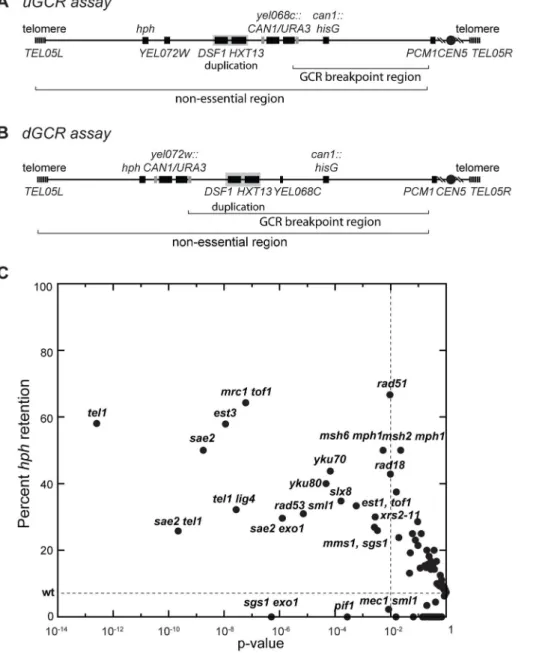
Two GCR assays on chrV that incorporated a telomeric hygromycin resistance marker (hph) (Figure 1A and B) [14] were used to characterize the GCR rate and the frequency of GCRs retaining hph in over 95 mutant strains [14,21]. The yel072w:: CAN1/URA3GCR (dGCR) assay primarily mediates GCRs by duplication-mediated rearrangements with chromosomes IV, X, and XIV; the GCRs derived using this assay frequently lost the telomeric portion of chrV that includes the hphmarker [14,21]. Consistent with this, 0 of 62 GCRs (0%) derived in the wild-type dGCR assay strain and 15 of 2435 GCRs (0.6%) formed in all tested dGCR assay strains retainedhph. In contrast, the frequency of hph retention was higher in the GCRs formed in the
yel068c::CAN1/URA3GCR (uGCR) assay, which mediates GCRs by single copy or ‘‘unique’’ genomic sequences. In the wild-type uGCR assay strain, 2 of the 27 GCR-containing isolates (7%) retainedhph, and 367 of 2670 GCRs (14%) formed in all tested uGCR assay strains retained thehph marker. Specific mutations significantly increased the frequency ofhph+GCRs relative to wild type (Figure 1C). These mutations included tel1D (58% hph+; p = 3610213, G-test), sae2D (50% hph+; p = 261029, G-test),
rad53D sml1D(31% hph+; p = 761026, G-test), and mrc1D tof1D
(64%hph+; p = 661028, G-test).
hph+uGCRs from thetel1Dstrain are either interstitial
deletions or inverted duplications
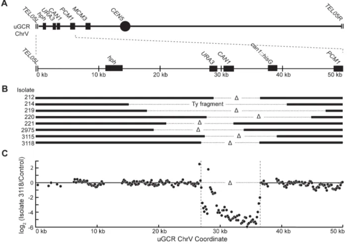
We characterized 18hph+GCRs isolated from thetel1DuGCR assay strain by pulsed-field gel electrophoresis (PFGE) and Southern blotting. Probes complementary tohphand to MCM3, which is an essential gene on chrV, hybridized to the same band in lanes with undigested chromosomes (Figures 2 and S1), indicating thathph was retained on chrV. The size of chrV was similar to wild-type in 8 isolates and was larger than wild-type in 10 isolates. Digestion of chrV by AscI generates three fragments from the starting chromosome: the left telomeric fragment contains hph
sequence, the internal fragment contains both hph and MCM3
sequence, and the right telomeric fragment has neither hph nor
MCM3sequence (Figure 2A). In all cases with a larger than wild-type chrV, the change in size appeared to be due to changes in the internalAscI fragment (Figure 2B and C; Figure S1).
Analysis of the 8 hph+ GCRs with a wild-type-sized chrV revealed that they all contained interstitial deletions. We used PCR to map and amplify the rearrangement breakpoints. Sanger sequencing of the PCR products revealed the presence of inter-stitial deletions that spanned theCAN1/URA3cassette (Figure 3A and B) and had short sequence identities at the breakpoint junc-tions (0–5 basepairs in length; Figure S2), consistent with previous observations [18]. In addition, isolate 214 contained an insertion of a,4 kb fragment of a Ty retrotransposon at the breakpoint.
Lack of copy number changes other than the interstitial deletion was verified by array comparative genomic hybridization (aCGH) of isolate 3118 (Figure 3C). Paired-end whole genome sequencing (WGS) of isolate 3118 (Table S1 and S2) confirmed the interstitial deletion by the identification of 572 read pairs (‘junction-defining’ read pairs) that had mapped inter-read distances of ,5.4 kb
as compared to the median mapped inter-read distance of 417 bp for all 11,333,616 uniquely mapping read pairs (Figure S3).
Author Summary
Additionally, alignment of 114 unmapped reads, which were paired with a read that mapped adjacent to the junction-defining read pairs (‘junction-sequencing’ reads), identified the same junction sequence observed by PCR amplification and Sanger sequencing (Figure S3).
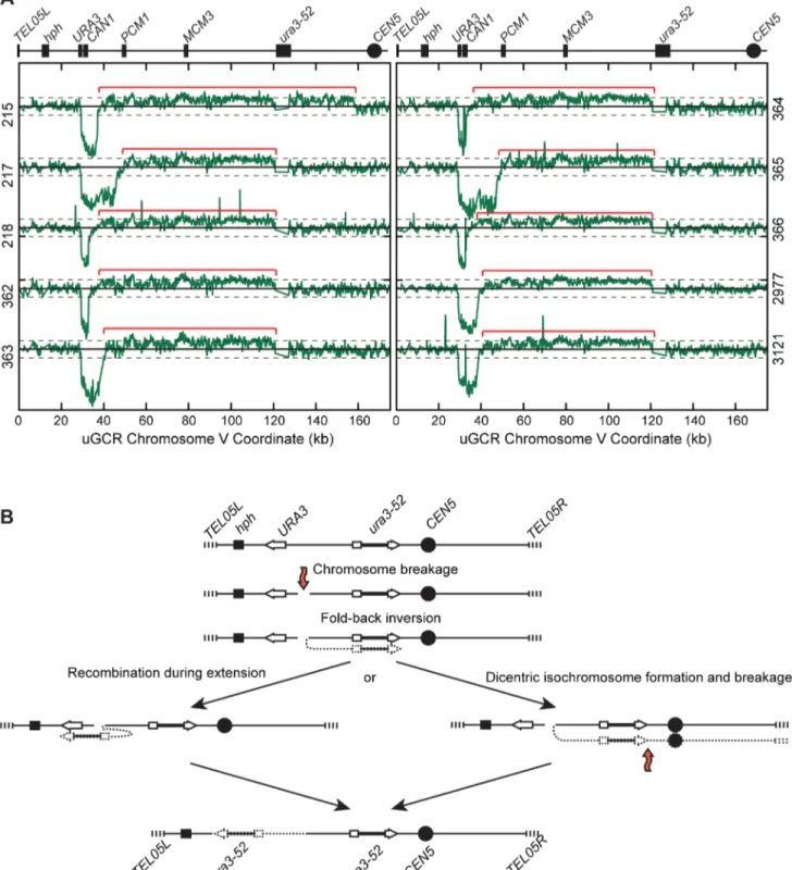
The 10hph+GCRs with a large chrV were inverted duplications associated with a second homology-mediated rearrangement. aCGH analysis revealed that in the strains containing these GCRs all of the copy number changes detected were restricted to chrV: the changes associated with these GCRs included a ,4–19 kb
chrV L deletion spanning theCAN1/URA3cassette, and a,80–
100 kb chrV L duplication extending from the GCR breakpoint region, which is bounded by theCAN1/URA3cassette andPCM1
(Figure 1A), to a centromeric repetitive element, which was most frequently the Ty-containingura3-52(Figure 4A). In each case, the aCGH data also indicated that the GCRs retained the hph -containing region of chrV fromTEL05Lto the telomeric half of
YEL068C, consistent with HR-mediated fusion between ura3-52
andURA3in theCAN1/URA3cassette. We verified theura3-52/ URA3fusion by PCR amplification and Sanger sequencing (Figure S4). WGS of 8 isolates (Table S1) identified and sequenced an inversion junction at the telomeric end of the chrV L duplica-tion (Figure S5; Table S2). If these juncduplica-tions were formed by folding back and priming of a single strand (Figure 4B), then the homologies for priming were 3–9 bases and the unpaired single-stranded hairpin ranged from 25 to 44 bases (Figure S5).
Figure 1. Biased distribution of GCRs retaininghph.(A and B) Schematic showing the positions of theCAN1/URA3cassette in the uGCR and
dGCR assays relative to the 4.2 kbHXT13-DSF1segmental duplication on chrV. The GCR breakpoint region (horizontal bracket) is the region in which rearrangements must occur to loseCAN1/URA3cassette but not the essential genePCM1. (C) Plot of the percent retention ofhphin the uGCR assay in various mutant backgrounds against the respective p-value for retention (G-test) using the wild-type distribution (2 of 27) as the expected distribution. These data include strains generated and analyzed in this study. Points to the left of the vertical dashed line correspond to mutations with p-values,0.01. The horizontal dashed line is the frequency ofhphretention in the wild-type uGCR assay strain.
Palindromes are typically difficult to amplify, which may explain the reduced number of junction-defining read pairs recovered for the inversion junctions relative to other rearrangement junc-tions introduced during strain construction (Table S2). The analyses of these inverted duplication GCRs were consistent with the changes observed by PFGE (Figure 2B and C; Figure S1), because the duplicated regions lackedAscI sites, and the rearranged chromosomes were capped by the AscI-containing left telomeric fragment.
Some hph2rearrangements isolated using thetel1D
uGCR assay are inverted duplications
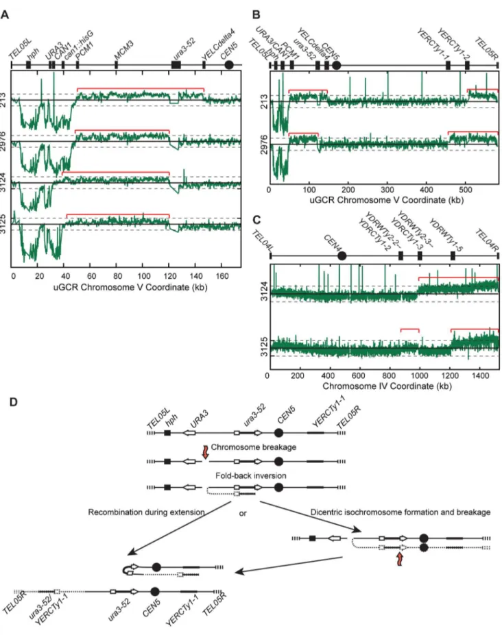
PFGE analysis of the 13hph2GCR-containing isolates from the
tel1D uGCR assay strain revealed that 9 contained a wild-type-sized chrV and 4 contained a large chrV (Figure S6A). PCR mapping [26] revealed that the 9 isolates with wild-type-sized
chrV had deletions that included the CAN1/URA3 cassette; sequencing the breakpoints of 4 of these GCRs confirmed that one was a translocation and 3 were de novo telomere additions (Figure S6B). In contrast, aCGH analysis of the 4 isolates with a larger than wild-type chrV (Figure 5A–C) was consistent with a chrV inverted duplication combined with rearrangements target-ing homologies unrelated toURA3: these GCRs contained a chrV L deletion from the telomere to the GCR breakpoint region (Figure 5A), a chrV L duplication from the GCR breakpoint region to a Ty-related repetitive element (Figure 5A), and an additional duplication of at least one other additional genomic region bounded by Ty-related elements and telomeres (Figure 5B and C). Isolate 3125 had two duplicated regions (between the inverted Ty pairs YDRWTy2-2/YDRCTy1-2 and YDRWTy2-3/ YDRCTy1-3and betweenYDRWTy1-5andTEL04R), which was consistent with a mechanism involving more than one round of
Figure 2. GCRs retaininghphbelong to two size classes.(A) Digestion of the uGCR chrV divides the uGCR chrV into left telomeric, internal, and
right telomeric fragments. Vertical arrows indicate theAscI cleavage sites and relevant chromosomal features are labeled. (B) Southern blot using an hphprobe of a pulsed-field gel (PFG) with DNA from the wild-type strain (RDKY6677) and 6 GCR-containing isolates (212, 214, 215, 217, 218, and 219) with and withoutAscI digestion. Thehphprobe hybridizes to the intact chromosome and the internal and left telomeric fragments. (C) Southern blot of a second PFG with the same samples as in panel B using anMCM3probe. TheMCM3probe hybridizes to the intact chromosome and the internal fragment.
HR-mediated rearrangements similar to GCRs obtained using other GCR assays [15,20]. The inversion junctions were identified and sequenced by analysis of WGS data from isolates 3124 and 3125 (Figure S5; Table S1 and S2). Thus, the hph2 inverted duplications differed from thehph+inverted duplications only with regard to the homologies involved in the resolution of the initial inversion chromosome (Figure 5D).
Detection of chrV L duplications by a multiplex ligation-mediated probe amplification (MLPA) assay
Because inverted duplications could form with or withouthph
retention, we developed an MLPA probe set [27,28] to identify chrV L duplications. MLPA results were validated by comparison with aCGH data for isolates 213, 217, 362, and 3178 (Figure S7). Using MLPA we verified that the 9hph2GCRs with a wild-type sized chrV from the tel1D uGCR assay strain lacked a chrV L duplication. The aggregate data indicated that 14 of 31 GCRs isolated in the tel1D uGCR assay strain contained chrV L dup-lications consistent with inverted dupdup-lications (Table 1), whereas the remaining 17 GCRs lacked chrV L duplications and were consistent with interstitial deletions,de novotelomere additions, or translocations.
NHEJ is required for efficient formation ofhph+
interstitial deletions
To investigate the mechanisms of GCR formation, we first tested the effect of alig4Dmutation, which causes an NHEJ defect [29]. In the dGCR assay,lig4Dcaused a modest increase in GCR rate (Table 2), and thetel1Dlig4Ddouble mutation caused a higher rate in the dGCR assay relative to each single mutation (p = 0.0003 and p = 0.0016, respectively, Mann-Whitney test). Thelig4Dmutation did not affect the GCR rate orhph+retention in the uGCR assay, but thetel1Dlig4Ddouble mutation modestly decreased the GCR rate and increased the frequency of hph
retention relative to the wild-type strain (p = 261028, G-test). The most striking change in the uGCR product spectrum of thetel1D lig4Dstrain relative to thetel1Dstrain was the lack of GCRs that did not contain a chrV L duplication that retainedhph(Table 1). Eighteen of 19hph+GCRs from thetel1Dlig4DuGCR assay strain belonged to the inverted duplication class of GCRs: these GCRs had a chrV that was larger than wild-type, fusion ofura3-52with
URA3(data not shown), and a chrV L duplication (Table 1). Thus, the major mechanism forming the interstitial deletion class ofhph+ GCRs is NHEJ, which is consistent with the short homologies found at the interstitial deletion breakpoints (Figure S2).
Figure 3.hph+GCRs associated with wild-type-sized chrV are interstitial deletions.(A) Diagram of the uGCR chrV and the features on the
first 50 kb containinghph, theCAN1/URA3cassette and the GCR breakpoint region. (B) Map of the retained (solid bar) and deleted (dotted line) regions for the 8hph+GCR isolates with wild-type-sized chrV. Interstitial deletions on chrV entirely (isolates 214, 219, 220, 2975, 3115, and 3118) or partially (isolates 212 and 221) spanned theCAN1/URA3cassette. All of the isolates are simple deletions, indicated by aDsymbol, other than 214, which is fused to a fragment of a Ty element. (C) The log base 2 ratio of the aCGH hybridization intensity for a portion of chrV L from isolate 3118 illustrating the agreement between aCGH and sequenced junctions. The coordinates are mapped to the ‘‘uGCR Chromosome V’’ of RDKY6677, which differs somewhat from the database S288c sequence due to modifications introduced onto chrV during strain construction. No data are present for thehphandcan1::hisGinsertions because these regions were not probed by the aCGH array.
Figure 4.hph+GCRs associated with chrV larger than wild-type contain duplicated chrV sequences.(A) The log base 2 ratio of the aCGH hybridization intensity for chrV L ofhph+isolates with larger than wild-type-sized chrV. The solid horizontal bar is at 0 and dashed lines are at21 and 1 (2-fold decreased and increased, respectively). Probes were mapped onto the ‘‘uGCR Chromosome V’’ coordinate system. Chromosomal features such ashph, theCAN1/URA3cassette, the ura3-52mutation, and the centromere (CEN5) are indicated at top. Red brackets indicate duplicated chromosomal regions that span from the GCR breakpoint region (between theCAN1/URA3cassette andPCM1) to a Ty-related element, most frequentlyura3-52. (B) Proposed mechanism for rearrangement formation (see Discussion). Orange arrows indicate DSBs.
HR is required for efficient formation ofhph+inverted
duplications
Because the inverted duplication class of GCRs involved homology-mediated rearrangements, we tested the effect of a
rad52Dmutation that eliminates HR. In the dGCR assay, arad52D
mutation suppressed the GCR rates [14], and the tel1D rad52D
double mutant had modestly increased GCR rates relative to both single mutants (Table 2). Therad52Dand thetel1Drad52Dmutants had higher GCR rates in the uGCR assay, but had no significant increase in the frequency ofhphretention relative to the wild-type strain (p = 1.0 and 0.5, respectively, G-test; Table 1). Analyses of the 13hph+GCRs from therad52Dtel1DuGCR assay strain was consistent with these GCRs belonging to the interstitial deletion class of GCRs: these GCRs had wild-type-sized chrV, noura3-52/ URA3 fusions (data not shown), and none of the 5 hph+isolates tested by MLPA had a chrV L duplication (Table 1). These data suggest that HR mediates a key step in the formation of the inverted duplication class of GCRs, likely by the formation of stable monocentric chromosomes (Figure 4B and 5D).
Interstitial deletions and inverted duplications are formed in the wild-type uGCR assay strain
Two of 27 GCRs formed in the wild-type uGCR assay strain retainedhph+(isolates 3178 and 3255). The GCR in isolate 3255 was an interstitial deletion: chrV was of wild-type size with no chrV L duplication, and WGS identified an interstitial deletion (Table 1; Figure S8A, B, and D; Table S1 and S2). The GCR in isolate 3178 was an inverted duplication: chrV was larger than wild-type due to a change in the size of the centralAscI fragment; it contained a chrV L duplication extending from the GCR breakpoint region toura3-52; aura3-52/URA3breakpoint junction was present that could be amplified by PCR; and an inversion junction was present that was identified by WGS (Table 1, S1, and S2; Figure S4, S5G, and S8A–C). The remaining 25hph2GCRs formed in the wild-type uGCR assay strain werehph2GCRs that lacked chrV L duplications (Table 1). Thus, both the interstitial deletion and inverted duplication classes of hph+ GCRs were observed with the wild-type uGCR assay strain, suggesting that deletion ofTEL1changes the efficiency rather than the pathways by which these GCRs are formed.
DNA hairpins are likely intermediates in the formation of inverted duplications
The structures of the inverted duplication GCRs were consistent with the formation of single-stranded hairpins (Figure S5), but do not rule out interchromosomal Break-Induced Replication (BIR) events occurring after DNA replication [30,31]. Because hairpin-capped duplexes are substrates for Sae2-promoted cleavage [32– 34], we determined the effect of deletingSAE2onhphretention in the uGCR assay. Half of the GCRs from thesae2DuGCR assay strain werehph+(14 of 28 isolates; p = 261029, G-test). All 14hph
+ isolates and 7 of 14 hph2 isolates contained GCRs that were consistent with the inverted duplication class of GCRs: all had a chrV that was larger than wild-type and had chrV L duplications as measured by MLPA (Table 1). Additionally, 13 of the 14hph+ isolates had aURA3/ura3-52fusion (data not shown). These results support the hypothesis that the formation of the inverted duplication class of GCRs involves a DNA hairpin intermediate. Deletion ofRAD52eliminated thesae2D-mediated increase in the
frequency ofhph+ GCRs in the uGCR assay (Table 2), which is consistent with the importance ofRAD52for the HR-dependent event that occurs after inversion formation and stabilizes the inverted duplication GCRs formed in thetel1DuGCR assay strain.
TEL1promotes the formation of thehph+inverted
duplication class of GCRs insae2Dmutants
Mec1 and Tel1 promote Sae2 activity by phosphorylation [35– 37]. To test ifTEL1andSAE2function in the same pathway in the formation ofhph+GCRs, we generatedtel1Dsae2Ddouble mutant strains. In the dGCR assay, the double mutant had a 2.5-fold lower GCR rate relative to thesae2Dsingle mutant (p = 0.0001, Mann-Whitney test) and a 2.3-fold higher GCR rate relative to the
tel1Dsingle mutant (p = 0.006; Table 2). In the uGCR assay, the double mutant had a 1.7-fold higher GCR rate relative to the
sae2Dsingle mutant, and the frequency ofhphretention was lower than seen in both the tel1D and sae2D single mutant strains (p = 1610213and p = 261028, respectively, G-test), but still was higher than wild-type (p = 2610210; Table 2). MLPA analysis of
hph+ and hph2 GCRs from the tel1D sae2D uGCR assay strain revealed that 50% (11 of 22) contained chrV L duplications, and the frequency of hph+ GCRs without chrV L duplications (probable interstitial deletions), like the case of the sae2D strain, was much lower than seen with thetel1Dstrain (Table 1). These data suggest thatTEL1is not required for the formation of chrV L inverted duplications in the sae2D uGCR assay strain, but does promote the formation of chrV L inverted duplications associated withhphretention.
Mutations affecting Sae2 phosphorylation sites do not cause increasedhphretention
We then tested the ability of different plasmid-borne phosphor-ylation-defective alleles ofSAE2to complement thesae2Dmutation (Table 3). In the dGCR assay, thesae21–9
andsae22,4,5,8,9
alleles, which eliminated multiple Mec1 and Tel1 phosphorylation sites [35], either did not or partially suppressed the increased GCR rate caused by deletingSAE2. In the uGCR assay, these sae2alleles partially complemented the increased GCR rate and decreased the
hph retention observed in the sae2D single mutant. Sae2 is also phosphorylated by the Cdc28 cyclin-dependent kinase at Ser267, and thesae2-S267Amutation is phenotypically similar to asae2D
single mutant [38]. The sae2-S267A allele did not suppress the higher GCR rate of the sae2D mutation in either GCR assay; however, the sae2-S267A allele did not cause increased hph
retention in the uGCR assay. The lack ofhphretention in strains containing these sae2 phosphorylation-defective alleles suggests that these alleles are not simply null mutations but additionally disrupthphretention potentially by affecting Tel1 signaling or by affecting the capture of the acentrichph-containing fragment.
Disruption of the Tel1 interaction with Mre11-Xrs2-Rad50, but not other Mre11 defects, causes increased
hphretention
Tel1 and Sae2 interact functionally with the Mre11-Rad50-Xrs2 (MRX) complex [39,40]. However, the mre11D single muta-tion caused increased GCR rates in both GCR assays without affecting the frequency ofhph-retention in the uGCR assay (Table 2). In addition, themre11DuGCR strain did not have an increased frequency of hph2 GCRs associated with chrV L duplications chromosomal regions. (C) The log base 2 ratio of aCGH hybridization intensity for all of chrIV for isolates 3124 and 3125. Red brackets indicate duplicated chromosomal regions. (D) Proposed mechanism for rearrangement formation (see Discussion). Orange arrows indicate DSBs.
(Table 1). Consistent with the differences in rate and types of GCRs formed, thetel1Dandmre11Dmutations were not epistatic; thetel1D mre11Ddouble mutant had higher GCR rates relative to both single mutants in both GCR assays and did not have increased hph
retention in the uGCR assay, similar to themre11Dsingle mutant (Table 2). These results suggest that Mre11 plays roles in main-taining genome stability that are independent of Tel1 and Sae2.
The stability of the MRX complex is more important than the Mre11 nuclease function in maintaining genome stability [41,42]; however, the nuclease-defective mre11-D56N and mre11-H125N
alleles are similar to the sae2D mutation in causing persistent Mre11 foci and in reducing recombination at inverted repeats [33,43]. We therefore investigated if nuclease-defective mre11
alleles might increasehph retention in the uGCR assay like the
sae2Dmutation. Plasmid-borne wild-typeMRE11and the meiotic-processing defective mre11S allele [44] complemented or largely complemented the mre11D defect, respectively, in both GCR assays (Table 3). In contrast, the mre11-2 allele, which causes defects in MRX complex formation [45], caused defects similar to those caused by themre11Dmutation, and the nuclease-defective
mre11-3 and mre11-H125N alleles [45,46] caused partial defects. None of the testedmre11 alleles tested significantly changed the frequency ofhph-retaining GCRs in the uGCR assay relative to the wild-type ormre11Dmutant strains (Table 3), and all of the GCRs analyzed from the nuclease-defectivemre11alleles lacked inverted duplications (Table 1). Thus, the GCRs accumulating in strains with a sae2D mutation differ from GCRs in the mre11D and nuclease-defectivemre11mutations, despite the similarity of these mutations in assays for inverted repeat-mediated recombination likely caused by defects in hairpin cleavage [33]. The differences in types and rates of GCRs formed in strains withmre11mutations indicate that defects inMRE11cause additional defects relative to defects inSAE2 and that the GCRs that are initially formed in these mre11 strains are not mediated by hairpin-mediated formation of inverted duplications.
Tel1 is recruited to DSBs through interaction with the C-terminus of Xrs2, and consequently the xrs2-11 allele, which encodes a truncated Xrs2 protein lacking the C-terminal 162 residues that does not interact with Tel1, is similar to a tel1D
mutation in some assays [39]. The xrs2-11 mutant had an increased GCR rate in the dGCR assay that was 4- to 5-fold higher than that of the wild-type andtel1Dstrains (Table 2). In contrast, the GCR rate in the uGCR assay in thexrs2-11mutant was not distinguishable from that of the wild type ortel1Dstrains, whereas the frequency ofhphretention in thexrs2-11uGCR assay strain was increased relative to wild-type (p = 0.003; G-test) but not significantly different from that caused by the tel1D mutation. These data suggest that Tel1 recruitment to the MRX complex is required to suppress the formation ofhph-retaining GCRs, despite the fact that other MRX defects cause higher GCR rates without increasinghphretention.
End resection promotes GCRs associated with chrV L duplications andhphretention
End resection during double strand break repair (DSB) repair in
S. cerevisiae is proposed to involve two steps [47–49]: the initial removal of a short oligonucleotide by the MRX complex in conjunction with Sae2 followed by extensive resection by either Exo1 alone or by Sgs1 in combination with Dna2. Deletion of both SAE2 and SGS1 causes synthetic lethality, which can be suppressed by deletingYKU70[50], but thesae2Dmutation is not lethal in combination with anexo1Dmutation. In the uGCR assay, thesae2Dexo1Ddouble mutant had a level of GCRs retaininghph
and having a chrV L duplication that was intermediate between
that of the wild-type and sae2D strains (Tables 1 and 2). Additionally, the double mutant had modestly reduced GCR rates relative to thesae2Dsingle mutant in both assays (p = 0.002 uGCR assay, p = 0.06 dGCR assay; Mann-Whitney) (Table 2). Elimination of both resection pathways in thesgs1Dexo1Ddouble mutant caused a substantial increase in GCR rate relative to the single mutants in both GCR assays (Table 2). The fact that the
sgs1Dexo1Ddouble mutant had the same GCR rate in both GCR
assays is consistent with the observation that the sgs1D exo1D
double mutant repairs DSBs primarily through the formation ofde novotelomeres [51] and with the significantly reduced frequency of
hphretention in the uGCR assay (Table 2; p = 561027, G-test). Together these results suggest that at least Exo1 contributes to the formation ofhph-retaining chrV L inverted duplications in sae2D
mutants, potentially by mediating resection to initiate a DNA hairpin structure.
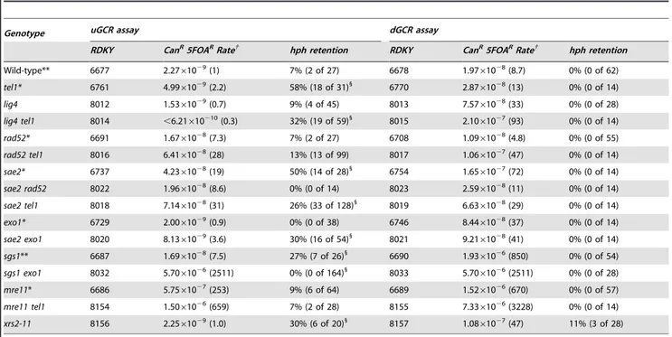
Table 2.GCR rates and percenthphretention intel1,sae2, and related mutants.
Genotype uGCR assay dGCR assay
RDKY CanR5FOARRate{ hph retention RDKY CanR5FOARRate{ hph retention
Wild-type** 6677 2.2761029(1) 7% (2 of 27) 6678 1.9761028(8.7) 0% (0 of 62)
tel1* 6761 4.9961029(2.2) 58% (18 of 31)1
6770 2.8761028(13) 0% (0 of 14)
lig4 8012 1.5361029(0.7) 9% (4 of 45) 8013 7.5761028(33) 0% (0 of 28)
lig4 tel1 8014 ,6.21610210(0.3) 32% (19 of 59)1
8015 2.1061027(93) 0% (0 of 14)
rad52* 6691 1.6761028(7.3) 7% (2 of 27) 6708 1.0961028(4.8) 0% (0 of 55)
rad52 tel1 8016 6.4161028(28) 13% (13 of 99) 8017 1.0661027(47) 0% (0 of 14)
sae2* 6737 4.2361028(19) 50% (14 of 28)1
6754 1.6561027(72) 0% (0 of 14)
sae2 rad52 8022 1.9661028(8.6) 0% (0 of 14) 8023 2.5961028(11) 0% (0 of 14)
sae2 tel1 8018 7.1461028(31) 26% (33 of 128)1
8019 6.6361028(29) 0% (0 of 14)
exo1* 6729 2.0061029(0.9) 0% (0 of 38) 6746 8.4461028(37) 0% (0 of 14)
sae2 exo1 8020 8.1361029(3.6) 30% (16 of 54)1
8021 9.2161028(41) 0% (0 of 14)
sgs1** 6687 1.6961028(7.5) 27% (7 of 26)1
6690 1.9361026(850) 0% (0 of 54)
sgs1 exo1 8032 5.7061026(2511) 0% (0 of 164)1
8033 5.7061026(2511) 0% (0 of 28)
mre11* 6686 5.7561027(253) 9% (6 of 64) 6689 1.5261026(670) 0% (0 of 57)
mre11 tel1 8154 1.5061026(659) 7% (2 of 28) 8155 7.3361026(3228) 0% (0 of 14)
xrs2-11 8156 2.2561029(1.0) 30% (6 of 20)1
8157 1.0861027(47) 11% (3 of 28)
* Rate data from [14].
** Rate andhphretention data from [14]. {
Rate of accumulating Canr5FOArprogeny. The number in parenthesis is the fold increase relative to the wild-type uGCR assay.
1
Retention ofhphin GCR-containing isolates is statistically significantly different than wild type (G-test). doi:10.1371/journal.pgen.1004277.t002
Table 3.GCR formation in plasmid-complemented strains.
Genotype uGCR Assay dGCR Assay
Strain Plasmid CanR5FOARrate{ hph retention CanR5FOARrate{ hph retention
sae2 SAE2 1.1061029(0.5) 20% (3 of 15) 8.7161028(38) 0% (0 of 13)
sae2 empty 8.7661028(39) 45% (9 of 20)1
1.9961027(88) 14% (4 of 24)
sae2 sae21–9 1.8761028(8.3) 24% (5 of 21) 2.0261027(89) 0% (0 of 33)
sae2 sae22,4,5,8,9 1.9161028(8.4) 14% (3 of 21) 1.0661027(47) 0% (0 of 31)
sae2 sae2-S267A 6.7961028(30) 15% (3 of 20) 1.7961027(79) 0% (0 of 13)
mre11 MRE11 8.14610210(0.4) 15% (3 of 20) 1.3461027(59) 0% (0 of 14)
mre11 empty 1.3361026(586) 3% (2 of 58) 4.8561026(2134) 0% (0 of 14)
mre11 mre11-2 1.1761026(516) 4% (2 of 45) 3.4361026(1512) 0% (0 of 14)
mre11 mre11-3 5.0361027(221) 8% (3 of 40) 1.3661026(598) 0% (0 of 14)
mre11 mre11-H125N 2.3661027(104) 10% (6 of 60) 4.8861027(215) 0% (0 of 14)
mre11 mre11S 1.7861028(7.7) 8% (4 of 49) 1.1561027(64) 0% (0 of 10)
{
Rate of accumulating Canr5FOArprogeny. The number in parenthesis is the fold increase relative to the wild-type uGCR assay.
1
Retention ofhphin GCRs formed in checkpoint-defective mutant strains
BecauseTEL1is involved in DNA damage checkpoint signaling [52–54], we analyzed other mutations affecting the DNA damage and replication checkpoints (Table 4). Most of the mutations tested did not cause increased frequency ofhphretention in the uGCR assay, except forrad53Dsml1D,tof1D, andmrc1Dtof1D. Thehph+ GCRs obtained from the rad53D sml1D strain were primarily inverted duplications (11 of 13) and the hph+ GCRs from the
mrc1Dtof1Ddouble mutant were primarily interstitial deletions (8 of 10) on the basis of the size of the rearranged chrV and the presence of a URA3/ura3-52 fusion junction detected by PCR (data not shown). Thus, the defects intel1Dmutants appear to be distinct from defects causing increasedhphretention in themrc1D tof1Dorrad53Dsml1Dmutants.
Retention ofhphin GCRs formed in strains containing mutations affectingde novotelomere addition
Strains withtel1mutations have short telomeres and can formde novo telomere additions, even if the efficiency appears to be decreased in some cases [16,17,22,55]. Because efficient de novo
telomere addition might be predicted to prevent the formation of both interstitial deletion and inverted duplication GCRs, we investigated strains with mutations affecting de novo telomere addition (Table 5). To test mutations affecting telomerase, we generated post-senescent type II survivorest1D and est3Dstrains after sporulating heterozygous est1D/EST1 or est3D/EST3 dip-loids. These strains had an increased frequency ofhphretention in the uGCR assay (p = 0.0006 and p = 261029, respectively, G-test; Table 5). Similarly, deletion of YKU70 and YKU80, which are required forde novotelomere addition and NHEJ but not telomere maintenance [29,56], increased the frequency ofhphretention to 44% and 40%, respectively (p = 761025 and p = 561025, respectively, G-test; Table 5). In contrast, deletion ofLIG4, which
is required for NHEJ but notde novotelomere addition [29,56], did not increase the frequency ofhphretention (Table 2).
Consistent with the effects of mutations eliminating de novo
telomere addition, mutations that increase the rate of de novo
telomere addition caused a reduced frequency ofhph retention. Deletion ofMEC1, which causes increased rates of GCRs that are mediated primarily by de novo telomere addition due to loss of inhibition ofCDC13[22,57], simultaneous deletion of SGS1and
EXO1, which results in high rates of healing of DSBs byde novo
telomere addition [51], and deletion of PIF1, which causes increased rates of GCRs mediated primarily byde novotelomere addition due to loss of inhibition of telomerase at sites ofde novo
telomere addition [16,58–60], caused a significantly reduced frequencies of retention of hph relative to that of the wild-type strain in the uGCR assay (mec1D sml1D p = 0.008; sgs1D exo1D
p = 561027;pif1Dp = 0.0002; Table 2 and 5). In addition,hph+ GCRs were not observed in the pif1D tel1D double mutant, reminiscent of the inability oftel1Dto suppress the increased GCR rate caused byde novotelomere addition in thepif1-m2mutant [16]. Consistent with the hypothesis thatde novotelomere additions were the primary type of GCR formed in thepif1Dtel1Ddouble mutant strains, the rates in the uGCR and dGCR assays were essentially the same, like that seen in thepif1Dsingle mutant and thesgs1D exo1D double mutant (Table 2 and 5). Together, these results suggest that de novo telomere addition suppresses hph retention, potentially by competing for broken ends that could otherwise undergo either NHEJ or resection leading to interstitial deletions or inverted duplications.
Discussion
The observed genotype-specific increase in retention of the telomerichphmarker in the uGCR assay was due to the formation of NHEJ-dependent interstitial deletions spanning theCAN1/URA3
cassette or inverted duplications that recaptured the telomeric end
Table 4.GCR rates and percenthphretention in checkpoint defective mutants.
Genotype uGCR assay dGCR assay
RDKY CanR5FOARRate{ hph retention RDKY CanR5FOARRate{ hph retention
Wild-type** 6677 2.2761029(1) 7% (2 of 27) 6678 1.9761028(8.7) 0% (0 of 62)
tel1* 6761 4.9961029(2.2) 58% (18 of 31)1
6770 2.8761028(13) 0% (0 of 14)
mec1 sml1* 6760 2.3461028(10) 2% (2 of 101)1
6769 1.5061027(66) 0% (0 of 13)
rad53 sml1* 6762 5.6061028(25) 31% (13 of 42)1
6771 3.0561027(134) 0% (0 of 14)
dun1** 6763 1.6361028(7.2) 0% (0 of 13) 6772 1.6161027(71) 0% (0 of 14)
chk1** 6764 1.7661028(7.8) 0% (0 of 22) 6773 1.9661027(86) 0% (0 of 61)
rad24** 6759 2.0061028(8.8) 0% (0 of 13) 6768 1.9761027(87) 0% (0 of 13)
mrc1* 6730 3.3561029(1.5) 19% (5 of 26) 6747 3.7561027(165) 0% (0 of 14)
mrc1-aq** 6766 1.5161029(0.7) 0% (0 of 7) 6775 1.2361027(54) 0% (0 of 7)
tof1* 6767 5.7161029(2.5) 33% (7 of 21)1
6776 4.2561027(187) 0% (0 of 14)
mrc1 tof1** 6779 6.4161028(28) 64% (9 of 14)1
6780 1.2661026(555) 0% (0 of 14)
mrc1-aq tof1** 6848 3.6961029(1.6) 0% (0 of 11) 6849 2.0661027(91) 0% (0 of 14)
hta-S129X` 8010 4.61610210(0.2) 12% (1 of 8) 8011 6.6361028(29) 0% (0 of 7)
* Rate data from [14].
** Rate andhphretention data from [14]. {
Rate of accumulating Canr5-FOArprogeny. The number in parenthesis is the fold increase relative to the wild-typeyel068c::CAN1/URA3assay.
`
hta-S129Xis the genotypehta1-S129X hta2-S129X. 1
of chrV using the homology betweenURA3 of theCAN1/URA3
cassette andura3-52located on chrV L. Previous studies identified inverted duplication GCRs involving dicentric isoduplication intermediates; however, these occurred at low rates [18] and were difficult to identify, because their identification required sequencing of their rearrangement breakpoints. The ability of the uGCR assay to capture these events combined with more facile product analysis provided a convenient genetic assay for use in studying the structural features and genetic requirements underlying these types of GCRs. Thetel1DuGCR assay strain had increased frequencies of forming both types of hph+ GCRs, whereas other mutant backgrounds with increased hph retention yielded primarily interstitial deletions (mrc1D tof1D) or inverted duplications (sae2D
andrad53Dsml1D). Remarkably, both types of rearrangements were observed in GCRs formed in the wild-type uGCR assay strain. These hph-retaining GCRs were suppressed by de novo telomere addition; mutations promoting de novo telomere addition (mec1D,
pif1D, andsgs1Dexo1D) suppressedhphretention, whereas mutations inhibiting de novo telomere addition (est1D, est3D, yku70D, and
yku80D) enhancedhphretention. In contrast, extensive competition between other implicated pathways likely precludes simple extrap-olation of the conclusions based on the phenotypes caused by individual mutations that result in increased frequencies of hph -retaining GCRs to the predicted effects of other mutations affecting the same or related pathways. Examples include the differences betweentel1Dandmec1D, betweentel1Dandrad53D, betweentel1D
andmre11D, betweensae2Dandmre11D, as well as the differences between mutations affecting different features of Mre11-Rad50-Xrs2. Despite this, our analysis allowed us to link an unusual signature of GCRs to specific genetic defects.
Current and previous results suggest that several mechanisms contribute to the hph+ GCRs observed. The GCRs could be initiated by one or more DSBs betweenhphandPCM1, the most telomeric essential gene, although a DSB-independent mechanism for generating similar products has been proposed for forks stalling in the context of large inverted repeats [61,62]. Interstitial deletions then appear to be formed by NHEJ-mediated rejoining of the two ends associated with potential processing of the ends at the DSB in some cases. Inverted duplications appear to be initiated by 59resection of a DSB followed by fold-back invasion of
the 39single stranded end (Figure S5 and S9A). Subsequently, one of three mechanisms operate (Figure S9B): 1) intramolecular BIR occurs up to the position of ura3-52 followed by HR-mediated template switching to the telomericURA3and continuation of BIR to the end of chrV; 2) intermolecular BIR extends the entire length of chrV yielding an isoduplication chromosome that then breaks during cell division and is resolved by secondary rearrangements to yield a stable monocentric chromosome [63]; and 3) the fold-back hairpin is covalently closed followed by replication to yield an isoduplication chromosome, which is further processed as described above in mechanism 2. Notably,hph+inverted duplications were much more prevalent thanhph2inverted duplications resolved by HR between a chrV Ty element and any of the other 254 Ty related elements in the genome. Strand switching during BIR (mechanism 1; [64,65]) combined with the possibility that the telomerichph+ fragment is recombinogenic because it contains a DSB could explain this bias. However, if an isoduplication chromosome is formed first (mechanisms 2 and 3), it must subsequently break during cell division before undergoing a secondary rearrangement(s) to capture a new telomere. Consequently, the telomeric hph -containing fragment might be diluted out by loss or segregation into the wrong progeny during cell division, thereby reducing the formation of hph+ recombinants; this would allowing other Ty-related sequences to serve as substrates for HR with the broken isoduplication chromosome at higher relative efficiencies relative to thehph-containing fragment. Together, these models predict that genetic alterations that either directly or indirectly facilitate hairpin formation, protect hairpins that have formed, promote the use of the
hph-containing fragment as a template, facilitate NHEJ, or suppress pathways that compete with these events will increase the formation of the types ofhph+GCRs seen in the present study.
The sae2D and tel1D uGCR assay strains accumulated high frequencies of hph+ inverted duplications that required HR for their formation. However, these results are not consistent with a simple model in whichhph+inverted duplications are suppressed primarily by Tel1-activated Sae2, which is phosphorylated by Tel1 and Mec1 [35], becausesae2phosphosite mutations did not caused increased levels of hph+ GCRs in the uGCR assay, the tel1D
mutant uGCR assay strain differed from the sae2D strain by accumulating interstitial deletion GCRs in addition to inverted
Table 5.GCR rates and percenthphretention in mutants affectingde novotelomere addition.
Genotype uGCR dGCR
RDKY CanR5FOARrate{ hph retention RDKY CanR5FOARrate{ hph retention
Wild-type** 6677 2.2761029(1) 7% (2 of 27) 6678 1.9761028(8.7) 0% (0 of 62)
tel1* 6761 4.9961029(2.2) 58% (18 of 31)1
6770 2.8761028(13) 0% (0 of 14)
est1 8000 ,1.0161029(,0.5) 33% (7 of 21)1
8001 1.9661028(8.7) 3% (1 of 37)
est3 8002 ,9.50610210(
,0.4) 58% (11 of 19)1
8003 1.8561028(8.1) 0% (0 of 14)
yku70 8004 ,5.32610210(
,0.2) 44% (7 of 16)1
8005 5.3361028(23) 0% (0 of 14)
yku80 8006 ,6.88610210(,0.3) 40% (8 of 20)1
8007 2.7361028(12) 0% (0 of 14)
mec1 sml1* 6760 2.3461028(10) 2% (3 of 133)1
6769 1.5061027(66) 0% (0 of 13)
pif1*** 6894 3.7361027(164) 0% (0 of 86)1
6936 3.6161027(159) 0% (0 of 14)
pif1 tel1 8008 1.5261026(669) 0% (0 of 14) 8009 1.4361026(629) 0% (0 of 14)
* Rate data from [14].
** Rate andhphretention data from [14]. *** Rate data from [21].
{
Rate of accumulating Canr5-FOArprogeny. The number in parenthesis is the fold increase relative to the wild-typeyel068c::CAN1/URA3assay.
1
duplication GCRs, and the tel1D sae2D double mutant uGCR assay strain had suppressed levels ofhph+ GCRs relative to both single mutants. The lack of interstitial deletion GCRs in thesae2D
uGCR assay strain would be consistent with Sae2 primarily promoting cleavage of DNA hairpins [33,34,66] and with Tel1 affecting multiple pathways, potentially including promotion ofde novotelomere additions, suppression of NHEJ, and/or suppression of hairpin cleavage by Sae2-MRX. In addition, GCRs formed in thetel1Dsae2DuGCR strain do have higher levels ofhph2GCRs, indicating that the retention or preferential use of the acentrichph -containing telomeric chrV fragment during BIR is dependent on Tel1 in the absence of Sae2. Consistent with this model,TEL1can more readily compensate for the deletion ofMEC1in strains with
sae2D mutations [54], presumably because of increased Tel1 signaling from DSBs that are not resected due to the uncleaved terminal hairpins that accumulate in sae2D mutants. Thus, our data would suggest that thesae2phosphosite mutations, unlike a
sae2Dmutation, may disrupt functions of Tel1 that promote use of the hph-containing telomeric chrV fragment during BIR. Our data, however, do not rule out a scenario in which moderate overexpression of the mutantsae2alleles from low copy number
ARS CENplasmids might be sufficient to overcome the effect of these phosphosite mutants. The fact thatTEL1andSAE2jointly suppress inverted duplications suggests an alternative explanation for the apparent requirement ofTEL1andSAE2in microhomol-ogy-mediated end joining (MMEJ; [67]): TEL1 and SAE2 may suppress pathways that compete with MMEJ for substrates rather than directly functioning in MMEJ.
Consistent with the differences in the effects of defects inTEL1
andSAE2on the rate and types of GCRs formed, genetic analyses revealed that the effects of mutations affecting related pathways are difficult to predict. For example, the increased hphretention seen in therad53Dsml1Dstrain, which is primarily due to inverted duplications and not interstitial deletions, argues that Tel1 has additional repair-related functions that can suppress the formation ofhph+GCRs. However, extrapolation of this result to other DNA damage checkpoint defective mutations is problematic. For example, the mec1D sml1D double mutant uGCR assay strain had a significantly increased accumulation ofhph2GCRs relative to the wild-type strain likely due to a failure to suppressde novo
telomere additions [22,57]. Similarly, both Tel1 and Sae2 function in conjunction with the MRX complexin vivo, and disruption of the Tel1-Xrs2 interaction caused increased formation of hph+ GCRs similar to that caused by the tel1D mutation. Thus, the recruitment of Tel1 to DSBs by MRX is likely required for suppressing hph retention. Yet, the mre11D mutation and mre11
point mutations that disrupt complex formation and nuclease activity result in much higher GCR rates than thetel1Dandsae2D
single mutations but did not result in increased accumulation of
hph+ GCRs in the uGCR assay, suggesting that these mre11
mutants cause defects in addition to defects in hairpin cleavage. In sum, our results indicate that GCR signatures observed here often reflect the properties caused by individual genetic defects rather than inactivation of entire pathways in which genes of interest function, except in the case of mutations that directly affectde novo
telomere addition; this likely limits our ability to predict the exact GCR signature caused by individual pathway defects.
Our results support the hypothesis that extensive competition between different DNA repair mechanisms determines the spec-trum of genome rearrangements that accumulate in cells and this spectrum can be altered by subtle changes in the efficiencies of different pathways. This competition likely underlies the fact that rearrangement spectra caused by mutations in related genes tend to differ. Therefore, the spectrum of genome rearrangements that
accumulate can provide insights into the underlying genetic defects in DNA repair pathways. For example, in a recent analysis of GCRs in human metastatic pancreatic cancers, 1 out of every 6 GCRs was a copy number change mediated by an inverted dup-lication that showed an association with hallmarks of telomere dysfunction and a dysregulated G1-to-S-phase transition in conjunction with an intact G2/M checkpoint [7]. These pheno-types are highly reminiscent of the phenopheno-types caused by defects in
TEL1 as described here and in previous studies [16,22–25]. Together, these results are consistent with the notion that defects in signaling by the ATM pathway, which involves the human homolog ofTEL1, may play important roles in the formation of the GCRs seen in a fraction of metastatic pancreatic cancer. Similarly, other defects such as defects inRBBP8, which encodes the human Sae2 homolog CtIP, might also play a role in the formation of the inverted duplications seen in metastatic pan-creatic cancer. However, additional experimentation will be required to determine if the genetic insights into the origin of genome instability signatures inS. cerevisiaecan be used to predict genetic changes with functional consequences in human cancer.
Materials and Methods
Construction and propagation of strains and plasmids GCR assays were performed using derivatives of RDKY6677 (yel068c::CAN1/URA3) or RDKY6678 (yel072w::CAN1/URA3) that in addition have the genotype MATa leu2D1 his3D200 trp1D63 lys2DBgl hom3-10 ade2D1 ade8 ura3-52 can1::hisG iYEL072::hph as previously described [14]. Mutant derivatives of these strains (Table S3) were constructed using standard PCR-based gene disruption methods or mating to strains containing mutations as described [11]. The xrs2-11 allele [39] was generated by inte-grating aHIS3marker at the 39end ofXRS2to introduce a stop codon and delete the codons encoding residues 693–854 of Xrs2. Post-senescent est1D and est3D survivors were generated by deleting one copy of eitherEST1orEST3in diploid versions of RDKY6677 and RDKY6678, sporulating the heterozygous diploids, and performing multiple sequential re-streaks of individ-ual spore clones on YPD agar media until growth of the mutants was equivalent to a wild-type control as described [68]. Type I and type II survivors were distinguished on the basis of Southern blotting ofXhoI digested genomic DNA as described [68] and by the fact that chromosomes of type I survivors do not properly enter PFGE gels [69].
Alleles of sae2 were introduced into the sae2::TRP1 deletion strains using pRS313-based ARS CEN plasmids containing the
HIS3marker [70]. Integration plasmids bearing the sae21–9 and
sae22,4,5,8,9alleles, pML468.6 and pML488.15, were kind gifts of Maria Pia Longhese (Universita` degli Studi di Milano-Bicocca). The SAE2-bearing fragments from pML468.6 and pML488.15 were subcloned into the EcoRI site of pRS313 and verified by sequencing to generate pRDK1698 and pRDK1699. The wild-typeSAE2-bearing plasmid, pRDK1700, was generated by PCR amplification ofSAE2from wild-type genomic DNA with primers 59-TGC AAT AGA GTC GTG AAT TCG TCT GAG TTA GCG TCT GAT TTT GAC TCT TTC TTC TTC TTT TTC GTC TT-39and TGC AAT AGA GTC GTG AAT TCC CTG GTA GTT AGG TGT CAT TTG TTT AAC GTC CGT TAA CTT CCC CTT TCT-39to generate an insert spanning the same genomic region as pRDK1698 and pRDK1699. Thesae2-S267A
plating on –HIS media, and GCR-containing progeny were selected on Can/5-FOA media lacking histidine.
Alleles ofmre11were introduced into themre11::HIS3 deletion strains using pRS314 ARS CEN plasmids containing the TRP1
marker. The wild-type MRE11 plasmid was generated by PCR amplifyingMRE11from wild-type genomic DNA with the primers 59-CTG AGG AAT TCG ATT TGG CTA AAC TAG GCT GAG GTA GGC TCG-39and 59-CTG AGC TCG AGG GTA TTG TTT CCC ACA AGG GGA CGG TTA ATG-39 and cloning the PCR product into pRS314 cut withEcoRI andXhoI. The resulting plasmid, pRDK1702, was verified by sequencing. The mre11-H125N and mre11S (mre11-P84S,T188I) plasmids, pRDK1703 and pRDK1704, were generated by site-directed mutagenesis and verified by sequencing. Themre11-2andmre11-3
plasmids, pRS314-mre11-2 and pRS314-mre11-3, were kind gifts of John Petrini (Sloan-Kettering Institute). For GCR rate deter-mination, the transformed query strains were grown in –TRP liquid media, viable cell determination was performed by plating on –TRP media, and GCR-containing progeny were selected on GCR media lacking tryptophan.
Determination of GCR rates and hphretention
GCR rates were determined using multiple independent biological isolates as previously described [71]. The frequency of
hphretention was determined by testing a single GCR-containing isolate from each of a number of individual independent cultures for growth on YPD media supplemented with 200mg/mL hygromycin B (Invitrogen).
Statistical analysis
The significance of the deviation of hph-retention for each genotype was measured using the maximum likelihood statistical significance G-test [72] as implemented for R by P. Hurd (http:// www.psych.ualberta.ca/,phurd/cruft/). Probabilities for the null
model that the observed distributions were generated by the same underlying rate were calculated using the two-tailed Mann-Whitney U-test (http://faculty.vassar.edu/,lowry/utest.html). A
significant differences was inferred when the probability of the null model was 0.01 or less.
PFGE gel and Southern blotting
DNA plugs for PFGE were prepared as described [73].Asc I-digested plugs were prepared by treating plugs with 50 units ofAsc
I (New England Biolabs) overnight at 37uC. Electrophoresis was performed using a Bio-Rad CHEF-DRII apparatus at 7 V/cm, with a 60 to 120 s switch time for 24 h. The gels were stained with ethidium bromide and imaged. The DNA in the gel was transferred to Hybond-XL membranes by neutral capillary blotting. The DNA was crosslinked to the membrane by UV irradiation in a StratalinkerTM (Stratagene) apparatus at maxi-mum output for 60 seconds. Probes were generated by random primer labeling ofMCM3andhphfragments with the Prime-It II kit (Stratagene). Probe hybridization was performed at 68uC for 2– 4 hr. The membrane was then washed extensively and imaged with a PhosphoImager (Molecular Dynamics, Inc.).
Multiplex ligation-mediated probe amplification analysis Primers targeted to the left arm of chromosome V (Figure S7A) were designed according to the recommendations on the MRC-Holland website (http://www.mrc-holland.com) with the length of each amplification product differing by 6 basepairs (Table S4). The reagents were purchased from MRC-Holland, and the amplification, fragment separation, and fragment detection steps
were performed essentially as described [28]. Data were collected on an ABI 3730XL sequencer using the POP7 polymer and GS500-LIZ sizing standard (Life Technologies). The raw data for each run were integrated using GeneMapper software (Life Technologies) and analyzed using a custom Python script that uses gnuplot (http://www.gnuplot.info) to plot the integrated area for each peak in wild-type controls against the respective peak in experimental samples (Figure S7B and C). Amplification detected by MLPA was verified by comparison with aCGH data for isolates 213, 217, 362, and 3178 (Figure S7D).
Array comparative genomic hybridization
Onemg of genomic DNA was prepared from GCR-containing isolates and the wild-type strain RDKY6677 using the Purgene kit (Qiagen) and concentrated to .100 ngmL21. The DNA from GCR-containing isolates was amplified and labeled with Cy5, and wild-type control DNA was amplified and labeled with Cy3. Subsequently, four mixtures containing GCR isolate/wild-type pairs were hybridized to a NimbleGen 4-plex chip. Data were analyzed using the SignalMap software (NimbleGen) and remapped from the chrV sequence of the reference genome to the coordinates of chrV in RDKY6677. Microarray data have been deposited at ArrayExpress (http://www.ebi.ac.uk/arrayexpress) with under the accession E-MTAB-2377.
Whole genome paired-end sequencing
Multiplexed paired-end libraries were constructed from 5mg of genomic DNA purified using the Purgene kit (Qiagen). The genomic DNA was sheared by sonication and end-repaired using the End-it DNA End-repair kit (Epicentre Technologies). Com-mon adaptors from the Multiplexing Sample Preparation Oligo Kit (Illumina) were then ligated to the genomic DNA fragments, and the fragments were then subjected to 18 cycles of amplifica-tion using the Library Amplificaamplifica-tion Readymix (KAPA Biosys-tems). The amplified products were fractionated on an agarose gel to select 600 bp fragments, which were subsequently sequenced on an Illumina HiSeq 2000 using the Illumina GAII sequencing procedure for paired-end short read sequencing. Reads from each read pair were mapped separately by bowtie version 0.12.7 [74] to a reference sequence that contained revision 64 of theS. cerevisiae
S288c genome (http://www.yeastgenome.org),hisGfromSamonella enterica, and thehphMX4marker (Table S1). Sequencing data have been deposited at NCBI Sequence Read Archive (http://www. ncbi.nlm.nih.gov/sra) under the accession SRP039033.
Rearrangement and copy number analysis of paired-end sequencing data
read mapped next to the junction-defining read pairs. Sequences of the junctions were generated by de novo alignment of the junction-sequencing reads associated with rearrangements defined by statistically significant junction-defining read pairs (Figure S3). The identified rearrangements included all known rearrangements in the strains that could be defined based on the average distance between the read pairs in the library (Table S2).
Supporting Information
Figure S1 PFGE analysis ofhph+GCR-containing isolates from thetel1DuGCR assay strain. (A and C) Southern blot using anhph
probe of a pulsed-field gel (PFG) of the wild-type strain (RDKY6677) and 6 GCR-containing isolates with and without
AscI treatment. (B and D) Southern blot of a second PFG with identical samples as in panel A or C using aMCM3probe. (PDF)
Figure S2 Analysis of interstitial deletions from thetel1DuGCR assay strain. Sequence of the junction (middle line) is displayed between the sequences of the two target regions. Bases between colons are identical in both joined fragments. Coordinates of the breakpoint mapped to the two targets are reported above and below the alignment for both the reference S288c genome sequence and the uGCR chrV. For isolate 214, the fusion is to a Ty element. For the 214 ‘‘left’’ junction, the Ty elements in the reference genome that best matched the junction sequence were
YERCTy1-1,YMLWTy1-2,YPLWTy1-1,YGRWTy1-1, YDRWTy1-5, YOLWTy1-1, YLRWTy1-2, YLRWTy1-3, YLRCTy1-1,
YLRWTy1-1, and YPRCTy1-4. For the 214 right junction, the related junction sequence mapped to a large number of Ty-related elements.
(PDF)
Figure S3 Searching and subsequent identification of rearrange-ments by the Pyrus programs. (A) For a novel rearrangement, depicted here as a translocation between chrA (white) and chrB (gray), junction-defining read pairs are read pairs for which both read pairs map (black arrows separated by a dashed line): one read pair maps to chrA and one read pair maps to chrB. To defined as belonging to the same rearrangement ‘event’, reads mapped to each target must additionally (i) have the same orientation as the other reads that map to that target and (ii) map within a short distance (defined based on the distribution of distances between read pairs) of other read pairs indicating the same event. Importantly, because junction-defining read pairs map to each target, these read pairs must span any novel junction and, in general, cannot sequence the junction. Junction sequencing reads, however, can be identified as non-mapping (red arrows) reads associated in read pairs with other reads that map uniquely to the two targets in the vicinity of junction-defining read pairs. Alignment of junction sequencing reads can identify the sequence. (B) Mapping of the 572 junction-defining read pairs and 114 junction-sequencing read pairs for the chrV interstitial deletion in isolate 3118. Junction-defining read pairs are sorted by the mapped position of telomeric marker. The position of the junction-sequencing reads (red arrows) is arbitrary. Note that the reads paired with the junction sequencing reads are in the vicinity and have the same orientation as the junction-defining read pairs. (C) The junction sequence derived from alignment of the junction-sequencing reads for the isolate 3118 interstitial deletion is displayed on the second line. The telomeric sequence alignment is on the top line and the centromeric alignment is on the bottom line. Bases of identity between the two targets are surrounded by colons. (D) The interstitial deletion junction sequence derived by
PCR amplification and Sanger sequencing for isolate 3118 is identical to the sequence derived by alignment of the junction-sequencing reads.
(PDF)
Figure S4 Examples of PCR mapping indicating the presence of aURA3/ura3-52fusion junction. (A) A primer located telomeric to theyel068c::CAN1/URA3insertion (fore) and a primer within the 39end ofURA3(rev1) amplify an identical fragment in the starting and GCR-containing strains. (B) The fore primer and a primer within the end of Ty element (rev2) only amplifies products in strains with aURA3/ura3-52junction. (C) The fore primer and a primer within the 59end ofURA3(rev3) amplify a,1.8 kb
frag-ment in the starting strain, but a large,,8 kb fragment in strains
with aURA3/ura3-52 junction, consistent with the presence of a Ty element. (D) The fore primer and a primer telomeric toura3-52
(rev4) only amplifies a large ,8 kb fragment in strains with a URA3/ura3-52junction.
(PDF)
Figure S5 Formation and sequences of the inversion junctions in the sequenced inverted duplication GCRs. Predicted mechanism of hairpin formation and the sequence of the inversion junction for the inverted duplication class of GCRs. The arrow indicates the position of the double strand break (DSB) under the simplest scenario in which the 39 end of the DSB is the 39 end used to initiate the inversion; however, more complicated scenarios with more distal initiating DSBs have been observed [75]. 59-.39
resection generates a single-stranded region that can mediate the formation of a single-stranded hairpin that can anneal and extend from the exposed single stranded region. Regions of homology involved in annealing are underlined. Duplicated palindromic sequence in the replicated product and the annotated junction sequence are displayed with arrows. Sequences derived from alignment of the junction sequencing reads are displayed at bottom. Panel A depicts the inversion junction for isolate 217; panelB depicts the inversion junction for isolate 218, 362, 364, and 366; panel Cdepicts the inversion junction for isolate 365; panelD depicts the inversion junction for isolate 2977; panelE
depicts the inversion junction for isolate 3124; panelFdepicts the inversion junction for isolates 3121 and 3125; and panelGdepicts the inversion junction for isolate 3255.
(PDF)
Figure S6 Analysis of hph2GCR-containing isolates from the
tel1DuGCR assay strain. (A) Southern blot using aMCM3probe of a PFG of the wild-type strain (RDKY6677) and 13hph2GCR isolates revealed that isolates 213, 2976, 3124, and 3125 had a rearranged chrV that was substantially larger than wild-type, whereas the other isolates had rearranged chrV that was similar to wild-type. (B) Sequences of some of the breakpoints from GCRs associated with a normal-sized chrV showed the GCRs involved translocations (isolate 213) orde novo telomere additions (isolates 3116, 3117, and 3120). For isolate 216, the junction sequence is displayed as in Figure S2 and the Ty elements in the reference genome that best matched the junction sequence were YJRWTy1-2andYGRWTy1-1.
(PDF)
![Table 5). Similarly, deletion of YKU70 and YKU80, which are required for de novo telomere addition and NHEJ but not telomere maintenance [29,56], increased the frequency of hph retention to 44% and 40%, respectively (p = 7 610 2 5 and p = 5 610 2 5 , respe](https://thumb-eu.123doks.com/thumbv2/123dok_br/18249293.342105/11.918.93.833.702.1015/similarly-deletion-telomere-maintenance-increased-frequency-retention-respectively.webp)
