Received on 12 April 2008; revised 15 July 2008.
Address for correspondence: Dr. José Júlio Costa Sidrim. Rua Jabaquara, 344; Castelão. Zip code: 60861-200, Fortaleza- CE, Brazil. Phone/ Fax: 55 (85) 3295 - 1736. E-mail: sidrim@ufc.br.
The Brazilian Journal of Infectious Diseases 2008;12(4):333-337. © 2008 by The Brazilian Journal of Infectious Diseases and Contexto Publishing. All rights reserved.
Successive Mycological Nail Tests for Onychomycosis: A Strategy to Improve Diagnosis Efficiency
Tereza Elizabeth Fernandes Meireles1, Marcos Fábio Gadelha Rocha1,2, Raimunda Sâmia Nogueira Brilhante1, Rossana de Aguiar Cordeiro1 and José Júlio Costa Sidrim1
1Medical Micology Specialized Center; Department of Pathology and Legal Medicine; Faculty of Medicine; Federal University of Ceará; 2Post-Graduation Program of Veterinary Science; Veterinary Faculty, State University of Ceará; Fortaleza, CE, Brazil
Onychomycosis is a fungal infection of nails caused by dermatophytes, yeasts and moulds, accounting for about 50% of onychopathies. A high frequency of onychomycosis caused by Candida species has been reported during the last few years in northeast Brazil, as well as in other regions of the world. A clinical diagnosis of onychomycosis needs to be confirmed through laboratory exams. We evaluated the importance of serial repetition of direct microscopic exams and fungal culture for the diagnosis of onychomycosis in the city of Fortaleza, Ceará, in northeast Brazil. We first made a retrospective study of 127 patients with onychomycosis, identifying the fungi that had been isolated from fingernails and toenails. We then made a prospective study of 120 patients, who were submitted to three successive mycological examinations. Ungual residues were scraped off and directly examined with a microscope and fungal cultures were made. In the retrospective study, in which only one sample was analyzed, the incidence of onychomycosis was 25.0%. In our prospective study, in which we had data from successive mycological examinations, 37.8% had onychomycosis. The most commonly isolated fungi in both studies were yeasts from the genera Candida, especially C albicans, C. parapsilosis and C. tropicalis. We found a high proportion of onychomycosis caused by
Candida species. We also concluded that serial repetition of direct microscopic examination and fungal culture, with intervals of 2-5 days improved the diagnosis of onychomycosis. We suggest that this laboratorial strategy is necessary for accurate diagnosis of this type of mycosis, especially when the standard procedures fail to diagnose fungal infection, despite strong clinical suspicion.
Key-Words: Diagnosis, onychomycosis, Candida, moulds and dermatophytes.
Onychomycosis is a chronic fungal infection of fingernails and/or toenails, caused by dermatophytes, yeasts and moulds, leading to gradual destruction of the nail plate [1-3]. It is not self healing and may be a source of more widespread fungal lesions of the skin [1]. Trichophyton is the most frequent dermatophyte genus found affecting nails. Trichophyton
rubrum is the cause of most onychomycosis cases, followed
by T. mentagrophytes and T. tonsurans [1,4-6].Some yeasts, such as Candida [1,4,5,7-9], Trichosporon [10] and Malassezia
[11] species, are also able to cause ungual infection, invading the ungual folds and the ungual bed, as well as the ungual sheet. Candida albicans predominates in most yeast-caused onychomycosis cases [1,8]. However, other Candida species, including C. tropicalis, C. parapsilosis, C. glabrata, C.
guilliermondi, C. krusei and C. famata, have also been
isolated in infected nails [1,7-9,12,13]. There have been reports of a high frequency of onychomycosis caused by Candida
species in northeast Brazil [8,9]. These moulds are saprophytic fungi, normally found in the soil, air and some plants; they are considered opportunistic fungi. Their pathogenic role in onychomycosis is still controversial; however, their isolation in nails is becoming more and more frequent [4,14,15].
Scopulariopsis spp., Fusarium spp., and Aspergillus spp., for example, have been identified in nails as primary pathogens of onychomycosis [1,4,16-18].
Diagnosis of onychomycosis is made by direct microscopic examination and fungal culture [1-3,19,20]. However, when only one sample is analyzed, the frequency of false-negative results is very high. Other laboratory methods, such as ungual biopsy, PCR, flow cytometry and immunohistochemical techniques, have been used to improve onychomycosis diagnosis; however, these methods are not usually available in common dermatology centers [1,21-23]. As a viable alternative in such labs, we investigated whether serial repetition of routine direct microscopy examination and fungal culture improves diagnosis efficacy.
Material and Methods
Patients
between each sample collection from the same nail varied from two to five days. We maintained a short interval to ensure isolation of the same fungus in the affected nails. During the sample collection interval the patients could not use any onychomycosis therapy, including alternative medicine. In addition, they were advised that the affected nails could not be scraped or clipped during that period.
Laboratory Methods
Each clinical specimen was processed with 40% KOH for a direct microscope examination; a part of the original sample was cultured on Sabouraud dextrose agar, Sabouraud dextrose agar with chloramphenicol, Sabouraud dextrose agar with chloramphenicol and cycloheximide and Dixon’s medium. The cultures on Sabouraud agar were incubated at 25-28oC and at
37oC on Dixon’s medium. These cultures were observed once
a week for up to three weeks. Identification of dermatophytes was made by observing the macro and micro characteristics of the colonies, microculture in potato agar and biochemical tests. Microculture in potato agar was also used in the identification of the moulds. The following procedures were used to identify the yeasts: microculture in corn meal agar with Tween 80, germination tube tests, as well as zymograms and auxanograms [9].
Diagnosis Criteria
The criteria employed for the diagnosis of onychomycosis in both stages of the study were culture identification of dermatophytes, even with no identification from direct microscopy examination, and positive microscopy examination associated with cultures of yeasts and moulds [17,20].
Statistical Analysis
The data were processed with the software SPSS 10.0. The χ2 test and the Fisher – Freemann – Halton exact test
were used for comparisons, with a significance level of 5%.
Results
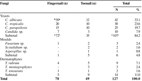
Among the 507 mycological records analyzed in the retrospective study, 127 (25.0%) diagnoses of onychomycosis were obtained in accordance with the diagnosis criteria. Among these, 107 (84.3%) were infected by Candida spp., 72 being in fingernails, caused mainly (p< 0.05) by C. albicans
(Table 1).
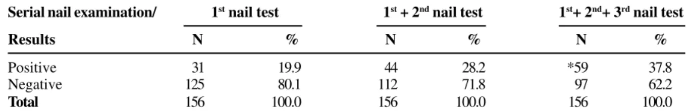
Among the 156 mycological nail examinations in the prospective study, there were only 31 (19.9%) positive diagnoses of onychomycosis in the mycological exams, 25 (80.6%) by yeasts and only one (3.2%) by dermatophytes. In the second analysis (n=125), 13 nail tests that were negative in the first analyses were positive in the second nail evaluation. These new positive diagnoses in the second mycological nail examination were by yeasts (5; 38.5%), moulds (5; 38.5%) and dermatophytes (3; 23.0%). Additionally, 15 mycological nail tests that were negative in the first and second tests were positive in the third mycological analysis (n=112), including
two (13.3%), five (33.3%) and eight (53.4%) by dermatophytes, moulds and yeasts, respectively. Fifty-nine diagnoses of onychomycosis were obtained with the three successive mycological nail tests, including 38 (64.4%, p < 0.05), 15 (25.4%) and six (10.2%) by yeasts, moulds and dermatophytes, respectively (Table 2).
Among the fungi isolated in the prospective study, C. albicans (15, 35.7%) and C. parapsilosis (4, 23.5%) were the most frequent yeasts in fingernails and in toenails, respectively. Among the moulds, Fusarium sp. (11, 18.6%) was the most commonly isolated fungus, especially in toenails. Dermatophytes were less frequently found, comprised of one case with T. tonsurans, two T.mentagrophytes and three T. rubrum (Table 3).
The incidence of onychomycosis in the retrospective study was 25%; however, the frequency increased with the repetition of the mycological nail analysis in the prospective stage. In the first mycological nail test, an incidence of 19.9% onychomycosis was obtained; in the second mycological nail test, considering the diagnoses in the first test and 13 diagnoses in the second mycological evaluation, the incidence observed was 28.2%. With the third repetition, considering all the diagnoses obtained in the three mycological analyses, the incidence obtained was 37.8% (p<0.05) (Table 4).
Discussion
Various studies of the incidence of onychomycosis, in which only one mycological nail examination of each patient was performed, report incidences of from 6.5% to 20% [6,24,25]. In our retrospective study, we found a slightly higher incidence of 25% onychomycosis. In the prospective study, we analyzed three successive mycological examinations over a short time period (2-5 day intervals between sample collections). Some cases that were negative in the first mycological examination were positive in the second or third analysis, increasing the final incidence of onychomycosis to 37.8%. Possibly, this is because multiple sampling gives better access to nail-bed debris, because it becomes easier to collect an adequate specimen after the first or second nail scraping, favoring isolation of the fungus.
Although a positive microscope exam associated with negative cultures has been observed in certain situations, we did not address this possibility, because the criteria employed for the diagnosis of onychomycosis were culture of dermatophytes, along with positive direct microscopy examination associated with positive cultures for yeasts and moulds [17,20].
[20,26,27]. We found that the accuracy of onychomycosis diagnosis was increased with the repetition of direct microscopy examination and fungal culture. Therefore, we recommend this strategy to improve the diagnosis and consequent treatment of this type of mycosis.
Onychomycosis is a common fungal infection of the nails, accounting for 50% of nail diseases, and this percentage has been progressively increasing [4,13,28]. Several studies have reported dermatophytes as the main etiological agent of onychomycosis, especially in temperate zones. Trichophyton
rubrum and T. mentagrophytes are the main dermatophytes
isolated from toenail infections [4-6]. However, reports of onychomycosis caused by yeasts [7-13] and moulds [14,15,24] have become more and more frequent.
We observed a low percentage of dermatophytes isolated from onychomycosis cases of patients from Fortaleza city, northeast Brazil. This differs from what was found in studies made in Canada [6], USA [25] and Europe [29,30]. Nevertheless, T. rubrum was the most commonly isolated dermatophyte, followed by T. mentagrophytes in both ours and these other studies.
The percentage of dermatophytes isolated from nails is usually low. For example, Pontes et al. [8] found that the main fungi involved in onychomycosis in João Pessoa city, northeast Brazil were Candida species (82%), followed by dermatophytes (13.4%). Also, Brilhante et al. [9] reported that the etiological agents most frequently found in cases of onychomycosis in Ceará (northeast Brazil) were Candida
species (74.42%), followed by dermatophytes (12.99%) and
Fusarium sp. (8.19%).
We found yeasts to be common; there were 107 (84.3%) positive diagnoses in our retrospective study, including 72 (p<0.05) and 35 onychomycosis cases from fingernails and toenails, respectively. In our prospective study, 64.4% of the diagnoses by yeasts were obtained from fingernails (p<0.05). Other researchers also observed frequent involvement of yeasts in fingernail onychomycosis [7,8,12].
Identification of the Candida species was based on phenotypical features, such as macro and micromorphological descriptions, as well as through zymograms and auxanograms [9]. Some micromorphological characteristics, especially round
Table 1. Diagnosis of onychomycosis in our retrospective study.
Fungi Fingernail (n) Toenail (n) Total
N %
Yeasts
C. albicans *30a 12 42 33.1
C. tropicalis 20 10 30 23.6
C. parapsilosis 15 10 25 19.7
Candida sp. 7 3 10 7.9
Subtotal *72b 35 *107c 84.2
Moulds
Fusarium sp. 1 2 3 2.4
Scytalidium sp. - 2 2 1.6
Aspergillus sp. - 1 1 0.8
Subtotal 1 5 6 4.8
Dermatophytes
T. rubrum 4 5 9 7.1
T. mentagrophytes - 3 3 2.4
T. tonsurans 1 1 2 1.6
Subtotal 5 9 14 11.0
Total 78 49 127 100.0
χ2=10.5; DF=2; *p=0.005. a(C. albicans versus other Candida species); b(Candida species in fingernails versus Candida species in toenails) and c(Candida speciesversus other fungi).
Table 2. Diagnosis of onychomycosis from our prospective study.
Serial repetition/ First mycological Second mycological Third mycological Total
examination examination examination
(n=156) (n=125) (n= 112)
N % N % N % N %
Yeasts 25 80.6 5 38.5 8 53.4 38 64.4
Moulds 5 16.1 5 38.5 5 33.3 15 25.4
Dermatophytes 1 3.2 3 23.0 2 13.3 6 10.2
Total 31 100.0 13 100.0 15 100.0 *59a 100.0
Fisher – Freeman – Halton exact test: *p= 0.04. a(first + second + third mycological examination vs.first mycological examination).
to oval cells (isolated blastoconidia), were more frequent than the blastoconidia form in association with pseudohyphae. Possibly nail characteristics favor the isolated blastoconidia form.
Candida albicans was the most frequently isolated yeast in both our retrospective and prospective studies. However,
C. parapsilosis was the most commonly isolated fungus in
toenail onychomycosis in our prospective study. Other researchers also reported that C. parapsilosis is the most common yeast in toenail infections [12,31]. The much greater proportion of Candida species compared to dermatophytes and moulds could be due to Candida contamination in the altered nail. However, this possibility appears to be unlikely, as there was no evidence of mixed cultures of Candida spp. plus dermatophytes or moulds.
Isolation of moulds from nail infections has been frequently observed, especially from toenails of patients from tropical countries. Some studies have reported onychomycosis by S. dimidiatum and S. brevicaulis [1]. In our prospective analysis, Fusarium sp., was the most frequent mould in toenail onychomycosis. Other research carried out in Brazil came to the same conclusion [15].
In conclusion, we found a high proportion of onychomycosis caused by Candida species in the city of Fortaleza, Ceará, northeast Brazil and we found that serial repetition of direct microscopic examination and fungal culture improves onychomycosis diagnosis efficiency. This strategy of repetition would allow for more accurate diagnosis of this mycoses.
References
1. Seebacher C., Brasch J., Abeck D., et al. Onychomycosis. Mycoses 2007;50:321-7.
2. Gupta A.K., Ricci M.J. Diagnosing onychomycosis. Dermatol Clin 2006;24:365-9.
3. Hay R. Literature review. Onychomycosis. J Eur Acad Dermatol Venereol 2005;19(Suppl 1):1-7.
4. Veer P., Patwardhan N.S., Damle A.S. Study of onychomycosis: prevailing fungi and pattern of infection. Indian J Med Microbiol 2007;25:53-6.
5. Vender R.B., Lynde C.W., Poulin Y. Prevalence and epidemiology of onychomycosis. J Cutan Med Surg 2006;10(Suppl 2):S28-33. 6. Gupta A.K., Jain H.C., Lynde C.W., et al. Prevalence and
epidemiology of onychomycosis in patients visiting physicians,
offices: a multicenter Canadian survey of 15,000 patients. J Am Acad Dermatol 2000;43:244-8.
Table 3. Isolated fungi in our prospective study, based on three successive mycological nail analysis.
Attacked nails/ Fingernails (n) Toenails (n) Total
Fungi N %
Yeasts
C. albicans *15a - 15 25.4
C. parapsilosis 8 4 12 20.3
C. tropicalis 8 1 9 15.3
C. glabrata - 1 1 1.7
Candida sp. - 1 1 1.7
Subtotal *31 b 7 *38 c 64.4
Moulds
Fusarium sp. 4 *7 d *11 18.6
Aspergillus sp. 1 2 3 5.1
Scytalidium sp. 1 - 1 1.7
Subtotal 6 9 15 25.4
Dermatophytes
T. rubrum 3 7.1 - 3 5.1
T. mentagrophytes 1 2.4 1 2 3.4
T. tonsurans 1 2.4 - 1 1.7
Subtotal 5 11.9 1 6 10.2
Total 42100.0 17 59 100.0
Fisher – Freemann – Halton exact test: *p= 0.001. a(C. albicans versus other Candida species); b(Candida species in fingernail versusCandida species in toenail); c(Candida speciesversus others fungi) and d(Fusarium sp. vs. other moulds).
Table 4. Incidence of onychomycosis, considering the positive results obtained in the three mycological examinations.
Serial nail examination/ 1st nail test 1st + 2nd nail test 1st+ 2nd+ 3rd nail test
Results N % N % N %
Positive 31 19.9 44 28.2 *59 37.8
Negative 125 80.1 112 71.8 97 62.2
Total 156 100.0 156 100.0 156 100.0
7. Ellabib M.S., Agaj M., Khalifa Z., et al. Yeasts of the genus Candida are the dominant cause of onychomycosis in Libyan women but not men: results of a 2-year surveillance study. Br J Dermatol 2002;146:1038-41.
8. Pontes Z.B., Lima Ede O., Oliveira N.M., et al. Onychomycosis in João Pessoa City, Brazil. Rev Argent Microbiol 2002;34:95-9. 9. Brilhante R.S.N., Cordeiro R.A., Medrano D.J., et al.
Onychomycosis in Ceará (Northeast Brazil): epidemiological and laboratory aspects. Mem Inst Oswaldo Cruz 2005;100:131-5. 10. Han M.H., Choi J.H., Sung K.J., et al. Onychomycosis and
Trichosporon beigelli in Korea. Int J Dermatol 2000;39:266-9. 11. Escobar M.L., Carmona-Fonseca J., Santamaria L., Onicomicosis
por Malassezia. Rev Iberoamer Micol 1999;16:225-9. 12. Gautret P., Rodier M.H., Kauffmann-Lacroix C., et al. Case report
and review. Onychomycosis due to Candida parapsilosis. Mycoses 2000;43:433-5.
13. Foster K.W., Ghannoum M.A., Elewski B.E. Epidemiologic surveillance of cutaneous fungal infection in the United States from 1999 to 2002. J Am Acad Dermatol 2004;50:748-52. 14. Ellis D.H., Marley J.E., Watson A.B., et al. Significance of
non-dermatophyte moulds and yeasts in onychomycosis. Dermatol 1997;194(Suppl 1):40-2.
15. Lopes J.O., Alves S.H., Mari C.R., et al. A ten-year survey of onychomycosis in the central region of the Rio Grande do Sul, Brazil. Rev Inst Med. Trop São Paulo 1999; 41: 147 – 9. 16. Escobar M.L., Carmona-Fonseca J. Onicomicosis por hongos ambientales
no dermatofíticos. Rev Iberoamer Micol 2003;20:6-10.
17. Gupta A.K., Ryder J.E., Baran R., et al. Non-dermatophyte onychomycosis. Dermatol Clin 2003;21:257-68.
18. Godoy P., Nunes E., Silva V., et al. Onychomycosis caused by Fusariun solani and Fusariumoxysporum in São Paulo, Brasil. Mycopaphologia 2004;157:287-90.
19. Scher R.K., Tavakkol A., Sigurgeirsson B., Hay R.J., et al. Onychomycosis: diagnosis and definition of cure. J Am Acad Dermatol 2007;56:939-44.
20. Ellis D.H. Diagnosis of onychomycosis made simple. J Am Acad Dermatol 1999;40:S3-S8.
21. Hull P.R., Gupta A.K., Summerbell R.C. Onychomycosis: an evaluation of three sampling methods. J Am Acad Dermatol 1998;39:1015-7.
22. Lawry M.A., Haneke E., Strobeck K., et al. Methods for diagnosing onychomycosis. Arch Dermatol 2000;136:1112-6.
23. Gianni C., Morelli V., Cerri A., et al. Usefulness of histological examination for diagnosis of onychomycosis. Dermatol 2001;202:283-8.
24. Elewski B.E. Onychomycosis: pathogenesis, diagnosis and management. Clin Microbiol Rev 1998;11:415-29.
25. Ghannoum M.A., Hajjeh R.A., Scher R., et al. A large-scale North American study of fungal isolates from nails: the frequency of onychomycosis, fungal distribution, and antifungal susceptibility patterns. J Am Acad Dermatol 2000;43:641-8.
26. Arrese J.E., Piérard-Franchimont C., Piérard G.E. Facing up to diagnosis uncertainty and management of onychomycosis. Int J Dermatol 1999;38(Suppl 2):1-6.
27. Borkowski P., Williams M., Holewinski J., et al. Onychomycosis: an analysis of 50 cases and a comparison of diagnostic techniques. J Am Podiatr Med Assoc 2001;91:351-5. 28. Faergemann J., Baran R. Epidemiology, clinical presentation and
diagnosis of onychomycosis. Br J Dermatol 2003;149(Suppl 65):1-4.
29. Haneke E., Roseeuw D. The scope of onychomycosis: epidemiology and clinical features. Int. J Dermatol 1999;38(Suppl 2):7-12.
30. Torres-Rodriguez J.M., López-Jodra O. Epidemiology of nail infection due to keratinophilic fungi. Rev Iberoamer Micol 2000;17:122-35.