ISSN 0100-879X
BIOMEDICAL SCIENCES
AND
CLINICAL INVESTIGATION
www.bjournal.com.br
www.bjournal.com.br
Volume 43 (11) 1010-1134 November 2010
Institutional Sponsors
The Brazilian Journal of Medical and Biological Research is partially financed by
Hotsite of proteomics metabolomics developped by:
Braz J Med Biol Res, November 2010, Volume 43(11)1076-1083
Y. Xu, Y.F. Song and Z.X. Lin
doi:
Transplantation of muscle-derived stem cells plus biodegradable fibrin
glue restores the urethral sphincter in a pudendal nerve-transected rat
model
Transplantation of muscle-derived
stem cells plus biodegradable fibrin glue
restores the urethral sphincter in a
pudendal nerve-transected rat model
Y. Xu
1,2, Y.F. Song
1,2and Z.X. Lin
31Fuzong Clinical College, Fujian Medical University, Fuzhou, Fujian, China 2Department of Obstetrics and Gynecology, Fuzhou General Hospital, Fuzhou, Fujian, China 3Fujian Institute of Hematopathy, Fujian Medical University Union Hospital, Fuzhou, Fujian, China
Abstract
We investigated whether fibrin glue (FG) could promote urethral sphincter restoration in muscle-derived stem cell (MDSC)-based
injection therapies in a pudendal nerve-transected (PNT) rat, which was used as a stress urinary incontinence (SUI) model.
MDSCs were purified from the gastrocnemius muscles of 4-week-old inbred female SPF Wistar rats and labeled with green fluorescent protein. Animals were divided into five groups (N = 15): sham (S), PNT (D), PNT+FG injection (F), PNT+MDSC injection (M), and PNT+MDSC+FG injection (FM). Each group was subdivided into 1- and 4-week groups. One and 4 weeks after injection into the proximal urethra, leak point pressure (LPP) was measured to assess urethral resistance function. Histology and immunohistochemistry were performed 4 weeks after injection. LPP was increased significantly in FM and M animals after
implantation compared to group D (P < 0.01), but was not different from group S. LPP was slightly higher in the FM group than
in the M group but there was no significant difference between them at different times. Histological and immunohistochemical
examination demonstrated increased numbers of surviving MDSCs (109 ± 19 vs 82 ± 11/hpf, P = 0.026), increased muscle/
collagen ratio (0.40 ± 0.02 vs 0.34 ± 0.02, P = 0.044), as well as increased microvessel density (16.9 ± 0.6 vs 14.1 ± 0.4/hpf,
P = 0.001) at the injection sites in FM compared to M animals. Fibrin glue may potentially improve the action of transplanted MDSCs to restore the histology and function of the urethral sphincter in a SUI rat model. Injection of MDSCs with fibrin glue
may provide a novel cellular therapy method for SUI.
Key words: Muscle-derived stem cells; Fibrin glue; Urinary incontinence; Cellular therapy; Pudendal nerve transection
Introduction
Correspondence: Y.F. Song, Fuzong Clinical College of Fujian Medical University, Fuzhou 350025, Fujian, China/Department of Obstetrics and Gynecology, Fuzhou General Hospital, Fuzhou 350025, Fujian, China. Fax: 0591-2493-7024. E-mail: [email protected]
Received May 6, 2010. Accepted September 27, 2010. Available online October 15, 2010. Published November 12, 2010.
Stress urinary incontinence (SUI) is defined as involun
-tary leakage of urine on effort or exertion or during sneezing
or coughing. It is a widespread health problem that adversely affects the quality of life of women (1,2). Its etiology is mul-tifactorial, including functional impairment of muscle and associated nerves that may occur as a result of advanced
age, hormonal status, or pelvic floor damage at childbirth.
Urethral injection therapy, such as bovine glutaraldehyde
cross-linked collagen injection, is widely accepted as a
less invasive SUI treatment modality with a relatively low
risk of adverse events as well as good short-term results (3). However, the efficacy of such treatment declines with
time, and repeated injections are required (4). Currently,
cell-based injection therapies are being investigated as
alternatives. Autologous adult stem cell therapy recently
demonstrated the potential for regenerative repair of the
deficient urethra for the treatment of SUI (5). Among adult
stem cells, muscle-derived stem cells (MDSCs) possess several characteristics that are ideally suited for cellular transplantation. MDSC therapy appears to be effective in the regeneration of urethral sphincter in SUI animals and patients (6-8). However, the current transplantation tech-nique involves the injection of cells suspended in saline,
cell culture medium, or bovine serum albumin (BSA), and
is plagued by limited cell retention and transplant survival
MDSCs plus fibrin glue restores urethral sphincter 1077
for clinical therapy is the lack of an adequate vascular
supply for larger volume implants (11). Cells at the center of an injected cell mass depend on an extensive blood
vessel network, which supplies nutrients and oxygen while
removing waste products (12). Many different strategies are currently being pursued to promote cell survival and retention. Tissue engineering approaches aim to repair lost or damaged tissue through the use of cell transplantation and biomaterial scaffolds, which provide a microporous
framework and are etched with ‘mini-networks’, imitating
vascular systems, that promote cell survival. To be clinically useful as cell transplant vehicles, such scaffolds need to be biocompatible without toxic side effects and clinical grade components need to be used to attach the cells to the
scaf-fold. A liquid support matrix that polymerizes to a gel would
be shaped more easily. In addition, an injection would be much less invasive than open implantation.
Fibrin glue (FG), a composite of fibrinogen and throm -bin, is a physiologically relevant matrix whose principal
component, fibrin, has a fundamental role in the process
of blood clotting and wound healing. Thrombin is an endo-protease that naturally functions as a blood-clotting factor
to convert fibrinogen to fibrin. Within seconds after fibrino
-gen and thrombin are mixed together, they form a gel-like
component (13,14). FG has been widely used to decrease intraoperative bleeding and to promote homeostasis. Fibrin
is a natural polymer known to support wound healing by
inducing angiogenesis and promoting cell attachment and proliferation, and thus may provide a more conducive
envi-ronment for accelerated tissue regeneration (15,16). While
FG has been applied as a scaffold in tissue engineering
(17,18), it is not known if it could promote effective
cell-based injection therapies in SUI.
In the present study, we explored the use of biodegrad-able FG scaffolds as an appropriate carrier to which primary rat MDSCs could be attached, and their use in vivo to support transplanted cells. Our model was designed to predict the potential of this technique for future clinical applications.
Material and Methods
Animals and experimental design
All experiments, including MDSC isolation and the denervation model, were performed on 75 normal inbred virgin female SPF Wistar rats (4 weeks old) weighing 80-100 g. The rats were randomly divided into five groups: sham (S), pudendal nerve-transected (PNT, D), PNT+FG injec
-tion (F), PNT+MDSC injec-tion (M), and PNT+MDSC+FG injection (FM). Groups were then subdivided into 1- (N = 5) and 4-week (N = 10) groups. The experimental protocol
was approved by the Fuzhou General Hospital Institutional
Animal Care and Use Committee.
Denervation of the bilateral pudendal nerve
Groups D, M, F, and FM underwent bilateral PNT (19).
Group S was the sham operation control. The rats were
given chloral hydrate anesthesia (400 mg/kg, ip), and a
dorsal midline incision in the skin and bilateral dorsal inci -sions in the muscle were performed over the ischiorectal fossa. Under an operating microscope, the sciatic nerve on
each side was identified and a 2-mm segment distal to its
origin from the vertebral column but proximal to the branch
of the pudendal nerve was transected. The muscle and skin
incisions were closed separately with 3-0 vicryl sutures.
After the operation, animals were given a cefazolin sodium injection (100 mg/kg, ip) once a day for 3 days.
MDSC isolation and purification
MDSCs were isolated and purified from the hind gas
-trocnemius of 4-week-old normal inbred virgin female SPF Wistar rats via a previously described modified preplate technique (20). According to the results of previous stud -ies on murine cells, early plates (preplates 1-2) contain a
majority of fibroblasts, and late plates (preplates 5-6) are
highly enriched with myogenic cells (20,21). The cells used
in the present experiment were taken from the last preplate,
preplate 6, which contains MDSCs.
Fibrin glue
The FG used in this study is commercially available (BD
Biosciences, USA). The two-component system remains
liquid for several seconds before solidifying into a semirigid
gel matrix. The first component consists of concentrated fibrinogen and aprotinin, a fibrinolysis inhibitor. The second
component is a mixture of thrombin and CaCl2. It was
de-livered through the supplied applicator, which held the two components in separate syringes and provided
simultane-ous mixing and delivery. The ratio of fibrinogen to thrombin
components was 1:1 (w/w).
MDSC labeling and injection surgeries
To facilitate the identification of the cells after their
transplantation, the MDSCs from the third passage were
infected with lentivirus encoding green fluorescent protein
(PGC-FU-GFP, ShangHai GeneChem Co., China) for 8 h in the presence of 8 µg/mL Polybrene at 20 multiplicities
of infection. The transducing efficiency was up to 95%. The transduced cells were expanded for 2 weeks before
implantation. The injection surgeries were performed 2
weeks after bilateral PNT. A low midline incision was made
to expose the urethra and different media were injected into the proximal urethra using a 100-µL Hamilton syringe, with microscopic guidance on each side of the urethra. Five experimental groups of female rats were established. The
cell injection group (M) was injected with 50 μL MDSCs
suspended in PBS (1 x 10⁶ cells/50 μL); the FG group (F)
was injected with 50 μL of the mixed FG via the Baxter
supplied Duploject applicator; the FG and cell injection
group (FM) was injected with 25 μL MDSCs suspended
μL fibrinogen component. The S group was used as the
positive control, which received no injection and the PNT group (D) was used as the negative control, which was
injected with 50 μL PBS.
Leak point pressure test
Leak point pressure (LPP) was determined in all
animals at 1 and 4 weeks after injection as described by Damaser et al. (22). Briefly, after anesthesia with chloral hydrate (400 mg/kg, ip), an abdominal midline incision of
0.5 cm was performed cephalad to the urethral meatus. A
circular purse-string suture (6-0 chromic gut) was placed in the anterior vesical wall and a puncture incision was then
made in the center of the purse-string stitch. A suprapubic catheter (PE-50 tubing) was inserted through this incision
and secured with the suture. The catheter was
subcutane-ously tunneled to exit the skin at the level of the neck and
the end was ligated. The bladder catheter was then
con-nected to both a flow pump (P-600, Atom Medical Corp., Japan) and a pressure transducer via a 3-way stopcock. The transducer, connected to an amplifier, polygraph and com
-puter, digitized the pressure data (UDS64-111, LABORIE Durado Urodynamic System, USA). The rat was placed in
the supine position and underwent a 30-min accommodation
period of filling (5 mL/h) and voiding. The bladder was then
palpated, submitted to the Crede maneuver for emptying,
and filled with saline to approximately half of its capacity.
While bladder pressure was recorded and digitized, gentle pressure was applied externally over the bladder (a gentle Crede maneuver) to slowly increase pressure until the rat
leaked saline through the urethra. At the first indication of leakage, the externally applied abdominal pressure was rapidly removed. The peak bladder pressure was taken
as the LPP. The test was repeated at least three times to produce a mean LPP value for each rat.
Histology
After LPP testing at 4 weeks after injection, all animals were sacrificed. The proximal urethra was removed, and five specimens in each group were fixed in 10% phosphate-buffered formalin, embedded in paraffin and sectioned into 5-µm thick slices. Paraffin sections were stained with
Masson trichrome according to the manufacturer protocol
(Sigma-Aldrich, USA). Images from the entire sections
were acquired using a digital camera system (Imaging
Micropublisher 5.0 PTV). To prevent variations in staining,
all samples were stained simultaneously using this proce-dure. In the images, cells in blood vessels and the smooth muscle layer, as well as the rhabdosphincter layer in the sections, stained red while collagens stained blue. Image
analysis was done as described above and quantified using
the Image-Pro Plus 6.0 image software. The software can automatically distinguish regions stained with different colors and accurately measure the areas of muscle and collagen to
yield a muscle/collagen ratio. Another five specimens from
groups FM and M were fresh frozen in Tissue Tek O.C.T. freezing medium (Sakura, USA) at -80°C and stored until
they were processed on a cryostat. They were sectioned
into 8-µm thick slices, and implanted cells were identified
with GFP while the nuclei were stained with
4,6-diamidino-2-phenylindole (DAPI, 1 µg/mL; Roche, USA). For image analysis, five randomly selected fields per tissue per ani -mal in the FM and M groups were photographed with an
Confocal Laser Microscope (LCSM510, Zeiss, Germany)
and the GFP-MDSCs were counted using the Image-Pro Plus image software.
Histochemical staining for factor VIII-related antigen and microvessel density count
For detailed immunohistochemical analysis of the
re-paired tissue, paraffin sections were probed with polyclonal rabbit anti-factor VIII-related antigen (FVIII-R Ag, F8, Zymed, USA). Briefly, all sections were deparaffinized in xylene and treated with 3% hydrogen peroxide in methanol for 10 min to block endogenous peroxidase activity. Enzyme pretreatment for antigen retrieval was necessary. Nonspecific binding of primary antibodies was reduced by blocking with normal
horse serum for 10 min. Sections were then incubated in
a humidified chamber at 4°C with the primary antibody
(1:100) for 24 h, followed by the second antibody using the
MaxVision™ HRP-Polymer anti-Rabbit IHC Kit, (Maxim Co.,
China). Control sections omitted the primary antibody and were treated with PBS alone. The sections were stained
with 3,3-diaminobenzidine tetrachloride solution (DAB) and
counterstained with hematoxylin. The endothelial cells in
blood vessels that stained brownish red were FVIII-R Ag+ cells. A microvessel was defined as any distinct FVIII-R Ag+ cell or cell cluster, with no requirement for a vessel
lumen. The number of microvessels was counted in 10
random fields (magnification: 20X). The average of the 10 high power fields (hpf) was calculated and the microvessel density was defined as microvessel/hpf using an Olympus CX40 microscope (Tokyo, Japan).
Statistical analysis
Data are reported as means ± SD. The software SPSS
11.0 for Windows (SPSS Inc., USA) was used for analysis. Comparison of continuous variables among the five groups was performed by one-way analysis of variance (ANOVA)
for within time-point analyses. The LSD method was used to specify differences among groups. Paired t-tests were
used for comparison between data at 1 and 4 weeks. A P value less than 0.05 was considered to be statistically significant.
Results
LPP testing
The LPPs of the FM, M, F, D, and S groups at 1 and 4
MDSCs plus fibrin glue restores urethral sphincter 1079
analysis indicated that the LPP of group D was lower than that of other groups at each time (P < 0.01). The LPPs of
group FM and M were significantly higher than that of group
D (P < 0.01) but there was no difference compared with group S at either time. The LPP of group FM was slightly higher than that of group M, although the difference was not
sig-nificant at either time. The LPP of group F was sigsig-nificantly higher than that of group D (P = 0.02) and significantly lower than that of group S (P = 0.025) at 1 week. At 4 weeks the LPP of group F was significantly lower than that of groups FM, M and S (P = 0.04, P = 0.09 and P = 0.01, respectively) but there was no difference compared to group D (P =
0.276). There was a sharp decrease of the LPP in group F
from 1 to 4 weeks (P = 0.002), but no significant difference between 1 and 4 weeks in the other groups.
Histology
The implanted GFP-MDSCs were detected in the muscle
layer of the proximal urethra at 4 weeks after the injection
in both groups FM and M. The GFP-MDSCs implanted in
combination with FG seem to localize in the host tissue and
fuse to form post-mitotic multinucleated myofibers, since the DAPI-stained nuclei showed varied sizes on the implanted
side. There was an increase of surviving GFP-MDSCs in situ in group FM compared to group M (P < 0.05; Figure
2A,B). Histological examination of the cross-section of the 4-week proximal urethra was performed on each section
after Masson trichrome staining. In the normal female rat
external urethral sphincter, the circular skeletal muscle fibers, a layer of striated muscle fibers that encircles the
smooth muscle layers, and the inner longitudinal as well as
middle circular layers were all stained red (Figure 3A). The
Figure 1. Leak point pressure (LPP) analysis 1 week and 4
weeks after injection. The LPP of the PNT+MDSC plus fibrin glue (FG) injection group (FM) and the PNT+MDSC injection group (M) was significantly higher than that of the PNT group (D) at 1 and 4 weeks (P < 0.01). There was no difference in LPP in either
group compared with that of the sham group (S) at either time. The LPP of group FM was slightly higher than that of group M
but there was no significant difference between the two groups at either time. The LPP of the PNT+FG injection group (F) was sig
-nificantly higher than that of group D (P = 0.002) and sig-nificantly lower than that of group S (P = 0.025) at 1 week, but there was no difference compared to that of group D at 4 weeks. Data are reported as means ± SD (N = 5 for each group). PNT = puden
-dal nerve transected; MDSC = muscle-derived stem cells. *P < 0.05 compared with group D; #P < 0.05 compared with group S (ANOVA + LSD test).
Figure 2. The implanted GFP-MDSCs were detected in the
muscle layer of the proximal urethra 4 weeks after injection in both the PNT+MDSC plus fibrin glue (FG) injection group (FM) and the PNT+MDSC injection group (M). A, A greater number
of surviving transplanted GFP-MDSCs localized at the injected sites and some of them had fused to each other to form
post-mi-totic multinucleated myofibers when rats were transplanted with MDSCs in combination with fibrin glue (white arrows). B, There was a smaller number of surviving transplanted GFP-MDSCs at the injected sites when rats were injected with MDSCs only.
Original magnification 200X. Scale bar = 20 µm. C, There was an increase of surviving GFP-MDSCs in situ in group FM compared
to group M (*P < 0.05, t-test). Data are reported as means ± SD
denervated proximal urethral sphincter showed atrophic
and thin skeletal and smooth muscle fibers (Figure 3B).
The denervated proximal urethral sphincter injected with
MDSC plus FG displayed an increased thickness of circular skeletal muscle at the injection sites (Figure 3C). The injec
-tion of MDSCs also displayed an increased thickness of muscle mass and the orientation of the new muscle fibers
was variable (Figure 3D). The injection of FG led to an
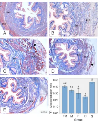
increased vessel density but few new muscle fibers could be found at the injection sites (Figure 3E). The muscle/ collagen ratios of the FM and M groups were significantly
higher than in group D (P < 0.01) but lower than in group S (P < 0.01). In group FM, the ratio was higher than in group
M (P = 0.044). In addition, there was no difference between
group F and group D.
Effects of MDSC plus fibrin glue transplantation on microvessel density
The microvessel density of the different groups
ob-served four weeks after injection is shown in Figure 4G. The microvessel density of the FM group was significantly higher than in the other groups (P < 0.01). After the injection of MDSCs plus FG, a large number of FVIII-R Ag-labeled
microvessels were found within the striated muscle layer (Figure 4B). There was a moderate increase in microvessel
density in the S, M and F groups (Figure 4A,C,D) compared with the D group (Figure 4E; P < 0.01). There was no sig
-nificant difference in microvessel density between the M
group and the F group.
Discussion
Several new materials are being investigated for
injec-tion therapy of SUI. Among these, MDSCs are especially
promising because the cells are muscle progenitor cells that have shown the capacity to differentiate into bone, cartilage, nerve, and endothelium (20,21). MDSCs have the potential to improve sphincter function by remodeling the damaged urethra, as well as acting to generate tissues
with bulking properties (23,24). However, MDSC injection
therapy has certain disadvantages including migration and absorption, as is the case for other commercial injection agents. This calls for the development of novel strategies,
like the tissue-engineering approach, to improve the reten -tion and survival of the transplanted stem cells.
Clinically, LPP determination is currently used to diag-nose SUI during a urodynamics exam. LPP by the Crede method represents the active component of the continence mechanism of the urethral sphincter in SUI rat models. Thus,
it probably better reflects the state of urethral resistance
function and has been used extensively in rat model
stud-ies (25-27). In the present study, we determined whether
FG can improve the effect of MDSCs on urethral function in the denervated urethral sphincter of rats by detecting
LPP 1 and 4 weeks after treatment. The results showed
that the urethral function in the MDSC+FG injection group and the MDSC injection group was significantly improved at both times although there were no significant differ -ences between groups. When FG was injected alone, LPP
was increased slightly at 1 week but was decreased at 4 weeks. The reduction in volume of the FG, which acted as
Figure 3. Masson trichrome staining of a female rat urethral
sphincter 4 weeks after injection. A, Normal anatomic structure of the urethral sphincter from a sham-operated control female rat (group S). B, Following denervation (group D), the circular skel -etal muscle and smooth muscle layer were thin and atrophic. C, The denervated proximal urethral sphincter of an animal injected
with MDSCs plus fibrin glue (group FM) displayed an increased thickness of circular skeletal muscle and more new muscle fibers (black arrow) on the external urethral sphincter layer. D, MDSCs injected into a denervated proximal urethral sphincter (group M)
led to increased skeletal muscle masses with variable fiber orien -tation in the external urethral sphincter layer. E, The denervated
proximal urethral sphincter injected with fibrin glue alone (group
F) displayed an increased vessel density but few new muscle
fibers could be seen at the injection site. F, The muscle/collagen
ratios were the highest in group S. They were significantly higher
in groups FM and M than in group D (P < 0.01), but lower than in group S (P < 0.01). The muscle/collagen ratio of group FM was
MDSCs plus fibrin glue restores urethral sphincter 1081
a bulking agent, was responsible for the decline of LPP as
it was gradually absorbed. These results demonstrate that the function of urethral sphincters was improved by the injection of MDSCs with or without FG while the injection
of FG alone led to a temporary improvement. The lack of difference in LPP between the MDSC+FG injection group
and the MDSC injection group may be attributed to the following reasons. First, in vivo, plasminogen and matrix metalloproteinases may be secreted from the encapsulated cells inside the FG, which contribute to its rapid
degrada-tion. Therefore, FG acting as a bulking agent may improve
LPP temporarily. Second, it remains to be determined if FG can improve the differentiation of injected MDSCs into
reinnervated myofibers for urethral sphincter contraction. Third, the lack of significance could simply be a type II error, with only 5 rats in each group, although the trends were
actually quite strong.
Our results also indicate that survival of transplanted
cells at the injection site 4 weeks after transplantation was
enhanced by injection of FG. The histological and immuno-histochemical analysis also showed an obvious increase of
the thickness of muscle mass and of the muscle/collagen
ratio, as well as a higher neovasculature density in the
MDSC+FG injection group. These data indicate that FG
might promote the differentiation of MDSCs toward a
myo-genic and endothelial lineage. In addition, fibrin-stabilizing
factor XIII contained in FG favors the migration of
undif-ferentiated stem cells on the highly cross-linked structure of
the glue and enhances cell proliferation. FG also contains arginine-glycine-asparagine motifs and binds to integrins, which facilitate cell adherence and proliferation.
In the present study, improved LPPs after injection of MDSCs suggest that MDSCs could induce the functional reinnervation of regenerated muscles. The feasibility of this
concept was first demonstrated by Chermansky et al. (28)
who showed that the striated muscle layer of the MDSC-injected urethra was contiguous with an increase in nervous tissue when compared with those of the cauterized urethra injected with only saline solution in a SUI rat model. Furuta et al. (29) further suggested that the physiological effects of
urethral sphincters were mediated not only by MDSC bulk -ing but also by autonomic nerves innervat-ing the urethral sphincters. MDSCs implanted into the proximal urethra
might enhance α1-ARs sensitivity by differentiating into
sympathetic neuronal cells or urethral smooth muscle cells
to restore the deficient middle urethral function induced by
transection of pudendal nerves. The transplanted MDSCs may also release NGF to promote axonal regeneration and functional recovery after nerve injury (30). However,
it is likely that transplanted MDSCs may promote both the
muscle and integrated nerve regenerative response of donor transplanted and host cells and that FG could promote such regenerative response.
FG in combination with an appropriate cell source has been used in a variety of tissue engineering applications, Figure 4. Histochemical analysis of the expression of factor
VIII-related (FVIII-R) antigen and neovascularization 4 weeks after
injection. Panels A-E show the capillaries with FVIII-R Ag+ cells
(black arrows) compared to the negative control (PNT group, pu
-dendal nerve transected; Panel F). After the injection of muscle-derived stem cells (MDSC) plus fibrin glue (FG), a large number of FVIII-R Ag-labeled microvessels were found within the stri -ated muscle layer (Panel B). Microvessel density was highest in
the PNT+MDSC plus FG injection (FM) group (Panel G). There
was a moderate increase in microvessel density in the sham
(S), PNT+MDSC injection (M) and PNT+FG injection (F) groups (Panels A,C,D) compared to the PNT (D) group (Panel E). *P <
References
1. Abrams P, Cardozo L, Fall M, Griffiths D, Rosier P, Ulmsten
U, et al. The standardisation of terminology in lower urinary tract function: report from the standardisation sub-committee of the International Continence Society. Urology 2003; 61: 37-49.
2. Hampel C, Wienhold D, Benken N, Eggersmann C, Thuroff
JW. Prevalence and natural history of female incontinence.
Eur Urol 1997; 32 (Suppl 2): 3-12.
3. Chapple CR, Wein AJ, Brubaker L, Dmochowski R, Pons ME, Haab F, et al. Stress incontinence injection therapy:
what is best for our patients? Eur Urol 2005; 48: 552-565.
4. Corcos J, Fournier C. Periurethral collagen injection for the treatment of female stress urinary incontinence: 4-year follow-up results. Urology 1999; 54: 815-818.
5. Furuta A, Jankowski RJ, Pruchnic R, Yoshimura N, Chancel -lor MB. The potential of muscle-derived stem cells for stress urinary incontinence. Expert Opin Biol Ther 2007; 7: 1483-1486.
6. Furuta A, Jankowski RJ, Honda M, Pruchnic R, Yoshimura
N, Chancellor MB. State of the art of where we are at using stem cells for stress urinary incontinence. Neurourol Urodyn
2007; 26: 966-971.
7. Kim YT, Kim DK, Jankowski RJ, Pruchnic R, Usiene I, de
Miguel F, et al. Human muscle-derived cell injection in a rat model of stress urinary incontinence. Muscle Nerve 2007; 36: 391-393.
8. Carr LK, Steele D, Steele S, Wagner D, Pruchnic R,
Jankowski RJ, et al. University of Toronto clinical trial of
muscle-derived stem cell injection in women with stress urinary incontinence. J Urol 2007; 177: 439.
9. Muller-Ehmsen J, Whittaker P, Kloner RA, Dow JS, Sakoda
T, Long TI, et al. Survival and development of neonatal rat cardiomyocytes transplanted into adult myocardium. J Mol Cell Cardiol 2002; 34: 107-116.
10. Reinecke H, Murry CE. Taking the death toll after cardiomyo -cyte grafting: a reminder of the importance of quantitative biology. J Mol Cell Cardiol 2002; 34: 251-253.
11. De Coppi P, Delo D, Farrugia L, Udompanyanan K, Yoo
JJ, Nomi M, et al. Angiogenic gene-modified muscle cells
for enhancement of tissue formation. Tissue Eng 2005; 11:
1034-1044.
12. Isner JM. Tissue responses to ischemia: local and remote responses for preserving perfusion of ischemic muscle. J Clin Invest 2000; 106: 615-619.
13. Mann KG, Brummel K, Butenas S. What is all that thrombin for? J Thromb Haemost 2003; 1: 1504-1514.
14. Sarpel U, Roayaie S, Schwartz ME, Labow DM. The role of fibrin sealants in hepatic surgery. Surg Technol Int 2007; 16: 31-36.
15. Bootle-Wilbraham CA, Tazzyman S, Thompson WD, Stirk CM, Lewis CE. Fibrin fragment E stimulates the prolifera -tion, migration and differentiation of human microvascular endothelial cells in vitro. Angiogenesis 2001; 4: 269-275.
16. Herrick S, Blanc-Brude O, Gray A, Laurent G. Fibrinogen.
Int J Biochem Cell Biol 1999; 31: 741-746.
17. Jockenhoevel S, Zund G, Hoerstrup SP, Chalabi K, Sachweh
JS, Demircan L, et al. Fibrin gel - advantages of a new scaf-fold in cardiovascular tissue engineering. Eur J Cardiothorac Surg 2001; 19: 424-430.
18. Cummings CL, Gawlitta D, Nerem RM, Stegemann JP. Properties of engineered vascular constructs made from
collagen, fibrin, and collagen-fibrin mixtures. Biomaterials
2004; 25: 3699-3706.
19. Hijaz A, Bena J, Daneshgari F. Long-term efficacy of a
vaginal sling procedure in a rat model of stress urinary incontinence. J Urol 2005; 173: 1817-1819.
20. Qu-Petersen Z, Deasy B, Jankowski R, Ikezawa M, Cum
-mins J, Pruchnic R, et al. Identification of a novel population
of muscle stem cells in mice: potential for muscle regenera-tion. J Cell Biol 2002; 157: 851-864.
21. Lee JY, Qu-Petersen Z, Cao B, Kimura S, Jankowski R,
Cummins J, et al. Clonal isolation of muscle-derived cells capable of enhancing muscle regeneration and bone heal-ing. J Cell Biol 2000; 150: 1085-1100.
22. Damaser MS, Broxton-King C, Ferguson C, Kim FJ, Kerns JM. Functional and neuroanatomical effects of vaginal dis-tention and pudendal nerve crush in the female rat. J Urol
2003; 170: 1027-1031.
23. Cannon TW, Lee JY, Somogyi G, Pruchnic R, Smith CP, Huard J, et al. Improved sphincter contractility after allogenic including maxillofacial bone, periodontal bone, bone, ear
cartilage, cartilage, cornea, heart, blood vessel, tendon, and ligament regeneration (31). It has proved to be a potentially suitable biological vehicle for cell transplantation due to its injectability, biocompatibility, biodegradability, and binding
capacity to cells. Unlike xenogenic gelatin and collagen, which may induce inflammatory responses, FG can avoid the potential risk of a foreign body reaction because it is
a naturally occurring molecule that can be produced from
proteins in the patient’s own blood (32). Another advantage
of using FG as a matrix is that it can be injected as a liquid and it will gel in situ. Injection of the fibrin/cell material
leads to the formation of tissue that is histologically more
mature. Our next step is to observe the therapeutic efficacy
of MDSCs combined with FG injection in a rat model of SUI
over a longer period of time.
The present short-term observation indicated that FG might potentially synergize with transplanted MDSCs to restore the histology and function of the urethral sphincter
and promote the efficacy of cell-based injection therapies in
a rat model of SUI. Thus, injection of MDSCs plus FG may provide a novel cellular therapy method for SUI.
Acknowledgments
Research supported by grants from the Key Science and Technology Planning Project of Fujian Provincial, China (grant #2009I0022), National Natural Science Foundation of China (grant #81070473) and Medical Science and
MDSCs plus fibrin glue restores urethral sphincter 1083
muscle-derived progenitor cell injection into the denervated rat urethra. Urology 2003; 62: 958-963.
24. Liu Z, Wu Y, Chen BG. Myoblast therapy: from bench to bedside. Cell Transplant 2006; 15: 455-462.
25. Conway DA, Kamo I, Yoshimura N, Chancellor MB, Can
-non TW. Comparison of leak point pressure methods in an
animal model of stress urinary incontinence. Int Urogynecol J 2005; 16: 359-363.
26. Cannon TW, Damaser MS. Effects of anesthesia on cystom
-etry and leak point pressure of the female rat. Life Sci 2001; 69: 1193-1202.
27. Ahn H, Lin DL, Esparza N, Damaser MS. Short-term time
-course of bilateral pudendal nerve injury on leak-point pres -sure in female rats. J Rehabil Res Dev 2005; 42: 109-114.
28. Chermansky CJ, Tarin T, Kwon DD, Jankowski RJ, Cannon
TW, de Groat WC, et al. Intraurethral muscle-derived cell
injections increase leak point pressure in a rat model of
intrinsic sphincter deficiency. Urology 2004; 63: 780-785.
29. Furuta A, Jankowski RJ, Pruchnic R, Egawa S, Yoshimura
N, Chancellor MB. Physiological effects of human muscle-derived stem cell implantation on urethral smooth muscle function. Int Urogynecol J Pelvic Floor Dysfunct 2008; 19: 1229-1234.
30. Dedkov EI, Kostrominova TY, Borisov AB, Carlson BM. Survival of Schwann cells in chronically denervated skeletal
muscles. Acta Neuropathol 2002; 103: 565-574.
31. Ahmed TA, Dare EV, Hincke M. Fibrin: a versatile scaffold
for tissue engineering applications. Tissue Eng Part B Rev
2008; 14: 199-215.
32. Ye Q, Zund G, Benedikt P, Jockenhoevel S, Hoerstrup SP, Sakyama S, et al. Fibrin gel as a three dimensional matrix in
cardiovascular tissue engineering. Eur J Cardiothorac Surg