ISSN 0100-879X
BIOMEDICAL SCIENCES
AND
CLINICAL INVESTIGATION
www.bjournal.com.br
www.bjournal.com.br
Volume 44 (7) 606-728 July 2011
Braz J Med Biol Res, July 2011, Volume 44(7) 682-687
doi: 10.1590/S0100-879X2011007500074
Supraorganized collagen enhances Schwann cell reactivity and
organization
in vitro
L.G. Maturana, R.G. Zanon, A. Pierucci, B.C. Vidal and A.L.R. Oliveira
Faculdade de Medicina de Ribeirão Preto Campus
Ribeirão Preto
Institutional Sponsors
The Brazilian Journal of Medical and Biological Research is partially financed by
analiticaweb.com.br S C I E N T I F I C
Supraorganized collagen enhances Schwann
cell reactivity and organization
in vitro
L.G. Maturana, R.G. Zanon, A. Pierucci, B.C. Vidal and A.L.R. Oliveira
Laboratório de Regeneração Nervosa, Departamento de Anatomia, Biologia Celular e Fisiologia e Biofísica, Instituto de Biologia, Universidade Estadual de Campinas, Campinas, SP, Brasil
Abstract
We investigated the reactivity and expression of basal lamina collagen by Schwann cells (SCs) cultivated on a supraorganized bovine-derived collagen substrate. SC cultures were obtained from sciatic nerves of neonatal Sprague-Dawley rats and seeded on 24-well culture plates containing collagen substrate. The homogeneity of the cultures was evaluated with an SC marker antibody
(anti-S-100). After 1 week, the cultures were fixed and processed for immunocytochemistry by using antibodies against type IV
collagen, S-100 and p75NTR (pan neurotrophin receptor) and for scanning electron microscopy (SEM). Positive labeling with antibodies to the citedmolecules was observed, indicating that the collagen substrate stimulates SC alignment and adhesion (collagen IV labeling - organized collagen substrate: 706.33 ± 370.86, non-organized collagen substrate: 744.00 ± 262.09; S-100 labeling - organized collagen: 3809.00 ± 120.28, non-organized collagen: 3026.00 ± 144.63, P < 0.05) and reactivity (p75NTR labeling - organized collagen: 2156.33 ± 561.78, non-organized collagen: 1424.00 ± 405.90, P < 0.05; means ± standard error
of the mean in absorbance units). Cell alignment and adhesion to the substrate were confirmed by SEM analysis. The present
results indicate that the collagen substrate with an aligned suprastructure, as seen by polarized light microscopy, provides an
adequate scaffold for SCs, which in turn may increase the efficiency of the nerve regenerative process after in vivo repair.
Key words: S-100; p75NTR; Type IV collagen; Collagen; Cell culture; Supraorganized collagen
Introduction
Correspondence: A.L.R. Oliveira, Departamento de Anatomia, Biologia Celular e Fisiologia e Biofísica, Instituto de Biologia, UNICAMP, Caixa Postal 6109, 13083-970 Campinas, SP, Brasil. Fax: +55-19-3521-6295. E-mail: alroliv@unicamp.br
Received January 11, 2011. Accepted June 2, 2011. Available online June 17, 2011. Published July 25, 2011.
Peripheral nerve regeneration is influenced by the chro -nology and synchronization of a chain of events following injury. The degeneration of the distal stump as well as the
sprouting of regenerating fibers are determined by the repair
technique, the level of the lesion and the regenerative po-tential of the axotomized neurons. The simple reconnection of the stumps may not provide all the necessary elements
for the most efficient regenerative process, so that the
use of different methods has been investigated. There is evidence that the regeneration process may be enhanced if cell grafts are introduced into the lesion microenvironment. In this way, stem cells from different sources and Schwann cells (SCs) combined with different substrates increase the speed, reduce the gap between stumps and the quality of the regeneration process (1-3).
SCs are vital elements during the process of peripheral nerve rearrangement following lesion, the so-called Wal-lerian degeneration. They support the axonal regrowth
from the proximal stump, serving as an efficient interface
to the extracellular matrix (ECM) (4). In this respect, they
express a number of laminins (4-7) and integrins (e.g., α1β1, α2β1, α6β1) (8) and secrete different neurotrophic factors,
including nerve growth factor (NGF), brain-derived neu-rotrophic factor (BDNF) and the glial-derived neuneu-rotrophic factor (GDNF) (9,10). Additionally, the SCs organize the so-called bands of Büngner, which provide guidance to the growth cones throughout the distal stump of a lesioned nerve (4,10,11).
The use of purified SCs as a graft inside a tubular pros
-thesis resulted in a more efficient nerve repair (1,2,12,13).
Supraorganized collagen provides a scaffold for Schwann cell culture 683
the ECM components, as well as the response of such cells when cultured on new substrates, including biomaterials (10-14) and supraorganized ECM-derived molecules.
Overall, collagens are the most abundant ECM elements in the peripheral nerve. They contribute to the structure and function of the nerve, including the connective tissue that is present in the epi-, peri- and endoneural tissue (4,5). Collagen is also present in the basal lamina synthesized
by SCs. An interesting feature of collagen fibrils is their
ability to become aligned, forming stable supramolecular
structures (9). In this way, the fiber architecture itself is a
limiting factor for such organization and, although the auto-assembly and macromolecular aggregational properties have not been fully investigated, they can be obtained in vitro under controlled conditions.
Naturally oriented proteins are known to positively influ -ence the axonal growth (14,15). They may also be degraded
or rearranged more efficiently in the ECM environment.
Interestingly, collagen extracted from bovine tendon
self-assembles after dialysis. The formation of oriented fibrils
can be enhanced by the use of an extrusion technique (16). In contrast, collagen from other sources, such as the rat tail
tendon, does not self-organize into helically oriented fibers
and chiral objects after extrusion. In this context, there is
evidence that supraorganized matrix may be of benefit for
cellular recognition and may facilitate the interaction of neuronal and glial cells, increasing axonal guidance.
On the basis of these considerations, the objective of the present study was to investigate the expression of ECM components by SCs when cultured on a naturally aligned collagen substrate. In this way, the expression of type IV
col-lagen, the low affinity receptor for neurotrophins (p75NTR)
and S-100 was assessed by immunocytochemistry and the interaction between the SCs and the substrate was studied by scanning electron microscopy (SEM).
Material and Methods
Collagen extraction and substrate preparation
Bovine calcanear tendon and rat tail collagen were extracted and separated using a patented technique (#P.I.
97015709, B.C. Vidal). Briefly, small fragments of defatted and
cleaned bovine and rat tendons were immersed in an aqueous solution of 5% acetic acid containing 0.01% HCl and 1 mg pepsin/g tendon at 7-10°C for 24 h. The solubilized collagen
was filtered and the fibers were reconstituted by adding NaCl solution to a final concentration of 5%. The fibers obtained
were then dialyzed (dialysis membrane with 25 Å porosity, cut off - 12,000-16,000 kDa; Inlab, Brazil) against distilled water in 6-mm diameter tubes at 5°C, with the outside bath water changed every 24 h(four times). One and a half liters of water were used for every 200 g collagen gel.
After lyophilization, the collagen substrate was cut into 5-mm3 fragments and sterilized with gamma radiation (15
kGy).
Culture of Schwann cells
The SC cultures were prepared using sciatic nerves from neonatal Sprague-Dawley rats (N = 10). The nerves were dissected out and freed from the epineurium and contaminating tissue. The tissue was then reduced into small fragments and incubated in collagenase and trypsin for 30 min at 37°C. The enzymes were inhibited with fetal
calf serum and the tissue was triturated, filtered, centrifuged, and resuspended in Dulbecco’s modified Eagle’s medium
with 10% fetal calf serum. The medium was enriched with forskolin (10 mM, Sigma, USA) and pituitary extract (10 µg/mL; Sigma). The cells thus obtained were then seeded onto 24-well culture plates containing the polymer
mem-branes. After 24 h of culturing, a further cell purification
was performed by adding 10 nM cytosine arabinoside for
48 h. The purity of the cultures was confirmedwith the anti-S-100 antibody (Dako, Denmark). The cultures, once
purified, were maintained for 1 week on the biopolymers
and the medium was changed every second day. All experi-ments were performed in triplicate and were approved by the Institutional Committee for Ethics in Animal Research, Universidade Estadual de Campinas (UNICAMP, protocol No. 1360-1).
Immunocytochemistry
After 1 week of culture, the cells were fixed in 4% para -formaldehyde (Merck, Germany) for 10 min and washed twice in 0.1 M phosphate buffer (PB), pH 7.4, for 5 min. Preincubation with 1% bovine albumin for 45 min was fol-lowed by overnight incubation at 4°C with primary antisera (anti-S-100, anti-p75NTR, and anti-type IV collagen; Santa Cruz Biotechnology Inc., USA). After three rinses in PB, the cultures were incubated for 45 min at room temperature with the conjugated donkey anti-goat or anti-rabbit second-ary antibodies Cy-2 or Cy-3 (Jackson Immunoresearch Laboratories Inc., USA) according to the primary antibod-ies. After rinsing in PB, the preparations were mounted in a mixture of glycerol/PB (3:1) and examined with a Nikon
inverted microscope equipped with epifluorescence and appropriate filter combinations for the fluorophores used. The immunofluorescence was quantified with a plate reader
(Synergy 2 Multi-Mode Microplate Reader, Biotek, USA). For this purpose, SCs were cultured on bovine or rat col-lagen (non-organized substrate, control) in 96-well special
optic plates designed for fluorescence assays (07-200-730, Fisher Scientific, USA). Blank as well as collagen only wells
were used as negative controls and the respective reading values were subtracted from the experimental results. All readings were performed in quadruplicate (N = 4 for each antibody analyzed). The readings were obtained using the
fluorescence detection method and the endpoint reading
option of the GEN5™ software (Microplate data collection
to ANOVA and differences between groups were analyzed by the Student t-test, with the level of significance set at
P < 0.05. Data are reported as means ± standard error of the mean.
Preparation for scanning electron microscopy The cultures were fixed in Karnovsky’s fixative (2% glu -taraldehyde and 1% paraformaldehyde in 0.1 M cacodylate buffer, pH 7.4; Fluka, Germany) for 15 min. A secondary
fixation with 1% osmium tetroxide was performed, followed
by washing with buffer and dehydration in ethanol (30, 50, 70, 90, and 100%, 15 min per step). The specimens were then taken to the critical point dryer (Balzers, CTD030, Liechtenstein) and coated with gold for 120 s at 20 mA using a sputter coater, resulting in an approximate coating thickness of 40 nm. The specimens were then viewed under an SEM (Jeol, JXA 840A, Japan) operated at 10 kV.
Results
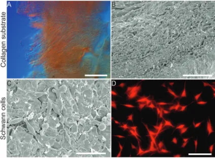
The collagen substrate was analyzed by polarized light microscopy and SEM. The general organization of the
fibrils is illustrated in Figure 1A. A parallel pattern could
be seen throughout different focus planes, indicating the suprastructural organization of the substrate. This was
confirmed by SEM, which also provided evidence of the presence of multiple layers of fibrils (Figure 1B). Combined
with the light microscopy observations, it was possible to conclude that such layers were arranged in
a three-dimensional helical fashion. SEM analysis of SCs reinforced the inte-gration with the organized collagen substrate (Figure 1C). Also, the cells tended to present a longitudinal organization, resembling the formation of the bands of Büngner in vivo.
Examining the SC culture, under phase contrast, it was possible to observe that SCs displayed their characteristic bipolar morphology with thin cell processes. The immunolabeling against S-100 revealed that the cultures were homogeneous, as seen in Figure 1D. Interestingly, SCs cultured on col-lagen presented longer cell projections, which were penetrating the substrate, indicating a three-dimensional organization of the cells.
Regarding ECM expression, observed by immunocytochemistry, it was possible to identify immunolabeling against type IV collagen both in the control and substrate-cultured cells (Figure 2J and L).
Neverthe-less, quantification did not show statistical
differences between the cultures seeded on bovine- or rat-derived collagen.
The analysis of S-100 and p75NTR im-munoreactivity is presented in Figure 2B, D,
F, and H and indicates a better alignment of cells cultured on the bovine substrate. Also, the quantitative data revealed
a statistically significant increase of immunoreactivity when
SCs were cultured on supraorganized bovine-derived col-lagen (Figure 2, bottom).
Discussion
The repair of a transected peripheral nerve in which the stumps are separated by a distance is a challenging medi-cal problem. Although the use of autografts is the option of choice, in many instances the size of the gap and the
caliber of the nerve require a significant amount of nerve
grafts, compromising sensory functions at the donor site.
Thus, the possibility of using artificial implants to bridge a
nerve defect has been pursued for some time. In particular, the tubulization technique has proved to be an alternative to nerve grafting over small gaps. Also, the use of ECM components within the tubular prosthesis made it possible to increase the gap between the stumps and still obtain a satisfactory guided axonal regeneration.
In order to further enhance the regeneration process, the use of three-dimensional scaffolds has become neces-sary. The three-dimensional organization of the implant may facilitate cell migration and tissue neoformation leading to a
faster repair of the lesion. Collagen-based fibrous scaffolds were produced by Yow et al. (17) and provided an efficient
substrate for the spatial organization and proliferation of
Figure 1.A, Representative samples of supraorganized collagen depicting the
parallel arrangement of the fibrils (bar = 100 µm). B, Collagen substrate observed by scanning electron microscopy. Note the multilayer organization as well as the porosity of the material (bar = 100 µm). C, Schwann cells cultured on the collagen substrate. Observe the adhesion shown by the cell projections over the surface of the material (bar = 50 µm). D, Representative example of a purified Schwann
Supraorganized collagen provides a scaffold for Schwann cell culture 685
Figure 2. Comparison between Schwann cell cultures performed on supraorganized (bovine collagen) and on non-organized
collagen (rat collagen). A and C, Substrate only (control) immunolabeled against S-100. B and D, Schwann cells cultured on bovine and rat collagen, respectively, and immunolabeled against S-100. Note the better alignment of SCs on the orga-nized collagen. E and G, Substrate only (control) immunolabeled against p75NTR (p75 pan neurotrophin receptor). F and
mesenchymal stem cells. These cells have been shown to enhance peripheral nerve regeneration after tubulization repair (3). Also, the concept that molecular alignment of collagen facilitates axonal growth and nerve regeneration has been proposed by Dubey et al. (15), Ceballos et al. (18), and Oliveira et al. (16).
Dubey et al. (15) and Ceballos et al. (18) used a
mag-netic field to force the arrangement of collagen fibrils, so
that a birefringent gel was obtained. In the present study, the molecular supraorganization was obtained naturally due to the maintenance of the self-assembly properties of the collagen after acid extraction and dialysis. This is particularly
beneficial because it results in a stable suprastructured
substrate, as seen by SEM analysis.
The efficiency of such naturally supraorganized collagen
substrate was assessed by analyzing SC immunoreactivity against S-100, p75NTR and type IV collagen. These three molecules are important elements regarding SC response to a nerve injury. In this way, the increased levels of the
low-affinity receptor for neurotrophins (p75NTR) observed
in supraorganized collagen cultures indicate an improved reactivity state that may further stimulate in vivo regenera-tion. The present results also demonstrate that SCs populate the three-dimensional scaffold, as shown by the presence of S-100-positive cells on different focal planes.
An important point to be emphasized is the fact that collagen is a natural component of the ECM of the nerve, so that its integration in such microenvironment is possibly
better than that of artificial biopolymers. Also, the degrada -tion and reabsorp-tion should be faster and less susceptible
to inflammatory reactions in the case of collagen. Another
important fact to be considered is that the better adhesion and integration to the substrate results in synthesis of neu-rotrophic factors, as already demonstrated in SC cultures (19) and supported here with regard to the immunoreactivity against p75NTR. These conditions also induce the formation of bands of Büngner, which are fundamental for the nerve regeneration process (14).
On the basis of the present data, we believe that the use of supraorganized collagen substrates enhances the response of SCs in vitro. The scaffold plus SCs produced in the present study may increase the peripheral nerve regeneration if used as an implant combined with a tubular prosthesis.
Acknowledgments
A.L.R. Oliveira is the recipient of a fellowship from CNPq (#300789/2009-2).
1. Abernethy DA, Thomas PK, Rud A, King RH. Mutual attrac-tion between emigrant cells from transected denervated nerve. J Anat 1994; 184 (Part 2): 239-249.
2. Madison RD, Archibald SJ. Point sources of Schwann cells result in growth into a nerve entubulation repair site in the absence of axons: effects of freeze-thawing. Exp Neurol
1994; 128: 266-275.
3. Pereira Lopes FR, Camargo de Moura Campos L, Dias Cor-rea J Jr, Balduino A, Lora S, Langone F, et al. Bone marrow stromal cells and resorbable collagen guidance tubes en-hance sciatic nerve regeneration in mice. Exp Neurol 2006; 198: 457-468.
4. Ide C. Peripheral nerve regeneration. Neurosci Res 1996; 25: 101-121.
5. Sunderland S. The anatomy and physiology of nerve injury.
Muscle Nerve 1990; 13: 771-784.
6. Timpl R, Brown JC. Supramolecular assembly of basement membranes. Bioessays 1996; 18: 123-132.
7. Badylak SF, Freytes DO, Gilbert TW. Extracellular matrix as a biological scaffold material: Structure and function. Acta Biomater 2009; 5: 1-13.
8. Milner R, Wilby M, Nishimura S, Boylen K, Edwards G, Faw-cett J, et al. Division of labor of Schwann cell integrins during migration on peripheral nerve extracellular matrix ligands.
Dev Biol 1997; 185: 215-228.
9. Zochodne DW. The microenvironment of injured and re-generating peripheral nerves. Muscle Nerve Suppl 2000; 9: S33-S38.
10. Schmidt CE, Leach JB. Neural tissue engineering: strategies
References
for repair and regeneration. Annu Rev Biomed Eng 2003; 5: 293-347.
11. Stoll G, Jander S, Myers RR. Degeneration and regeneration of the peripheral nervous system: from Augustus Waller’s
observations to neuroinflammation. J Peripher Nerv Syst
2002; 7: 13-27.
12. Evans GR, Brandt K, Katz S, Chauvin P, Otto L, Bogle M, et al. Bioactive poly(L-lactic acid) conduits seeded with Schwann cells for peripheral nerve regeneration. Biomateri-als 2002; 23: 841-848.
13. Schlosshauer B, Muller E, Schroder B, Planck H, Muller HW. Rat Schwann cells in bioresorbable nerve guides to promote and accelerate axonal regeneration. Brain Res 2003; 963: 321-326.
14. Ribeiro-Resende VT, Koenig B, Nichterwitz S, Oberhoffner S, Schlosshauer B. Strategies for inducing the formation of bands of Büngner in peripheral nerve regeneration. Bioma-terials 2009; 30: 5251-5259.
15. Dubey N, Letourneau PC, Tranquillo RT. Guided neurite elongation and Schwann cell invasion into magnetically aligned collagen in simulated peripheral nerve regeneration.
Exp Neurol 1999; 158: 338-350.
16. Oliveira ALR, Vidal BC, Langone F. Naturally supraorga-nized collagen increases axonal regeneration after tubuliza-tion repair. Braz J Morphol Sci 2005; 22: 143-148. 17. Yow SZ, Quek CH, Yim EK, Lim CT, Leong KW.
Supraorganized collagen provides a scaffold for Schwann cell culture 687
18. Ceballos D, Navarro X, Dubey N, Wendelschafer-Crabb G, Kennedy WR, Tranquillo RT. Magnetically aligned collagen
gel filling a collagen nerve guide improves peripheral nerve
regeneration. Exp Neurol 1999; 158: 290-300.