UPDATE ARTICLE
Animal models as tools to study the pathophysiology of
depression
Helena M. Abelaira, Gislaine Z. Re´us, Joa˜o Quevedo
Laboratory of Clinical Neurosciences, National Science and Technology Institute for Translational Medicine (INCT-TM), Center of Excellence in Applied Neurosciences of Santa Catarina (NENASC), Graduate Program in Health Sciences, Health Sciences Unit, Universidade do Extremo Sul Catarinense (UNESC), Criciu´ma, SC, Brazil.
The incidence of depressive illness is high worldwide, and the inadequacy of currently available drug
treatments contributes to the significant health burden associated with depression. A basic
understanding of the underlying disease processes in depression is lacking; therefore, recreating
the disease in animal models is not possible. Popular current models of depression creatively merge
ethologically valid behavioral assays with the latest technological advances in molecular biology.
Within this context, this study aims to evaluate animal models of depression and determine which has
the best face, construct, and predictive validity. These models differ in the degree to which they
produce features that resemble a depressive-like state, and models that include stress exposure are
widely used. Paradigms that employ acute or sub-chronic stress exposure include learned
helplessness, the forced swimming test, the tail suspension test, maternal deprivation, chronic mild
stress, and sleep deprivation, to name but a few, all of which employ relatively short-term exposure to
inescapable or uncontrollable stress and can reliably detect antidepressant drug response.
Keywords:
Face validity; construct validity; predictive validity; animals models;
antidepres-sants; depression
Introduction
Depression is a psychiatric disorder that has a poorly
understood neurobiology. Many patients do not respond
to existing treatments. Much research has been
under-taken, and its progress is coupled to the development of
animal models of human disease. As in all clinical
conditions, the rapprochement between the disease and
the corrective actions of drugs in laboratory animals is
essential for developing effective therapies. Criteria have
been established to define depressive mood disorders,
such as depressed mood and/or anhedonia, changes in
appetite, sleep disturbances, fatigue, and so on. These
are presented in Table 1.
1Animal models are important tools for investigating the
etiology of depression, as well as progress in the
development of effective therapeutic targets for its
treatment. Although animal models greatly help our
understanding of psychiatric disorders, they do have
some limitations. For example, animals cannot observe
feelings of sadness, guilt, or suicidal thoughts, symptoms
mainly limited to humans.
2The major obstacle is the
restricted availability of validated animal models. Firstly,
the ideal animal model should offer an opportunity to
understand molecular, genetic, and epigenetic factors
that may lead to depression.
3By using animal models,
the underlying molecular alterations and the causal
relationship between genetic or environmental alterations
and depression can be examined, which would afford a
better insight into the pathology of depression.
3Some
criteria have been established for the validation of an
animal model. These criteria are currently accepted and
are presented in Table 2.
Based on these criteria for validity, many animal
models of depression have been and are being
devel-oped, including those based on genetic engineering,
brain damage, and environmental manipulations. Table 3
Correspondence: Gislaine Z. Re´us, Laborato´rio de Neurocieˆncias, Programa de Po´s-Graduac¸a˜o em Cieˆncias da Sau´de, Unidade Acadeˆmica de Cieˆncias da Sau´de, Universidade do Extremo Sul Catarinense, CEP 88806-000, Criciu´ma, SC, Brazil.
E-mail: [email protected]
Table 1
Areas of impairment in depression and
corresponding diagnostic criteria
Area of
impairment DSM-IV diagnostic criteria
Mood Depressed mood
Motivation Markedly diminished interest or pleasure Food Weight loss or gain or decrease or increase in
appetite
Sleep Insomnia or hypersomnia
Motor Psychomotor retardation or agitation Energy Fatigue or loss of energy
Self-esteem Feelings of worthlessness or excessive or inappropriate guilt
Cognition Diminished ability to think or concentrate, or indecisiveness
Hope Recurrent thoughts of death, recurrent suicidal ideation
shows some existing models of depression and whether
they meet each of the criteria for validity presented in
Table 2.
The question therefore remains whether we can know if
a mouse is ‘‘depressed’’. In reality, few models of
depression fully fit these validating criteria, and most
models currently used rely on either the actions of known
antidepressants or responses to stress.
4Notably, it is not
necessary for an ‘‘ideal’’ animal model of depression to
exhibit all of the abnormalities of depression-relevant
behaviors, just as the patients do not manifest every
possible symptom of depression. In fact, anhedonia is the
core symptom of depression
4and most of the current
models only mimic anhedonia.
It should also be noted that there is a difference
between a model and a test. A model can be defined as
an organism (non-human) or a particular state of an
organism that reproduces aspects of human pathology,
providing a certain degree of predictive validity. A test, on
the other hand, provides only an endpoint – a behavioral
or physiological measure (read-out) designed to assess
the effect of a genetic, pharmacological, or environmental
manipulation.
5Thus, this work aims to study the animal
models of depression and determine which has the best
face, construct and predictive validity.
Animal models of depression: environmental
manipulations
Chronic mild stress
One of the major symptoms of depression in humans is
anhedonia, a reduction in interest or pleasure in daily
activities. Moreover, chronic mild stress (CMS) causes
many other symptoms of depression, such as decreases
in sexual, aggressive, and investigative behaviors, as well
as a decrease in locomotor activity.
3Furthermore,
repeated presentation of the same stressor usually leads
to adaptation, which can, however, be excluded by
presenting a variety of stressors in an unpredictable
sequence. According to these characteristics, many
researchers use an animal model of CMS to study the
neurobiology of depression, as well as to elucidate new
therapeutic targets for treatment.
The first CMS paradigm was introduced by Katz et al.
This model provides the basis for most of the currently
used paradigms. Initial protocols included 3 weeks of
exposure to electric shocks, immersion in cold water,
immobilization, reversal of the light/dark cycle, and a
variety of other stressors.
6This series of stressors could
cause an increase in plasma corticosteroid levels and a
reduction in sucrose preference,
6which suggests that
chronic stress may cause anhedonia.
In fact, rats subjected to different stressors (e.g., water
deprivation, food deprivation, exposure to cold
tempera-tures, isolation, etc.) over long periods (3 weeks–3
months) exhibited behavioral deficits, such as changes
in sleep and anhedonic behavior. With respect to
molecular parameters, which are related to the construct
validity of this animal model, changes were observed in
the
hypothalamic-pituitary-adrenal
(HPA)
system.
Usually, animals subjected to a stress protocol exhibit
increased levels of corticosterone and an increase in
adrenal gland weight.
7Changes in lipids and proteins, as
well as decreases in the activity of antioxidant enzymes
and increases in pro-inflammatory cytokines, were
observed in animals subjected to these experimental
protocols.
8,9In recent years, many researchers have focused on the
importance of neurotrophins, which are involved in
cellular plasticity. One of the most important
neurotro-phins is brain-derived neurotrophic factor (BDNF), which
is a target of prime importance in the neurobiology and
treatment of depression. In fact, it has been reported that
levels of this neurotrophin are decreased in serum and in
postmortem brain samples from patients with depression.
Moreover, antidepressants have shown a positive effect
on BDNF, increasing its expression.
10Reduced BDNF
levels, as well as attenuated neurogenesis, have been
found in the brains of animals exposed to a stress
Table 2
Criteria for the validity of an animal model of depression
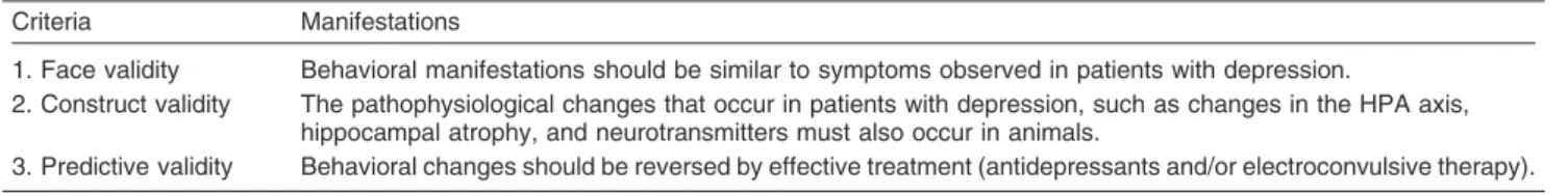
Criteria Manifestations
1. Face validity Behavioral manifestations should be similar to symptoms observed in patients with depression.
2. Construct validity The pathophysiological changes that occur in patients with depression, such as changes in the HPA axis, hippocampal atrophy, and neurotransmitters must also occur in animals.
3. Predictive validity Behavioral changes should be reversed by effective treatment (antidepressants and/or electroconvulsive therapy). HPA = hypothalamic-pituitary-adrenal.
Table 3
Animal models of depression and its criteria of validity
Criteria
Model Face Construct Predictive
Forced swim and tail suspension tests - - +
Chronic stressors (chronic mild stress, social isolation and learned helplessness) + + +
Maternal deprivation + + +
Injuries (olfactory bulbectomy) + + +
protocol.
11However, the opposite results – i.e., an
increase in BDNF levels in stressed animals – have been
reported by other authors, suggesting the existence of a
compensatory mechanism in response to stress.
7It is important to note that many of the changes found in
animals exposed to stress procedures and that serve as
criteria for face validation (such as anhedonia) and for
construct validation (such as changes in the HPA axis
and in neurotrophin levels) are reversed by various
classes of clinically effective antidepressants (e.g.,
fluoxetine and imipramine),
12demonstrating the
predic-tive validity of this animal model. However, the CMS
model has two major drawbacks. One is the practical
difficulty in carrying out CMS experiments, which are
labor-intensive, space demanding, and have long
dura-tion. The other is that the procedure itself can be difficult
to establish in a new laboratory setting, making replication
across laboratories challenging.
13Learned helplessness
Certain types of human depression are precipitated by
stressful life events, and vulnerable individuals
experien-cing these stressors may develop clinical depression. In
this aspect, stress can be used to induce depression-like
symptoms in rodent animals. One of the well-validated
animal models is learned helplessness, in which a
depressive-like state is induced by uncontrollable and
unpredictable electrical foot-shock stress.
14Following an uncontrollable and inescapable stress,
such as exposure to unavoidable electric shocks, the
animals develop a state of ‘‘helplessness’’ such that when
re-exposed to the same shocks, now with an easy escape
route, the animals will either display an increased escape
latency or completely fail to escape.
14Following one or
more sessions of inescapable shock, rats have been
shown to develop persistent changes including weight
loss, alterations in sleep patterns and HPA axis activity,
and a loss of spine synapses in the hippocampal
regions.
2,15Reduced weight, increased motor activity, reduced
libido, cognitive deficits, and changes in sleep have been
observed in helpless animals. In most cases, use of the
behavioral model of learned helplessness causes
ani-mals to present depressive-like behavior, as is observed
clinically in human patients.
16Therefore, this model
presents good face validity. In fact, animals subjected to
this model respond to tricyclic antidepressants, selective
serotonin reuptake inhibitors (SSRIs), monoamine
oxi-dase
inhibitors,
and
electroconvulsive
therapy.
17Response to these antidepressant drugs was observed
between 2-3 days after initiation of treatment.
Neurobiological changes have also been observed
after induction of learned helplessness. Depletion of
norepinephrine and serotonin, as well as changes in the
NE
b
norepinephrine and 5-HT1B
serotonin receptors in
the hippocampus, were reported. However, chronic
administration of antidepressants reversed the changes
in the NE
b
and 5-HT1B
receptors. In addition, the HPA
axis appears to play an important role in this animal
model. Indeed, an increase in the vulnerability to learned
helplessness has been observed in animals after
antagonism
of
the
glucocorticoid
receptors.
Furthermore, high levels of glucocorticoids and
homo-cysteine – which are found in human patients with
depression – have been reported in rats in an animal
model of learned helplessness.
18One advantage of learned helplessness as a model is
that its symptoms are parallel to those of major
depression, and most can be reversed by multiple acute
(subchronic) treatment with antidepressants (typically for
3-5 days).
16In addition, the cognitive (e.g., learning) and
other
behavioral
outcomes
(e.g.,
neurovegetative
abnormalities) seem to be correlated, thus enhancing
our understanding of depressive symptoms in humans.
The excellent face and predictive validities of learned
helplessness make it an interesting model for exploration
of the pathophysiology of depression.
19Furthermore, the
model can also be generally used to measure the escape
performance of mice with different mutations, showing
which target genes for depression may affect vulnerability
to development of a depressive-like state. However, the
major drawback of this model is that most of its
depression-like symptoms do not persist long enough
following cessation of the uncontrollable shock
stimu-lus.
15In addition, different protocols are used in different
laboratories.
Maternal deprivation
Early adverse life experiences represent one of the major
risk factors for the development of mental disorders such
as major depression. The early postnatal period is
characterized by considerable plasticity of the developing
nervous system. As such, the early postnatal
environ-ment is critical in its capacity to influence adult behavior.
Preclinical studies have provided direct evidence that
early life stress leads to heightened responsiveness to
stress and alterations in the HPA system throughout the
lifespan.
20Among the paradigms used to study early
adverse life events, long maternal separation in rodents
mimics early life neglect/loss of parents in humans, and
has been presented as one of the most potent natural
stressors during development. Maternal separation was
developed to examine the consequences of early adverse
experiences on behavior and neurobiology, and this
model has been described as a model of vulnerability to
drug dependence, anxiety, stress-induced illness, and
depression.
21In particular, maternal separation was proposed to
represent an important animal model for investigation of
the pathophysiology and treatment of major depression.
For example, treatment with antidepressants was able to
normalize
anxiety-like
behavior,
endocrine
stress
response, and preference for ethanol in adult male rats
subjected to maternal separation.
neurotrophin-3 [NT-3]).
22,23In contrast, treatment with
antidepressants in animals deprived of maternal care was
able to reverse depressive-type behaviors, promoting an
increase in neurotrophin levels and neurogenesis.
2Sleep deprivation
Sleep has important homeostatic functions, and sleep
deprivation is a stressor that has consequences for the
brain as well as for many body systems. Although sleep
deprivation is not yet a well-established model of
depression, many studies show that it alters important
pathways related to stress.
Increased levels of messenger RNA for interleukin-1b
(a pro-inflammatory cytokine) and for cortisol have been
shown in rodents after sleep deprivation. The procedure
of this study consisted of handling the animals gently to
prevent them from sleeping. Furthermore, 72 hours of
sleep deprivation in mice was induced using the platform
method, which is accomplished by placing the animal on
a platform submerged in water so that, when the animal
falls asleep, it falls into the water and must then climb
back onto the platform, thus forcing it to stay awake. This
study showed that after 72 hours of sleep deprivation,
there was an increase of oxidative stress in the
hippocampus. A decrease in cell proliferation has also
been observed after 96 hours of sleep deprivation.
9Some
classical tests used to assess cognitive parameters in
animals, such as the inhibitory avoidance and water maze
tests, show deficits in learning and memory of rodents
subjected to sleep deprivation, as well as aggressive
behavior and hyperactivity.
24Even though many studies using mice have shown
depressive-like behavior after sleep deprivation, it is
important to note that studies in patients with depression
have shown effects of antidepressants on selective
slow-wave sleep deprivation.
25,26The underlying mechanisms
are still unknown, however,
27and may be related to the
fact that the beneficial effects of sleep deprivation on
depressive-like behaviors require an astrocyte-dependent
signaling pathway.
Some neurotransmitters, such as dopamine and
serotonin, are altered following sleep deprivation, and
these alterations are associated with behavioral changes.
Sleep deprivation for short or long periods also altered
gene expression of several transcription factors and
genes that encode neurotransmitters and proteins
involved in metabolic processes and cellular plasticity.
28Anhedonic behavior has been shown in rats subjected to
paradoxical sleep deprivation.
29Although the sleep
deprivation protocol still has its limitations as an animal
model of depression, it meets some of the criteria for a
valid animal model, such as good face and construct
validity.
Changing photoperiod
More recently, it was proposed that manipulation of the
light/dark cycle could characterize a new animal model of
depression.
30In this model, nocturnally active mice are
exposed to long periods of artificial light (22 hours per
day) for a period of 2 weeks. Exploring the interactions
between these mechanisms and mood changes in diurnal
animals may provide new insight into depression. Recent
studies demonstrate that diurnal Fat sand rats and Nile
grass rats show depression-like behavior when
main-tained under short-photoperiod (SP) conditions compared
with animals maintained under neutral photoperiod (NP)
conditions. Moreover, these behaviors were ameliorated
by treatment with bright light.
31These animals also developed anhedonic behavior and
increased motor activity. Consistent with these behavioral
changes, increased levels of corticosterone and a
decrease in BDNF levels in the hippocampus were also
found, thus demonstrating face and construct validity.
However, the predictive validity of this model should be
examined, as treatment with the antidepressant
imipra-mine was only able to prevent some of these behavioral
and physiological changes.
32Diurnal mice develop
depressive-type behavior when subjected to
experimen-tal conditions with a decreasing photoperiod. Treatment
with the antidepressants bupropion and imipramine
reversed depressive behavior in these animals as shown
by the FST, but not anhedonic behavior.
Other studies using the dark phase of a 12:12 light/dark
cycle showed that rats exhibited depressive-like
beha-vior.
33,34On the other hand, brief or long exposures to
light treatment have an antidepressant effect on the
FST.
33,34Taken together, these studies reveal a
relation-ship between light control and depression.
Animal models of depression: injuries
Olfactory bulbectomy
Bilateral olfactory bulbectomy (OB) results in endocrine,
behavioral,
immune
system,
and
neurotransmitter
changes that mimic many of the symptoms seen in
human patients with major depression.
35The rat olfactory
system forms a part of the limbic region, which includes
the amygdala and hippocampus. These are responsible
for functions such as memory and emotion, and are
known to have altered morphology and activity in patients
with depression. After OB, there is a marked
degenera-tion of neurons in the olfactory bulb, but also in other
areas such as the hippocampus, cortex, amygdala, locus
ceruleus, and raphe nuclei. In all likelihood, these focal
brain changes lead to the dysfunction in serotonergic and
noradrenergic systems that is observed after
bulbect-omy.
36changes, both behavioral and cellular, are reversed by
several classes of antidepressants used in the clinic.
1With both behavioral changes and changes in
neuro-transmitters and in the immune system occurring after
OB, we can conclude that this may be a good animal
model of depression, since it has both face and construct
validity. Moreover, different classes of therapeutically
effective antidepressants reverse the behavioral and
molecular changes caused by OB, thus showing that this
model presents predictive validity.
Chronic, but not acute, administration of
antidepres-sants largely corrects most of the behavioral, endocrine,
immune, and neurotransmitter changes that occur
follow-ing bulbectomy. Thus, the olfactory bulbectomized rat is
not only a model for detecting antidepressant activity but
also one for exploring the inter-relationships between
these systems, which are also dysfunctional in patients
with major depression.
36Animal models of depression: chemical
manipulations
Stimulation of the immune system
Recently, several studies have drawn special attention to
the
role
of
the
immune
system
in
depression.
Immunologic abnormalities in depression have been
described for over two decades,
37,38but it is still unclear
whether these abnormalities play a role in the
pathogen-esis of depression.
Cytokines are inflammatory mediators that interact with
pathways related with depression, including
neurotrans-mitter metabolism, neural plasticity, and neuroendocrine
functions. Depressed patients exhibit high levels of
proinflammatory mediators, such as interleukins (IL-1,
IL-2 and IL-6) and tumor necrosis factor (TNF). Moreover,
treatment with antidepressants has been shown to
decrease levels of IL-4 in patients with depression.
Table 4 lists the major cytokines and their respective
functions.
1Animal models of depression involving the immune
system have been the target of criticism, but the fact
remains that there is still great difficulty in understanding
the complexity of the communication between the
immune system and the brain. Nevertheless, a few
studies have addressed this issue.
1In rodents, the
administration of endotoxin, a cell wall component of
Gram-negative cells, led to behavioral changes similar to
those seen in humans, such as anhedonia and sleep
disorders, which are parameters of face validity.
Neuroendocrine changes, which help determine
con-struct validity, were also found. Treatment with the
antidepressants fluoxetine and desipramine
37reduced
anhedonia in these animals (Table 5).
Animal models of depression: genetically
modified animal models
Tryptophan hydroxylase (TPH) is the rate-limiting enzyme
in 5-HT biosynthesis. The discovery of a neuronal
isoform, TPH2, by Walther & Bader
38opened up a new
way to reliably map 5-HT neurons in the brain by means
of immunohistochemistry and in situ hybridization.
Results confirmed wide expression, including cell bodies
in DRN and MRN.
39,40In fact, it is now established that
TPH2 is the predominant isoform in the rodent brain.
41,42However, early studies reported that TPH expression,
Table 4
Cytokines implicated in depression and their main
functions
Cytokine Main functions
Interleukin-1b(IL-1) Fever
HPA axis activation Lymphocyte activation Prostanoid synthesis Endothelial activation IL-6 synthesis
Interleukin-4 (IL-4) Inhibits production of TNF and IL-1 Stimulates B cells
Interleukin-6 (IL-6) Fever
Acute phase protein synthesis T and B cell differentiation and activation
Interleukin-8 (IL-8) Inflammation Neutrophil chemotaxis Interleukin-10 (IL-10) Inhibits inflammation
Inhibits production of IL-1, IL-6, TNF and IFN-c
Tumor necrosis factor (TNF) Fever
Endothelial activation Neutrophil activation
Migration of dendritic cells to lymph nodes
Interferon-a(INF-a) Induction of viral resistance Natural killer cell activation Macrophage activation Interferon-c(INF-c) Macrophage activation
T cell differentiation
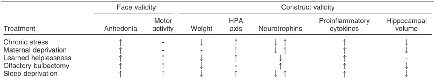
Table 5
Different animal models of depression and their effects on parameters of face and construct validity
Face validity Construct validity
Treatment Anhedonia
Motor
activity Weight
HPA
axis Neurotrophins
Proinflammatory cytokines
Hippocampal volume
Chronic stress q - Q q Q q q Q
Maternal deprivation q - - q Q q q Q
Learned helplessness q q Q q Q q
-Olfactory bulbectomy q q Q - q q Q
Sleep deprivation q q Q q Q q q Q
turnover, and distribution in DRN are complex, in line with
the existence of different subpopulations of 5-HT neurons
in this area.
43Changes in
TPH2
gene and/or protein
expression in the brain have been reported in various
mood disorders and have been validated in animal
models.
44-46TPH2
variants have also been extensively
reported to be associated with major depression.
47-49However, whether these changes might reflect alterations
in possible modulatory effects of other neurotransmitter
systems on 5-HT systems is still largely unknown. To
address this question directly,
TPH2
mRNA expression
was measured in three different transgenic mouse models,
all related to pathological states of depression and anxiety,
but caused by mutations affecting different
neurotransmit-ter systems. One of the models is the 5-HT transporneurotransmit-ter-
transporter-deficient mouse (5- HTT
-/-).
50,51By mediating 5-HT
reuptake in the nerve terminal (and other parts of the
5-HT neuron), 5-5-HTT fine-tunes the magnitude and duration
of serotoninergic signaling,
52,53which makes it the target
for many antidepressant drugs, including the SSRIs.
54These animals exhibit major adaptive changes in 5-HT
neurotransmission when compared with their wild-type
controls. It has been shown that lack of 5-HTT depletes
5-HT and its metabolite 5-hydroxyindoleacetic acid by
60–80% in several brain areas, such as the brainstem,
striatum, hippocampus, and frontal cortex.
50,55Functional
desensitization of 5-HT1A, 5-HT1B autoreceptors has
been reported in the DRN of 5-HTT
-/-mutants as a
consequence of high extracellular 5-HT levels in the vicinity
of the serotonergic cells in the DRN. However, in terms of
their target areas such as hippocampus and forebrain,
increased or unaltered expression of these receptors has
been reported.
55,56Autoradiographic labeling of 5-HT2A
receptors has also revealed a 30–40% reduction in the
density of these receptors in the cerebral cortex and lateral
striatum of 5-HTT
-/-animals in comparison with wild-type
mice.
57In addition, anxiolytic- and antidepressant-like
responses have been observed in 5-HTT
-/-mice in
behavioral test paradigms such as the elevated plus maze,
tail suspension test (TST), and FST.
58-60The second model consists of mice heterozygous for the
vesicular glutamate transporter 1 gene (VGLUT1
+/-).
61As
the first of three vesicular glutamate transporters
(VGLUT1, VGLUT2 and VGLUT3),
62VGLUT1 has been
shown to have a high level of expression in glutamatergic
neurons in the cerebral cortex.
63By concentrating
glutamate in synaptic vesicles, VGLUT1 mediates
gluta-mate release from synaptic terminals and facilitates
efficient glutamatergic transmission.
64,65Recent studies
have demonstrated that VGLUT1
+/-mice exhibit deficient
glutamate transmission,
66depressive- like behavior and
neurochemical changes, which are related to depression
and anxiety.
67,68Aberrations in glutamate synthesis and its
dysregulation also appear to play a relevant role in major
depression.
69In keeping with this, recent postmortem
studies showing decreased cortical VGLUT1 in depressed
subjects
70together with clinical findings of an excitatory
inhibitory
imbalance
in
the
cortex
of
depressed
patients
71,72suggest that decreased VGLUT1 levels may
have clinical implications.
Finally, mice with targeted disruption of the gene
encoding the cannabinoid 1 receptor (CB1R
-/-)
73were
used as an additional model of mood disorders. The
endocannabinoid system is a major neuromodulatory
system that contributes to the control of emotional
behavior.
74Pharmacological and genetic blockade of
the CB1R induces a behavioral state analogous to
depression in experimental animals.
75Thus, CB1R
-/-mice exhibit depressive-like symptoms, such as reduced
responsiveness to reward stimuli
74,76and enhanced
anxiety levels and sensitivity to stress.
77,78Moreover,
the chronic absence of CB1R activity induces alterations
in 5-HT-dependent negative feedback. In particular,
enhanced extracellular 5-HT levels in the prefrontal
cortex decreased 5-HTT binding site density and caused
functional desensitization of the 5-HT1A autoreceptors.
Reduced 5-HT2C receptor expression in different brain
regions has also been described in these mutants.
79In
addition, according to Mato et al.,
80mice lacking
CB1R
exert impaired post-synaptic serotonergic signaling,
suggesting that CB1R
-/-mice are useful models to reveal
more regarding the nature of cannabinoid/5-HT
interac-tions in mood disorders.
Although genetically modified animals can provide
good models of human diseases, mood disorders such
as depression are more difficult to represent, as these
disorders are associated with changes in several genes,
which are specific for each patient.
Predictive models of antidepressant activity
Forced swimming test
One of the tests most commonly used by researchers to
investigate new antidepressant drugs is the FST, first
described by Porsolt et al.
81This test was developed as
an animal model of depression that aimed to measure the
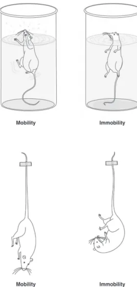
effects of antidepressant compounds in mice. In this test,
the animal is placed in a water-filled cylinder which it is
unable to exit. Initially, the animal will try to escape, but
eventually it adopts a posture of immobility, a passive
behavior characterized by the absence of movements
except for those necessary for the animal’s snout to
remain above the water level (Figure 1A). The test for rats
consists of two swimming exposures. The first exposure
is for 15 minutes and the second is performed 24 hours
after the first, with an exposure period of 5 minutes. The
test for mice consists of a single 6-minute exposure, with
the first 2 minutes serving as a habituation period and the
last 4 minutes consisting of the test itself, which yields the
duration of immobility.
horizontal and vertical movements of the animals as they
try to scale the cylinder walls with their paws. The effects
of antidepressants on FST behavior are relatively
specific, since they do not increase spontaneous motor
activity, unlike psychostimulants such as amphetamine
and cocaine.
83Besides the effects of antidepressant
drugs, the FST can also be used to evaluate the type of
depressive behavior; for example, it has been
demon-strated that animals subjected to a protocol of maternal
deprivation exhibit increased immobility time in the FST.
23Tail suspension test
Since its introduction almost 20 years ago, the TST has
become one of the most widely used models for
assessing antidepressant-like activity in mice. The test
is based on the fact that animals subjected to the
short-term, inescapable stress of being suspended by their tail
will develop an immobile posture.
15The FST is similar to
the TST, but differs in that it can be used on both rats and
in mice, whereas the TST can only be undertaken on
mice. In the TST, the tails of the mice are attached and
suspended by an adhesive tape (Figure 1B). The time
spent immobile by the animal during a period of 6 minutes
is interpreted as a measure of depressive-like behavior.
Various antidepressant medications reverse this
immobi-lity and promote the occurrence of escape-related
behavior.
Importantly, both the TST and FST are considered
predictive models of antidepressant activity, not animal
models of depression. Accordingly, they lack face and
construct validity.
Conclusion
Although there are many animal models of depression,
including some that have predictive, face, and construct
validity within the same model, many limitations constrain
their utility. It is remarkable that all animal models of
depression have contributed to a better understanding of
the neurobiology of this disorder, and offer new
pharma-cological targets for treatment. However, the
develop-ment of a model that represents most symptoms of
depression and meets all criteria for animal model validity
would be ideal.
Acknowledgements
This study was supported in part by grants from Conselho
Nacional de Desenvolvimento Cientı´fico e Tecnolo´gico
(CNPq-Brazil – JQ and GZR), Instituto Ce´rebro e Mente
(JQ), and UNESC (JQ). JQ is a recipient of productivity
grants from CNPq-Brazil. GZR and HMA receive
student-ship grants from Coordenac¸a˜o de Aperfeic¸oamento de
Pessoal de Nı´vel Superior (CAPES).
Disclosure
The authors report no conflicts of interest.
References
1 DellaGioia N, Hannestad J. A critical review of human endotoxin administration as an experimental paradigm of depression. Neurosci Biobehav Rev. 2010;34:130-43.
2 Nestler EJ, Hyman SE. Animal models of neuropsychiatric disorders. Nat Neurosci. 2010;13:1161-9.
3 Chen SK, Tvrdik P, Peden E, Cho S, Wu S, Spangrude G, et al. Hematopoietic origin of pathological grooming in Hoxb8 mutant mice. Cell. 2010;141:775-85.
4 Krishnan V, Nestler EJ. The molecular neurobiology of depression. Nature. 2008;455:894-902.
5 Urani A, Chourbaji S, Gass P. Mutant mouse models of depression: candidate genes and current mouse lines. Neurosci Biobehav Rev. 2005;29:805-28.
6 Katz RJ, Roth KA, Carroll BJ. Acute and chronic stress effects on open field activity in the rat: implications for a model of depression. Neurosci Biobehav Rev. 1981;5:247-5
7 Fortunato JJ, Reus GZ, Kirsch TR, Stringari RB, Fries GR, Kapczinski F, et al. Effects of beta-carboline harmine on behavioral and physiological parameters observed in the chronic mild stress model: further evidence of antidepressant properties. Brain Res Bull. 2010;81:491-6.
8 Lucca G, Comim CM, Valvassori SS, Reus GZ, Vuolo F, Petronilho F, et al. Effects of chronic mild stress on the oxidative parameters in the rat brain. Neurochem Int. 2009;54:358-62.
9 You Z, Luo C, Zhang W, Chen Y, He J, Zhao Q, et al. Pro- and anti-inflammatory cytokines expression in rat’s brain and spleen exposed to chronic mild stress: involvement in depression. Behav Brain Res. 2011;225:135-41.
10 Mao QQ, Xian YF, Ip SP, Tsai SH, Che CT. Long-term treatment with peony glycosides reverses chronic unpredictable mild stress-induced depressive-like behavior via increasing expression of neurotrophins in rat brain. Behav Brain Res. 2010;210:171-7. 11 Ibarguen-Vargas Y, Surget A, Vourc’h P, Leman S, Andres CR,
Gardier AM, et al. Deficit in BDNF does not increase vulnerability to stress but dampens antidepressant-like effects in the unpredictable chronic mild stress. Behav Brain Res. 2009;202:245-51.
12 Szymanska M, Budziszewska B, Jaworska-Feil L, Basta-Kaim A, Kubera M, Leskiewicz M, et al. The effect of antidepressant drugs on the HPA axis activity, glucocorticoid receptor level and FKBP51 concentration in prenatally stressed rats. Psychoneuroendocrino-logy. 2009;34:822-32.
13 Willner P. Validity, reliability and utility of the chronic mild stress model of depression: a 10-year review and evaluation. Psychopharmacology (Berl). 1997;134:319-29.
14 Seligman ME, Beagley G. Learned helplessness in the rat. J Comp Physiol Psychol. 1975;88:534-41.
15 Cryan JF, Mombereau C, Vassout A. The tail suspension test as a model for assessing antidepressant activity: review of pharmacolo-gical and genetic studies in mice. Neurosci Biobehav Rev. 2005;29:571-625.
16 Takamori K, Yoshida S, Okuyama S. Availability of learned helplessness test as a model of depression compared to a forced swimming test in rats. Pharmacology. 2001;63:147-53.
17 Malberg JE, Duman RS. Cell proliferation in adult hippocampus is decreased by inescapable stress: reversal by fluoxetine treatment. Neuropsychopharmacology. 2003;28:1562-71.
18 Setnik B, de Souza FG, d’Almeida V, Nobrega JN. Increased homocysteine levels associated with sex and stress in the learned helplessness model of depression. Pharmacol Biochem Behav. 2004;77:155-61.
19 Vollmayr B, Simonis C, Weber S, Gass P, Henn F. Reduced cell proliferation in the dentate gyrus is not correlated with the development of learned helplessness. Biol Psychiatry. 2003;54:1035-40.
20 Heim C, Nemeroff CB. The role of childhood trauma in the neurobiology of mood and anxiety disorders: preclinical and clinical studies. Biol Psychiatry. 2001;49:1023-39.
21 Anisman H, Zaharia MD, Meaney MJ, Merali Z. Do early-life events permanently alter behavioral and hormonal responses to stressors? Int J Dev Neurosci. 1998;16:149-64.
22 Ruedi-Bettschen D, Zhang W, Russig H, Ferger B, Weston A, Pedersen EM, et al. Early deprivation leads to altered behavioural, autonomic and endocrine responses to environmental challenge in adult Fischer rats. Eur J Neurosci. 2006;24:2879-93.
23 Reus GZ, Stringari RB, Ribeiro KF, Cipriano AL, Panizzutti BS, Stertz L, et al. Maternal deprivation induces depressive-like behaviour and alters neurotrophin levels in the rat brain. Neurochem Res. 2011;36:460-6.
24 McEwen BS. Sleep deprivation as a neurobiologic and physiologic stressor: allostasis and allostatic load. Metabolism. 2006;55:S20-3. 25 Goldstein MR, Plante DT, Hulse BK, Sarasso S, Landsness EC,
Tononi G, et al. Overnight changes in waking auditory evoked potential amplitude reflect altered sleep homeostasis in major depression. Acta Psychiatr Scand. 2012;125:468-77.
26 Landsness EC, Ferrarelli F, Sarasso S, Goldstein MR, Riedner BA, Cirelli C, et al. Electrophysiological traces of visuomotor learning and their renormalization after sleep. Clin Neurophysiol. 2011;122:2418-25.
27 Hines DJ, Schmitt LI, Hines RM, Moss SJ, Haydon PG. Antidepressant effects of sleep deprivation require astrocyte-dependent adenosine mediated signaling. Transl Psychiatry. 2013;3:e212.
28 Sei H, Saitoh D, Yamamoto K, Morita K, Morita Y. Differential effect of short-term REM sleep deprivation on NGF and BDNF protein levels in the rat brain. Brain Res. 2000;877:387-90.
29 Andersen ML, Hoshino K, Tufik S. Increased susceptibility to development of anhedonia in rats with chronic peripheral nerve injury: involvement of sleep deprivation? Prog Neuropsychopharmacol Biol Psychiatry. 2009;33:960-6.
30 Becker A, Bilkei-Gorzo A, Michel K, Zimmer A. Exposure of mice to long-light: a new animal model to study depression. Eur Neuropsychopharmacol. 2010;20:802-12.
31 Krivisky K, Ashkenazy T, Kronfeld-Schor N, Einat H. Antidepressants reverse short-photoperiod-induced, forced swim test depression-like behavior in the diurnal fat sand rat: further support for the utilization of diurnal rodents for modeling affective disorders. Neuropsychobiology. 2011;63:191-6.
32 Song C, Leonard BE. The olfactory bulbectomised rat as a model of depression. Neurosci Biobehav Rev. 2005;29:627-47.
33 Molina-Hernandez M, Tellez-Alcantara P. Long photoperiod regimen may produce antidepressant actions in the male rat. Prog Neuropsychopharmacol Biol Psychiatry. 2000;24:105-16.
34 Schulz D, Aksoy A, Canbeyli R. Behavioral despair is differentially affected by the length and timing of photic stimulation in the dark phase of an L/D cycle. Prog Neuropsychopharmacol Biol Psychiatry. 2008;32:1257-62.
35 Hellweg R, Zueger M, Fink K, Hortnagl H, Gass P. Olfactory bulbectomy in mice leads to increased BDNF levels and decreased serotonin turnover in depression-related brain areas. Neurobiol Dis. 2007;25:1-7.
36 Irwin MR, Miller AH. Depressive disorders and immunity: 20 years of progress and discovery. Brain Behav Immun. 2007;21:374-83. 37 Miller AH, Ancoli-Israel S, Bower JE, Capuron L, Irwin MR.
Neuroendocrine-immune mechanisms of behavioral comorbidities in patients with cancer. J Clin Oncol. 2008;26:971-82.
38 Walther DJ, Bader M. A unique central tryptophan hydroxylase isoform. Biochem Pharmacol. 2003;66:1673-80.
39 Zhang X, Beaulieu JM, Sotnikova TD, Gainetdinov RR, Caron MG. Tryptophan hydroxylase-2 controls brain serotonin synthesis. Science. 2004;305:217.
40 Clark JA, Flick RB, Pai LY, Szalayova I, Key S, Conley RK, et al. Glucocorticoid modulation of tryptophan hydroxylase-2 protein in raphe nuclei and 5-hydroxytryptophan concentrations in frontal cortex of C57/Bl6 mice. Mol Psychiatry. 2008;13:498-506. 41 Gutknecht L, Waider J, Kraft S, Kriegebaum C, Holtmann B, Reif A,
et al. Deficiency of brain 5-HT synthesis but serotonergic neuron formation in Tph2 knockout mice. J Neural Transm. 2008;115:1127-32.
42 Gutknecht L, Kriegebaum C, Waider J, Schmitt A, Lesch KP. Spatio-temporal expression of tryptophan hydroxylase isoforms in murine and human brain: convergent data from Tph2 knockout mice. Eur Neuropsychopharmacol. 2009;19:266-82.
43 Weissmann D, Chamba G, Debure L, Rousset C, Richard F, Maitre M, et al. Variation of tryptophan-5-hydroxylase concentration in the rat raphe dorsalis nucleus after p-chlorophenylalanine administra-tion. II. Anatomical distribution of the tryptophan-5-hydroxylase protein and regional variation of its turnover rate. Brain Res. 1990;536:46-55.
44 Hiroi R, McDevitt RA, Neumaier JF. Estrogen selectively increases tryptophan hydroxylase-2 mRNA expression in distinct subregions of rat midbrain raphe nucleus: association between gene expression and anxiety behavior in the open field. Biol Psychiatry. 2006;60:288-95.
45 Bach-Mizrachi H, Underwood MD, Tin A, Ellis SP, Mann JJ, Arango V. Elevated expression of tryptophan hydroxylase-2 mRNA at the neuronal level in the dorsal and median raphe nuclei of depressed suicides. Mol Psychiatry. 2008;13:507-465.
46 Bonkale WL, Austin MC. 3,4-Methylenedioxymethamphetamine induces differential regulation of tryptophan hydroxylase 2 protein and mRNA levels in the rat dorsal raphe nucleus. Neuroscience. 2008;155:270-6.
47 Zill P, Baghai TC, Zwanzger P, Schule C, Eser D, Rupprecht R, et al. SNP and haplotype analysis of a novel tryptophan hydroxylase isoform (TPH2) gene provide evidence for association with major depression. Mol Psychiatry. 2004;9:1030-6.
Swedish, isolated population. Arch Gen Psychiatry. 2006;63:1103-10.
49 Haghighi F, Bach-Mizrachi H, Huang YY, Arango V, Shi S, Dwork AJ, et al. Genetic architecture of the human tryptophan hydroxylase 2 Gene: existence of neural isoforms and relevance for major depression. Mol Psychiatry. 2008;13:813-20.
50 Bengel D, Murphy DL, Andrews AM, Wichems CH, Feltner D, Heils A, et al. Altered brain serotonin homeostasis and locomotor insensitivity to 3, 4-methylenedioxymethamphetamine (‘‘Ecstasy’’) in serotonin transporter-deficient mice. Mol Pharmacol. 1998;53:649-55.
51 Murphy DL, Lesch KP. Targeting the murine serotonin transporter: insights into human neurobiology. Nat Rev Neurosci. 2008;9:85-96. 52 Lesch KP, Mossner R. Inactivation of 5HT transport in mice: modeling altered 5HT homeostasis implicated in emotional dysfunc-tion, affective disorders, and somatic syndromes. Handb Exp Pharmacol. 2006417-56.
53 Canli T, Lesch KP. Long story short: the serotonin transporter in emotion regulation and social cognition. Nat Neurosci. 2007;10: 1103-9.
54 Owens MJ, Nemeroff CB. The serotonin transporter and depression. Depress Anxiety. 1998;8:5-12.
55 Fabre V, Beaufour C, Evrard A, Rioux A, Hanoun N, Lesch KP, et al. Altered expression and functions of serotonin 5-HT1A and 5-HT1B receptors in knock-out mice lacking the 5-HT transporter. Eur J Neurosci. 2000;12:2299-310.
56 Mannoury la Cour C, Boni C, Hanoun N, Lesch KP, Hamon M, Lanfumey L. Functional consequences of 5-HT transporter gene disruption on 5-HT(1a) receptor-mediated regulation of dorsal raphe and hippocampal cell activity. J Neurosci. 2001;21:2178-85. 57 Rioux A, Fabre V, Lesch KP, Moessner R, Murphy DL, Lanfumey L,
et al. Adaptive changes of serotonin 5-HT2A receptors in mice lacking the serotonin transporter. Neurosci Lett. 1999;262:113-6. 58 Holmes A, Yang RJ, Lesch KP, Crawley JN, Murphy DL. Mice
lacking the serotonin transporter exhibit 5-HT(1A) receptor-mediated abnormalities in tests for anxiety-like behavior. Neuropsychophar-macology. 2003;28:2077-88.
59 Holmes A, Murphy DL, Crawley JN. Abnormal behavioral pheno-types of serotonin transporter knockout mice: parallels with human anxiety and depression. Biol Psychiatry. 2003;54:953-9.
60 Renoir T, Paizanis E, El Yacoubi M, Saurini F, Hanoun N, Melfort M, et al. Differential long-term effects of MDMA on the serotoninergic system and hippocampal cell proliferation in 5-HTT knock-out vs. wild-type mice. Int J Neuropsychopharmacol. 2008;11:1149-62. 61 Wojcik SM, Rhee JS, Herzog E, Sigler A, Jahn R, Takamori S, et al.
An essential role for vesicular glutamate transporter 1 (VGLUT1) in postnatal development and control of quantal size. Proc Natl Acad Sci U S A. 2004;101:7158-63.
62 Takamori S, Rhee JS, Rosenmund C, Jahn R. Identification of a vesicular glutamate transporter that defines a glutamatergic pheno-type in neurons. Nature. 2000;407:189-94.
63 Hisano S. Vesicular glutamate transporters in the brain. Anat Sci Int. 2003;78:191-204.
64 Fremeau RT, Jr, Kam K, Qureshi T, Johnson J, Copenhagen DR, Storm-Mathisen J, et al. Vesicular glutamate transporters 1 and 2 target to functionally distinct synaptic release sites. Science. 2004;304:1815-9.
65 Wilson NR, Kang J, Hueske EV, Leung T, Varoqui H, Murnick JG, et al. Presynaptic regulation of quantal size by the vesicular glutamate transporter VGLUT1. J Neurosci. 2005;25:6221-34. 66 Balschun D, Moechars D, Callaerts-Vegh Z, Vermaercke B, Van
Acker N, Andries L, et al. Vesicular glutamate transporter VGLUT1
has a role in hippocampal long-term potentiation and spatial reversal learning. Cereb Cortex. 2010;20:684-93.
67 Tordera RM, Totterdell S, Wojcik SM, Brose N, Elizalde N, Lasheras B, et al. Enhanced anxiety, depressive-like behaviour and impaired recognition memory in mice with reduced expression of the vesicular glutamate transporter 1 (VGLUT1). Eur J Neurosci. 2007;25:281-90. 68 Garcia-Garcia AL, Elizalde N, Matrov D, Harro J, Wojcik SM, Venzala E, et al. Increased vulnerability to depressive-like behavior of mice with decreased expression of VGLUT1. Biol Psychiatry. 2009;66:275-82.
69 Krystal JH, Sanacora G, Blumberg H, Anand A, Charney DS, Marek G, et al. Glutamate and GABA systems as targets for novel antidepressant and mood-stabilizing treatments. Mol Psychiatry. 2002;7:S71-80.
70 Uezato A, Meador-Woodruff JH, McCullumsmith RE. Vesicular glutamate transporter mRNA expression in the medial temporal lobe in major depressive disorder, bipolar disorder, and schizophrenia. Bipolar Disord. 2009;11:711-25.
71 Sanacora G, Gueorguieva R, Epperson CN, Wu YT, Appel M, Rothman DL, et al. Subtype-specific alterations of gamma-amino-butyric acid and glutamate in patients with major depression. Arch Gen Psychiatry. 2004;61:705-13.
72 Bhagwagar Z, Wylezinska M, Jezzard P, Evans J, Ashworth F, Sule A, et al. Reduction in occipital cortex gamma-aminobutyric acid concentrations in medication-free recovered unipolar depressed and bipolar subjects. Biol Psychiatry. 2007;61:806-12.
73 Ledent C, Valverde O, Cossu G, Petitet F, Aubert JF, Beslot F, et al. Unresponsiveness to cannabinoids and reduced addictive effects of opiates in CB1 receptor knockout mice. Science. 1999;283:401-4. 74 Maldonado R, Valverde O, Berrendero F. Involvement of the
endocannabinoid system in drug addiction. Trends Neurosci. 2006;29:225-32.
75 Hill MN, Gorzalka BB. Is there a role for the endocannabinoid system in the etiology and treatment of melancholic depression? Behav Pharmacol. 2005;16:333-52.
76 Sanchis-Segura C, Cline BH, Marsicano G, Lutz B, Spanagel R. Reduced sensitivity to reward in CB1 knockout mice. Psychopharmacology (Berl). 2004;176:223-32.
77 Martin M, Ledent C, Parmentier M, Maldonado R, Valverde O. Involvement of CB1 cannabinoid receptors in emotional behaviour. Psychopharmacology (Berl). 2002;159:379-87.
78 Aso E, Ozaita A, Valdizan EM, Ledent C, Pazos A, Maldonado R, et al. BDNF impairment in the hippocampus is related to enhanced despair behavior in CB1 knockout mice. J Neurochem. 2008;105:565-72.
79 Aso E, Renoir T, Mengod G, Ledent C, Hamon M, Maldonado R, et al. Lack of CB1 receptor activity impairs serotonergic negative feedback. J Neurochem. 2009;109:935-44.
80 Mato S, Aso E, Castro E, Martin M, Valverde O, Maldonado R, et al. CB1 knockout mice display impaired functionality of HT1A and 5-HT2A/C receptors. J Neurochem. 2007;103:2111-20.
81 Porsolt RD, Le Pichon M, Jalfre M. Depression: a new animal model sensitive to antidepressant treatments. Nature. 1977;266:730-2. 82 Castagne V, Moser P, Roux S, Porsolt RD. Rodent models of