U
NIVERSIDADE DEL
ISBOAF
ACULDADE DEC
IÊNCIAS DEPARTAMENTO DE BIOLOGIA VEGETAL
U
NDERSTANDING THE INTERACTIONS BETWEEN RETROVIRAL VECTORS
AND THE GENOME OF HUMAN KERATINOCYTES
A
NAR
ITAM
ICAELOM
ENDESM
ASTER INA
PPLIEDM
ICROBIOLOGYU
NIVERSIDADE DEL
ISBOAF
ACULDADE DEC
IÊNCIAS DEPARTAMENTO DE BIOLOGIA VEGETAL
U
NDERSTANDING THE INTERACTIONS BETWEEN RETROVIRAL VECTORS
AND THE GENOME OF HUMAN KERATINOCYTES
A
NAR
ITAM
ICAELOM
ENDEST
HESIS COORDINATORS:
F
ULVIOM
AVILIOCENTRO DI MEDICINA RIGENERATIVA STEFANO FERRARI,UNIVERSITY OF MODENA AND REGGIO EMILIA
M
ARGARIDAG
AMA-C
ARVALHOCENTER FOR BIODIVERSITY,FUNCTIONAL AND INTEGRATIVE GENOMICS,FACULTY OF SCIENCES,UNIVERSITY OF
LISBON AND FACULTY OF MEDICINE,UNIVERSITY OF LISBON
MASTER IN APPLIED MICROBIOLOGY
1
INDEXABSTRACT...3
RESUMO...5
INTRODUCTION...8
- A brief review of retroviruses and retroviral vectors...9
- Retroviral integration features: state of the art...12
- Proposed mechanisms for integration site selection...13
METHODS AND MATERIALS...19
- Virus production and cell culture...19
- Transduction of cells...19
- Cloning and analysis of viral insertion sites...20
- Sequence analysis...20
- Gene expression profiling...21
- Functional gene analysis...21
RESULTS...22
- Retroviruses integrate preferably inside genes...22
- MLV is attracted to TSS proximal regions while HIV is repelled...23
- Retroviruses target mostly active genes...25
- MLV’s clusters are more packed while HIV’s expand through a wider chromatin region...27
- MLV integration positively associates with epigenetic markers of promoters and enhancers..28
- MLV has a superior integration in evolutionary conserved sequences...30
- MLV has a higher integration in oncogenes and common insertion sites...31
- MLV targets regions rich in transcription factor binding sites with a cell-specific pattern...32
- Ingenuity Pathways Analysis suggests functions for retroviruses’ target genes...35
- Retroviruses choose early replicating regions of the genome to integrate...39
DISCUSSION...40
2
“Retroviruses are unique among infectious agents, both in the way they interact with the host cell and organism and in the consequences of this interaction—not only to the life of the infected host, but also in some cases to the host's descendants. No other infectious agent of higher eukaryotes regularly integrates its genetic information into the host genome; no other regularly acquires host genes into its genome, no other can infect the germ line of its host; and no other has played such an important part in so many aspects of modern biology.” (in Retroviruses)
3
ABSTRACTRetroviruses are a unique family of infectious agents, its diverse life cycle and survival strategies have been an important object of study to modern biology. The integration pattern of gamma-retroviruses (RV) and lentiviruses (LV) in the human genome follows a non-random very distinct distribution, but the precise factors involved in choice of the integration sites are still unknown.
To further study this question, RV (MLV) and LV (HIV) integration sites were mapped after infection of primary human keratinocytes with vectors derived from these viruses. 3020 and 1601 unique sites for MLV and HIV, respectively, were retrieved and analysed for correlation with several genomic sites. Moreover, a comparative analysis between the new dataset and previously obtained datasets on lymphocytes T and haematopoietic stem cells Cd34+ was performed to understand the involvement of the cellular program in viral integration choices.
The outcome of this project suggests substantial differences in the molecular mechanisms tethering RV and LV PICs to human chromatin. Both vectors’ integration locates preferably inside genes and target mostly active genes with a higher frequency in lymphocytes, these characteristics showed a higher frequency in HIV’s integrations. MLV is attracted to TSS proximal regions with a higher frequency in lymphocytes, while HIV is repelled by these regions independently of the cell-type. MLV positively associates with epigenetic markers of promoters and enhancers and has a higher integration in evolutionary conserved non-coding sequences in mammals (CNCs). Additionally it targets regions rich in transcription factor binding sites (TFBS) with a cell-specific pattern, revealing a possible preference for functional TFBS rather then a general feature preference. HIV associates with epigenetic markers for the body of transcribed genes and has no preference for CNCs. HIV showed a more widely distributed integration, with clusters that spread through much larger regions of the genome and, therefore, target more genes then MLV’s clusters which have a more packed distribution. Analysis of gene function revealed some cell-specificity in MLV’s targets although common genes probably related to the transcription machinery are also highly targeted. HIV targets genes involved essentially in general cell-functions. Furthermore we predict that retroviruses choose early replicating regions of the genome to integrate.
Since primary human keratinocytes are clinically relevant in gene therapy for correction of inherited skin diseases, safety parameters related to both vectors were also analysed. The results of this study predict that HIV has a lower probability of generating insertional gene activation events that could lead to oncogene activation. Furthermore, since MLV has a more cell-specific integration pattern, there’s a different probability of integrating near specific oncogenes according to the cell-type.
Findings of this work reveal cellular specific patterns of integration as well as common integration preferences, clarifying the cellular machinery’s role in integration site-selection
4
viral components to host proteins and suggest a higher involvement of the cellular transcriptional machinery in MLV’s integration in comparison to HIV’s.5
RESUMOOs retrovírus fazem parte de uma família única de agentes infecciosos; as particularidades do seu ciclo de vida, a forma como interagem com o hospedeiro e as consequências desta interacção têm sido objecto de estudo fulcral da biologia moderna. O impacto dos retrovírus na sociedade actual é inegável e a busca pela sua compreensão tem direccionado esforço e recursos da comunidade científica.
O processo de integração retroviral no genoma humano é de primordial importância uma vez que, dele depende a sobrevivência do vírus no hospedeiro. A integração dos retrovírus segue um padrão não-aleatório que varia entre as diversas famílias virais, podendo-se facilmente identificar uma família retroviral pelas suas preferências na escolha do local de integração (Bushman, 2005). Apesar dos mecanismos envolvidos nas reacções de corte e ligação do genoma viral ao do hospedeiro serem já bastante conhecidos, os factores envolvidos na escolha do local de integração são ainda uma questão em aberto.
Para tentar compreender melhor esta questão, mapeei 3020 locais únicos de integração de moloney leukaemia virus (MLV) e 1601 locais de integração de human immunodeficiency virus (HIV) no genoma de queratinócitos humanos primários. Sendo MLV da família oncoretroviridae e HIV da família lentiviridae, procurei estabelecer as semelhanças e diferenças no processo de escolha do local de integração de cada um. A par de analisar as preferências integrativas neste tipo celular, realizei uma análise comparativa com dados anteriores obtidos em células estaminais hematopoiéticas (HSC) Cd34+ e linfócitos T de forma a tentar compreender o envolvimento do programa celular nas escolhas integrativas.
Os resultados deste trabalho demonstram que MLV, membro da família gamma-retroviridae, e HIV, da família lentiviridae, apresentam padrões de integração não aleatórios muito distintos. Os vírus em estudo apresentam diferenças substanciais nos mecanismos moleculares que atraem os complexos de pré-integração viral (PICs) para a cromatina humana revelando estratégias evolutivas muito diversas. Ambos os vectores se integram preferencialmente dentro de genes e maioritariamente em genes activos, com uma frequência mais elevada em linfócitos. Estas características demonstraram-se mais exacerbadas nas integrações de HIV.
MLV é atraído por regiões próximas de transcription start sites (TSS), com uma frequência mais elevada em linfócitos, enquanto HIV é repelido por estas mesmas zonas do genoma com igual frequência em todos os tipos celulares. Ambos os vírus se integram preferencialmente dentro de genes em comparação com o controlo, no entanto, esta preferência é bastante mais acentuada em HIV, em particular em linfócitos, apresentando mais de 80% das integrações em genes em comparação com 50% das integrações de MLV. Genes com expressão activa são candidatos preferenciais para a integração viral, incluindo 70% dos hits únicos. Esta preferência é privilegiada nas integrações dentro de genes de HIV, sendo mais elevada em Cd34+HSC
6
quando comparada com queratinócitos. MLV, mas não HIV, revela alguma preferência por zonas do genoma conservadas não-codificantes (CNCs) entre mamíferos e relaciona-se positivamente com regiões ricas em transcription factor binding sites, com um padrão particular para cada tipo celular. Esta particularidade sugere uma preferência por TFBS activos mais do que uma preferência geral pela vizinhança destas regiões genómicas.Foram também analisados diversos marcadores epigenéticos para os diversos tipos celulares. Esta análise revelou uma forte associação de MLV a regiões com promotores e enhancers e uma associação negativa a zonas com marcadores geralmente associados a cromatina inactiva ou repressão da expressão génica. Por outro lado, HIV associa-se positivamente a marcadores epigenéticos para o corpo de genes transcritos e negativamente para marcadores de regiões com promotores e enhancers, assim como zonas de cromatina inactiva.
Clusters de integração viral foram definidos estatisticamente, tendo em conta o tamanho da amostra e a distância entre uma integração e a integração consecutiva. Verificou-se que MLV apresenta clusters com maior densidade de integração, enquanto os clusters de HIV se expandem através de uma área mais vasta da cromatina. Consequentemente, os clusters de HIV incluem um maior número de genes relativamente a MLV.
Através da utilização do programa Ingenuity analisei os genes alvo de ambos os vírus relativamente à sua função em todos os tipos celulares. Verifica-se que MLV tem como alvo genes específicos do tipo celular assim como genes relacionados com funções mais gerais. HIV integra-se esintegra-sencialmente em genes com funções de regulação da expressão génica.
Seguidamente, coloquei a hipótese de que as integrações retrovirais se localizariam em zonas dos cromossomas que se replicam cedo durante o ciclo replicativo, uma vez que, estas zonas se relacionam com zonas de cromatina activa assim como com genes de elevada expressão em determinado tipo celular. Apesar de considerar os resultados com alguma cautela, tendo em conta a reduzida informação disponível em relação ao tempo de replicação, é previsível uma forte correlação com zonas dos cromossomas que replicam cedo, por parte de ambos os vírus.
Uma vez que os vírus estudados são aplicáveis ao nível da terapia génica de doenças epiteliais hereditárias, verifiquei alguns parâmetros de segurança relativamente a ambos os vectores virais. Após confronto de uma lista de common insertion sites e oncogenes com os genes alvo de MLV e HIV, prevê-se que os lentivírus sejam vectores mais seguros no entanto, dadas as limitações das análises, não foi possível concluir com bastante fiabilidade sobre esta questão.
A partir do seu padrão de integração, é possível que os gamma-retrovirus tenham desenvolvido um mecanismo que tira proveito da maquinaria celular transcripcional para promover a sua própria expressão, ligando integração do provírus com regiões de elevada actividade no genoma, ricas em TFBS, TSS, promotores, enhancers e com uma conformação epigenética característica de uma cromatina activa. Isto sugere um modelo no qual factores de transcrição
7
(TFs) ubíquos ligados aos complexos de pré-integração (PICs) de retrovírus, interagem com componentes gerais dos complexos de ligação a enhancers, por exemplo co-reguladores, complexos de remodelação da cromatina ou complexos mediadores, em vez de TFs ou famílias de TFs específicas. A ligação dos PICs a fábricas transcripcionais, onde promotores e regiões regulatórias são relocalizadas através de mecanismos específicos do tipo celular, pode ser a causa dos clusters específicos com alta frequência de integração de MLV assim como do targeting preferencial de genes associados a redes regulatórias específicas do tipo celular. Trabalhos recentes dão suporte a esta hipótese, propondo uma interacção directa entre integrase e remodelação da cromatina, reparação de DNA e factores transcripcionais. De um ponto de vista evolutivo, esta cooperação pode ser interpretada como o desenvolvimento dos mecanismos através dos quais os retrotransposões escolhem regiões alvo específicas através da ligação a proteínas da célula hospedeira. Um mecanismo ligando selecção de locais alvo a regulação génica pode ter evoluído para maximizar a probabilidade de que os gammaretrovírus sejam transcritos no genoma da célula-alvo e, possivelmente para induzir a expansão das células infectadas através de desregulação insercional de reguladores de crescimento específicos do tipo celular.Por outro lado, os lentivírus desenvolveram uma estratégia totalmente diferente, interagindo com muito menos interferência com a cromatina e maquinaria celular do hospedeiro, isto é, não provocando situações de desregulação insercional uma vez que não escolhem a proximidade de regiões regulatórias para se integrar. Esta estratégia prolonga o tempo de vida das células hospedeiras e consequentemente do hospedeiro, aumentando a probabilidade de disseminação viral para novos hospedeiros.
Este trabalho contribui assim significativamente para aprofundar as questões da integração retroviral no genoma humano, assim como para inferir sobre a segurança da utilização de
vectores virais em doenças hereditárias da pele.
8
INTRODUCTIONRetroviruses are obligate intracellular parasites whose replication depends absolutely on their hosts. In its integrated form, as provirus, the retroviral genome mimics a cellular gene, the interaction of the viral genome with the host machinery involved in gene regulation and expression is, therefore, extensive. This interplay has important consequences, resulting in an adaptation of different retroviruses to exploit the complex cellular host machinery in different ways and in the co-evolution of hosts to acquire new resistance mechanisms. From the virus point of view, it must choose strategies that allow the host to optimize viral production and its transmission to fresh hosts, avoiding undesirable side effects like blocking the normal function of a receptor but, not necessarily having a benign effect from the host’s perspective.
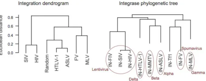
Choice of the integration site is an essential step in retroviral life cycle once it influences the following replication process and consequently the virus’ fate. In fact, clustering of different viruses on the basis of their integration preferences shows a high degree of overlaping with phylogenetic trees based on the sequence similarity of their integrases, which, in turn, are in good agreement with traditional trees based on genomic sequences (Derse, Crise et al. 2007). This emphasises that the survival of a retrovirus cannot but depend on the site in the host cell genome it integrates in.
Figure 1. Retroviruses’ phylogenetic trees based on integrase sequence and integration preferences.
(A) Dendrogram based on location of integration in relation to 69 genomic features. Unsupervised
hierarchical clustering, with euclidean distance and average linkage was used to generate the dendrogram. (B) Phylogenetic tree with integrase sequences, including P31822 (FIV), P03365 (MMTV), and AAA35339 (Tf1) integrases, showing that the three integrases have been placed into different clusters. (Derse, Crise et al.2007)
While theDNA breaking and joining reactions mediatingintegration are well understood, integration site selection is still a largely unknown molecular mechanism. A deeper understanding of the subject would have a double impact both on basic retrovirology as well as on its clinical
9
applications (new targets for antiretroviral drugs and site directed gene-therapy).Early studies considered integration a random process since there were no obvious patterns in the integration sequences, and disruption or activation of genes was thought to be a rare event. Nevertheless, some factors presumably involved in the enhancement or reduction of integration were identified, such as DNA bending induced by nucleosomal assembly, steric hidrance of DNA binding proteins and DNA physical structure. However, the assumption of random integration was reconsidered after reported high frequency events of oncogene activation in a gene therapy clinical trial of X-linked severe combined immunodeficiency (X-SCID) using retroviral vectors. 5 out of 8 patients developed leukaemia as the result of insertional activation of the oncogene LMO2 in 2 independent clinical trials, giving proof that the integration site-selection process is far from being random (Hacein-Bey-Abina, Le Deist et al. 2002; Hacein-Bey-Abina, von Kalle et al. 2003; McCormack and Rabbitts 2004).
Since complete human genome sequencing has become available, it has been possible to study in statistically rigorous manner retroviral integration by sequencing the junctions of provirus and the host’s genome in genome-wide studies. Several large scale, high-throughput methods were designed and, more recently, massive parallel sequencing techniques were adapted to increase the output of integration studies (Margulies, Egholm et al. 2005; Wang, Ciuffi et al. 2007; Wang, Garrigue et al. 2008). Once acquired, integration data can be correlated with several genomic features in an attempt to find patterns and preferences of site-selection. Since no significant differences in terms of integration distribution were found between wild-type viruses and the vectors derived from them, the latter, for practical and safety matters, are most commonly used in insertion studies. The results of these large-scale surveys have been so far very revealing, not only uncovering genomic features systematically and specifically associated with retroviral insertions, but also pointing out that each retrovirus has a unique, characteristic pattern of integration within the human genome. Nevertheless, these results still fail on providing an explanation to all viral integrations and a more absolute answer as to which mechanisms are in fact involved in the virus choice to integrate. Therefore, retroviruses still remain largely unpredictable and more studies are needed to uncover its biology.
- A brief review of retroviruses and retroviral vectors
Retroviruses comprise a large family of enveloped RNA virus with virions of 80-100 nm in diameter and an outer lipid envelope that incorporates and displays the viral glicoproteins. Their RNA (7-12 kb) is linear, single-stranded, nonsegmented, and of positive polarity. The hallmark of the family is its replicative strategy, which includes as essential steps after cell entry, reverse transcription of the virion RNA into linear double-stranded DNA and the subsequent integration of viral DNA into the cell’s chromatin.
10
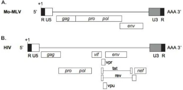
Retroviruses are broadly divided into simple and complex, distinguishable by the organization of their genomes. All retroviruses contain 4 major coding domains with information for virion proteins: gag, for the matrix, capsid and nucleoprotein structures; pol, for the reverse transcriptase and integrase enzymes; pro, for the protease enzyme; and env, for the viral envelope protein. In addition to the basic coding domains, complex retroviruses also encode several accessory genes, derived from multiple splicing. Both ends of all viral genomes contain terminal noncoding sequences, composed of 5’ and 3’ unique sequences (U5 and U3 regions), and of two direct repeats (R) where the transcription start site and the polyadenylation signal are located (Coffin, 1997).Figure 2. Simple and complex retroviral genomes. (A) Moloney murine leukaemia virus genomic RNA is
only made up of four elementary coding regions, gag, pro, pol, and env. Terminal noncoding R, U5 and U3 regions are depicted; transcription start site (+1) and polyadenylation signal (AAA) are specified. (B) Human immunodeficiency virus is a complex retrovirus, with six accessory, partially overlapping genes (vif, vpr, tat,
rev, vpu and nef) in addition to the four basic coding domains (Cattoglio, PhD dissertation).
Based on evolutionary relatedness, retroviruses are further classified into seven genera. Moloney murine leukaemia virus (Mo-MLV) and human immunodeficiency virus-type 1 (HIV-1), object of this thesis, belong to the genus of gamma-retroviruses (also known as oncoretroviruses) and of lentiviruses, respectively. Murine leukaemia viruses (Mo-MLV), are known to induce tumours in murine hosts and other vertebrates. Members of this family can either be endogenous or exogenous. In its replicative form, MLV has a single-stranded positive sense RNA genome that replicates via a DNA intermediate by the process of reverse transcription. HIV is a member of the Lentiviridae family, the name lentivirus means slow virus, so called because these viruses take a long time to cause exposed disease. Most lentiviruses target cells of the immune system and thus disease is often manifested as immunodeficiency. Primary targets of HIV are activated CD4+ T4
11
helper lymphocytes but the virus can also infect several other cell types including macrophages. Its genome, like MLV’s, is made of positive sensed RNA but, as a complex retrovirus, it is also composed of accessory genes namely vif, vpr, tat, rev, vpu and nef, in addition to the four basic coding domains (gag, pro, pol and env).These genes encode small proteins and overlap with the structural genes (especially ENV) but are in different reading frames. Mutants in the TAT and REV genes show that both proteins are necessary for virus production (Meredith, 2009).Life cycle of retroviruses and its essential interactions with the host can be summarized in five main steps. First, the host cell must express a specific receptor on its surface to provide a site for the virus to bind and trigger the entry process, mediated by the viral Env protein. Second, the cell must supply deoxynucleotides in adequate concentration for the virion reverse transcriptase to transform the RNA genome into DNA. Third, there must be a means for the viral DNA to access host chromosomes as targets for viral integration; integration may also require the aid of host repair enzymes. Fourth, host machinery and components are necessary to express viral RNA and carry out the processing (polyadenylation and splicing) and transport of both viral genomic and messenger RNA to the cytoplasm. Finally, host cell machinery is necessary for the synthesis, folding, modification, and transport of viral proteins to the membrane assembly sites (Coffin, Hughes and Varmus 1997).
The integration process, catalyzed by the viral integrase protein, takes place in the context of the preintegration complex (PIC), a nucleoprotein agglomerate formed after double stranded DNA synthesis that contains, in addition to the virus genetic material, the viral proteins integrase, nucleocapsid, virion protein R and matrix, as well as specific cellular proteins (Coffin, 1997). After completion of viral DNA synthesis, the integrase removes two nucleotides from the 3’ end of both strands of viral DNA, adjacent to a conserved CA dinucleotide, generating recessed 3’-hydroxyl groups; in the subsequent cleavage-ligation reaction, the processed 3’-hydroxyl ends are joined to protruding 5’ ends of the target DNA. Complete integration is achieved when cellular enzymes repair gaps at each host-virus DNA junction, resulting in a 4 to 6 base pair repeat in the host DNA, flanking each proviral end.
Retroviruses used in this study are retroviral vectors also applied for therapeutic purposes in the correction of genetic diseases, therefore, even though they keep the same integration pattern as wild type viruses, they differ in that they lack their basic coding elements. In this case, retroviral proteins are provided in trans by packaging cells so that the vectors are replication-incompetent, only the minimal viral elements required for high efficiency transfer are retained. The cis-acting elements essential for retroviral replication, together with the viral proteins (retrotranscriptase, integrase and protease) packaged within the virions make viral replication cycle possible. Essential elements still maintained in the viral genome include a promoter and a polyadenylation signal for viral genome production in the packaging cell, a packaging signal for incorporation of vector RNA into virions, signals required for reverse transcription and short
12
repeats at the termini of viral LTRs necessary for integration. All the intervening genomic material can be replaced with the sequence of interest, to accommodate up to 10 kb of heterologous DNA, in our case a reporter gene.- Retroviral integration features: state of the art
Several features have been investigated for a role in integration site-selection, one of the first and most intuitive was the sequence at target DNA sites.
Although the viral sequence required by integrase is already known (a dinucleotide CA is invariably positioned exactly 2 bp from both ends of the viral termini and the significant roles of the sequences internal to the CA dinucleotide extending for up to 15 bp also play a significant role), the sequences present at target DNA sites seem very diverse. In a recent study, Wu et al. discovered a statistically weak palindromic consensus at integration target sites while examining a large number of sequences from several retroviruses, including HIV-1, SIV, MLV, and ASLV. This consensus is weakly conserved but distinguishable between different retroviruses. The most probable hypothesis is that this conserved sequence is needed to meet the spatial or energy requirements of the integration complex, rather than the most favourite sequence at each base.
Although the host cell DNA sequences hosting integration events show some detectable similarity to one another, this similarity is very modest thus, retroviral DNA integration is not tightly sequence-specific (Stevens and Griffith 1996; Carteau, Hoffmann et al. 1998; Wu, Li et al. 2005; Berry, Hannenhalli et al. 2006).
Taking advantage of the large number of genome browsers and databases available, integration sites can be correlated positively or negatively with any genomic feature annotated, by comparison of the viral integration frequency near that feature to the frequency expected from random integration. Several genomic features were shown to have a correlation with integration site-selection: coding and non-coding transcription units, CpG islands, centromeric regions, repetitive elements, fragile sites, transcription factor binding sites (TFBS).
Another interesting approach is to correlate viral integration with cell-specific features, like gene expression, epigenetic modifications, and others that reflect cell-specific transcriptional regulatory pathways. Unfortunately, only a limited number of datasets for some cell-types is already available, therefore, one should be cautious in choosing the appropriate dataset to use for correlation with integration data.
For their clinical relevance, Mo-MLV and HIV-1 are the most studied retroviruses, having also the most largely and extensively studied integration pattern.
Mo-MLV showed to have a preference for active genes and particularly for regions with a role in transcription regulation like transcription start sites, CpG islands, DNase I hypersensitive sites and transcription factor binding sites. With a distinctively different pattern from Mo-MLV,
HIV-13
1, seems to have a negative correlation with regions involved in transcriptional regulation. It shows to have a preference to target genes in highly active regions of chromatin. These distinct patterns of integration imply a very different viral strategy for survival. One way to interpret these data is that, Mo-MLV chooses to integrate in regions that assure its transcription and further processing. The deregulation of some classes of genes resultant of Mo-MLV integration confers some growth and/or survival advantage for the virus, leading to their in vivo amplification. On the other hand, HIV-1 prefers to integrate in chromatin regions that, even though assure its survival and proliferation, since these are highly conserved and active, are not involved in transcriptional regulation and thus not prone to gene deregulation. This strategy implies a longer period of survival for the host and, consequently, a higher chance of viral propagation to a large number of hosts.- Proposed mechanisms for integration site selection
It has been proposed that integration is favoured in regions of open chromatin once MLV tends to integrate near DNase I hypersensitive-sites, maybe because these regions might be more accessible to the pre-integration complex (PIC) (Figure 3). This hypothesis has been supported by evidence of preferential integration in transcriptionally active chromatin and disfavoured integration in the centromeric heterochromatic region. However, only MLV strongly favoured integration near to these sites and the diversity of genomic targets both in MLV and among other retroviruses implies more complex and distinct mechanisms involved in integration site-selection for each family.
A different model invoked the effect of cell cycle in the mechanism for integration targeting (Figure 4). HIV can infect cells regardless of the cell-cycle phase, while MLV requires the host cell to pass through mitosis. The transcriptional state of a cell is known to vary with the cell cycle, so
Figure 3. Proposed mechanisms that direct integration site selection by retroviruses- accessibility of target DNA. According to this model, chromosomal DNA is
relatively inaccessible to integration complexes when packed in nucleosomes and other proteins. Exposure of target sequences promotes integration. (Bushman, Lewinski et al. 2005)
14
the organization of chromosomal DNA encountered by HIV and MLV complexes should differ. However, HIV constructs with cell-cycle restricted infectivity showed a very different integration pattern in relation to MLV, even though the pattern was also different from the wild type HIV. These results suggested that cell cycle does play some role in retroviral targeting but other factors seem to dominate. Additionally, studies of HIV integration targeting in non-dividing cells and in dividing cells didn’t show significant differences, providing more evidence against this targeting model.Another candidate mechanism for integration is the tethering of PIC to the genome by cellular or viral proteins (Figure 5). Through binding of the PIC to this (or these) supposed protein(s), the complex would be directed to a specific location in the genome where it would eventually integrate. Several cellular proteins have been isolated as physically bound to viral PICs, some common to both HIV and MLV and others specific, also for some of them, association occurs via direct interaction with the viral integrase. Some of these proteins studied so far will be described in the next paragraphs.
Kalpana et al. 1994 used the HIV-1 integrase protein as “bait” to screen for cellular proteins that might participate in viral DNA integration. Their screen yielded a cDNA clone encoding a protein that binds specifically to integrase both in vivo and in vitro. The integrase-binding protein, designated integrase-interacting protein 1 (Ini1), displays a high degree of sequence similarity to the yeast protein Snf5, implicated in the transcriptional activation of a number of genes. They also found that, at certain concentrations, Ini1 increased the efficiency of integration (Kalpana, Marmon et al. 1994).
Farnet et al. 1997, presented data indicating that the high mobility group protein HMGA1 (previously known by HMGI(Y)) is required for function of HIV-1 preintegration complexes (PICs) isolated from infected cells. Integration activity was lost from PICs following treatment with 600mM KCl. This activity could be restored, however, by addition of extracts from uninfected SupT1 cells, suggesting that a host activity might be required. Screening for the complementing protein yielded
Figure 4. Proposed mechanisms that direct integration site selection by retroviruses - timing of nuclear entry during the cell cycle. To
enter the nucleus, MLV requires the passage of cells through mitosis, whereas HIV can enter cells at different stages of the cell cycle. If the state of chromosomes differs at different points in the cell cycle, this could influence integration targeting (Bushman, Lewinski et al. 2005).
15
HMGA1. Analysis of protein composition by western blotting revealed that HMGA1 was present in PICs, but depleted from PICs by high salt treatment. Purified HMGA1 alone was not sufficient to carry out integration when mixed with purified HIV-1 cDNA, supporting a model in which HMGA1 is required as an accessory factor for the function of HIV-1 PICs. This work shows that the function of HMGA1 in integration seems to be related to one of the steps of integration process, possibly the covalent strand transfer step, rather than to PIC targeting in the genome. Lin et al. 2003, also studied the HMG 1 family proteins in Mo-MLV, finding a similar role to the one found in HIV-1. The outcome of this study also suggested that binding of multivalent HMGA1 monomers to multiple cDNA sites compacts retroviral cDNA, thereby promoting formation of active integrase-cDNA complexes. HMGA1 has not been found to bind to integrase directly, consistent with models in which HMGA1 acts by binding to the cDNA (Farnet and Bushman 1997).Barrier-to-autointegration factor (BAF) is a homodimeric protein with 89 amino acid residues and is highly conserved among species. This protein was found to protect viral DNA from autointegration both in Mo-MLV and HIV. Furthermore, it was found that BAF can promote the association of PICs with target DNA. In vitro studies have revealed that BAF bridges double-stranded DNA with no detectable sequence specificity. DNA bridging results in intramolecular compactation at low DNA concentrations and intermolecular aggregation at high DNA concentrations. Therefore BAF’s activity may have two different outcomes: intramolecular bridging may compact the viral DNA into a rigid structure, making it less accessible as a target for autointegration and anchoring of PICs to other DNA may promote intermolecular integration in target DNA (Suzuki and Craigie 2002; Lin and Engelman 2003).
The human protein Rad18 is also known to interact with integrase. This interaction results in an increased stabilization of integrase, which in its natural form is a particularly unstable protein. The re-localization of integrase and its co-localization with Rad18 in a subset of cells suggests an additional function for this association. Human Rad18 contains a putative SAP-box, a domain recently recognized to mediate the binding of certain proteins to specific A/T-rich DNA regions known as the scaffold attachment regions (SAR). Interestingly, PARP-1, Ku antigens, and HMGA1, which are involved in retroviral integration, have all been found to be SARbinding proteins. An intriguing possibility is that the molecules relevant for HIV-1 integration cluster together, perhaps in the vicinity of SARs, achieving in this way the coordination required for these complex reactions (Mulder, Chakrabarti et al. 2002).
Human embryonic ectoderm development protein (EED), a member of the superfamily of WD-40 repeat proteins, and of the Polycomb group proteins, has been identified as a cellular partner of the matrix (MA) protein of HIV-1. EED was also found to interact with the integrase both in vitro and in vivo in yeast. EED-binding site(s) are located in the C-terminal domain of the integrase, between residues 212 and 264. In EED, two putative discrete integrase-binding sites were mapped to its N-terminal moiety, at a distance from the MA-binding site, but EED-integrase
16
interaction also required the integrity of the EED last two WD repeats. EED showed an apparent positive effect on integrase-mediated DNA integration reaction in vitro, in a dose-dependent manner (Violot, Hong et al. 2003).Poly(ADP-ribose)polymerase-1 (PARP-1) is a nuclear protein mainly associated with chromatin, and is proposed to be involved in the process of DNA repair, including the repair of gapped intermediates generated during retroviral integration. This protein accumulates at the active mammalian centromere on metaphase chromosomes, and is associated with centromeric DNA and proteins, suggesting that PARP-1 and poly(ADP-ribosyl)ation reaction may be involved in the regulation of centromere function. Kameoka’s et al. results suggested that poly(ADP-ribosyl)ation, although it’s not required for efficient HIV-1 integration, seems to be necessary during integration near the centromere region. Even though a low frequency of integration has been shown in centromeric DNA, these rare events represent an important retroviral mechanism of survival since integration in these sites leads to the establishment of a latent infection that can be reactivated (Kameoka, Nukuzuma et al. 2005).
The most studied cellular tethering factor so far, is the lens epithelium derived growth factor (LEDGF) or p75, an ubiquitously expressed nuclear protein, tightly associated with chromatin throughout the cell cycle. It is a transcriptional coactivator involved in stress response, autoimmune disease, cancer and HIV replication. This protein was shown to be a strong interactor of the HIV-1 integrase and to stimulate its catalytic activity in vitro. It binds at its C-terminus to lentiviral IN protein dimers while the N-terminal half binds to chromatin (Llano, Vanegas et al. 2006; Engelman and Cherepanov 2008). When LEDGF/p75 was depleted from cells using RNA interference, integration in transcription units was diminished, documenting a role in integration targeting (Ciuffi, Mitchell et al. 2006). LEDGF/p75-responsive genes were identified by transcriptional profiling and found to be favored integration targets for both HIV (Ciuffi, Llano et al. 2005) and another lentivirus, feline immunodeficiency virus (Kang, Moressi et al. 2006). Correlation analysis with genomic features revealed an association with active chromatin markers, such as H3 and H4 acetylation, H3K4 monomethylation and RNA polymerase II binding. Interestingly, some associations did not correlate with HIV-1 integration indicating that not all LEDGF/p75 complexes on the chromosome are prone to HIV-1 integration (De Rijck, Bartholomeeusen et al.). Moreover, in cells depleted for LEDGF/p75, HIV integration was still favoured in transcription units. Thus additional factors may be involved in guiding HIV integration (Ciuffi, Llano et al. 2005).
17
Although essential for PIC integration activity, interaction with cellular proteins doesn’t explain by itself the integration characteristics of Mo-MLV. Integrase has an essential role targeting sites for MLV integration. Proof of this principle comes from experiments using an HIV vector packed with a MLV integrase, which gains preference for MLV integration sites (regulatory regions with transcription start sites (TSSs), CpG islands and TFBSs). Additionaly, the high integration near TFBSs is dramatically reduced when the U3 transcriptional enhancer is deleted from the Mo-MLV LTRs. These results indicate that the integrase and the U3 enhancer are the major viral determinants of Mo-MLV selection of regulatory regions in the genome. A plausible explanatory model is that cellular transcription factors binding the Mo-MLV U3 enhancer cooperate with the integrase in directing PICs towards regions actively engaged by the transcriptional machinery. Accordingly, some of the TFBSs enriched around Mo-MLV integrations are consensus motifs for transcription factors already known to bind the U3 enhancer and drive proviral expression after integration (e.g., members of the ETS family and the bivalent YY1 transcription factor).
This thesis analyzes a collection of retroviral integration sites retrieved from primary human keratinocytes at an early time point after infection, when clonal selection in culture is very unlikely to have occurred. Epithelial keratinocytes are exposed to retroviral infection, for example during oral-sexual contact and breast-feeding, making the epithelium a potential site of primary retroviral infection and dissemination. Even though these cells do not express the common receptor and co-receptors found on permissive cells, integration is shown to occur and persist in daughter cells after keratinocytes’ division (Vacharaksa, Asrani et al. 2008). After integration the life cycle of the virus aborts and no newly assembled virus particles are detectable, although, captured infectious viruses are harboured for at least 48h and transferred to blood mononuclear cells (Vacharaksa, Asrani et al. 2008). Epithelial cells can, therefore, be actively providing a route to systemic retroviral infection, making them an interesting target to study viral integration mechanisms.
The general goal of this work was to describe the integration preferences of retroviruses in the genome of these clinically relevant cells, perform a comparative analyses with previously
Figure 5. Proposed mechanisms that direct integration site selection by retroviruses - tethering by cellular proteins.
This model proposes that specific interactions between integration complexes and cellular proteins bound locally on target DNA promote integration at nearby sites. (Bushman, Lewinski et al. 2005)
18
obtained results for other cell-types and possibly provide new insights into the molecular mechanisms responsible for differential retroviral integration targeting. Furthermore, we intended to evaluate the safety parameters related to the use of these viral vectors in protocols of gene therapy for genetic skin diseases, the main research line followed in the laboratory.19
METHODS AND MATERIALS1. Virus production and cell culture
The vectors used were gamma-retroviral MFG.GFP and lentiviral derivated pRRLsin-18.ppt.K14.GFP.WPRE. Briefly, MFG.GFP vector expressing the EGFP cDNA under the control of the Moloney leukaemia virus LTR was constructed in the MFG backbone and integrated in the amphotropic Gp+envAM12 packaging cell line. The viral supernatant used for keratinocytes infection was collected from one clone of Am12MFG GFP packaging cell line. The VSV-G-pseudotyped pRRLsin-18.ppt.K14.GFP.WPRE lentiviral vector, containing a K14 promoter-driven GFP expression cassette, was produced by transient co-transfection of 293T cells with a second generation packaging system. Viral supernantant collected from transfected 293T cells was then concentrated by ultracentrifugation in order to increase the viral titer. Transfection efficiency was evaluated by analysis of GFP expression by flow cytometry 48 hours after infection.
2. Transduction of cells
For retroviral transduction subconfluent primary keratinocytes (taken from male newborns foreskin) were trypsinized and plated (2x10⁵ cells) in multi-6 wells plates, without feeder layer, in
Figure 6. General overview of the methods
used to analyze retroviral integration sites in the human genome (Bushman, Lewinski et al. 2005)
20
the presence of KBM (Lonza) medium supplemented with glutamine (2%) insulin (5 μg/ml) adenite (0.18mM) hydrocortisone (0.4 μg/ml) and EGF. Retroviral transduction was performed by spinoculation (3 rounds at 1,800 rpm for 45min) in the presence of 8 μg/ml polybrene.For lentiviral transduction subconfluent primary skin keratinocytes were trypsinized and 105 cells resuspended into 2 ml of Kno medium containing the retroviral virus at MOI of 8-10 in the presence of 8ug/ml polybrene. The transduction mixture containing keratinocytes, virus and polybrene was then plated on lethally irradiated 3T3-J2 cells. The medium was replaced after 5 hours. Transduced keratinocytes were grown to confluence, trypsinized and re-plated onto new feeder-layers for further analysis. Transduction efficiency was evaluated by analysis of GFP expression by flow cytometry 48 hours and two weeks after infection.
3. Cloning and analysis of viral insertion sites
Genomic DNA was extracted and digested with MseI. 3’ LTR vector genome junctions were amplified by linker-mediated PCR (LM-PCR) (Cattoglio et al., 2007), adapted to the GS-FLX Genome Sequencer (Roche/454 Life Sciences, Branford, CT) pyrosequencing platform. For each transduction, we performed 2 restriction digestions, 6 linker ligations and 18 nested PCRs, with nested primers specific for the linker and the 3’ LTR containing a bead-capture tag and a sequencing tag. A 4-nucleotide multiplex tag was also added to the 3’ LTR nested primer downstream of the sequencing tag to discriminate between different samples (see Supplemental methods). Pooled LM-PCR amplicons were quantified (NanoDrop Technologies, Wilmington, DE), checked by an Agilent Bioanalyzer (Agilent Technologies, Palo Alto, CA), size-fractionated by SPRI beads (Agencourt Bioscience Corporation, Beverly, MA), and sequenced according to the GS-FLX manufacturer's instructions.
4. Sequence analysis
All UCSC Known Genes having their transcription start site (TSS) at ±50 kb from an integration or random site were annotated as targets, as done on previous works (Cattoglio et.al, 2007). In case of multiple transcript variants, we arbitrarily chose the isoform with the nearest TSS to an integration or random site. For each site, we annotated the genomic features (CpG islands, conserved non-coding sequences, conserved TFBSs) whose hg18 coordinates overlapped for at least 1 nucleotide with the ±50 kb interval around the insertion site. We used UCSC tracks (http://genome.ucsc.edu) for both CpG islands (27,639 items) and conserved TFBSs (3,807,783 items). Genomic coordinates of 82,335 mammalian CNCs were described (Kim and Pritchard 2007). To generate a matched control dataset, we randomly extracted 20,000,000 sites from the human genome and discarded sites with the nearest MseI recognition site (TTAA) at <20 bp (the minimum requirement for a blast search) or >500 bp (the maximum estimated length for efficient
21
454 bead loading). The resulting 14,260,000 sequences were processed through the same bioinformatic pipeline used for integration sequences, ending with a library of 11,655,601 unique sites.5. Gene expression profiling
The expression profile of primary keratinocytes was determined by microarray analysis. RNA was isolated from 1-2 x 10⁶ primary human keratinocytes, transcribed into biotinylated cRNA and hybridized to Affymetrix HG-U133A Gene Chip arrays according to the Affymetrix instructions. Scanned images were processed by the Affymetrix GCOS suite, and transcript levels determined with the GCOS absolute analysis algorithm. To correlate retroviral integration and gene activity, expression values from the keratinocytes microarrays were divided into four classes, i.e. absent, low (below the 25th percentile in a normalized distribution), intermediate (between the 25th and the 75th percentile) and high (above the 75th percentile).
6. Functional gene analysis
Genes were analysed by the network-based Ingenuity pathways analysis tool (Ingenuity Systems, www.ingenuity.com). Gene identifiers were uploaded into the application, and mapped to their corresponding Focus Gene in the Ingenuity Pathways Knowledge Base. Networks were algorithmically generated based on the direct or indirect interaction between Focus Genes. The functional analysis of each network identified the biological functions and/or diseases that were most significant to the genes in the network (Bonferroni correction).
22
ResultsThe retroviral vector used for this study is an MFG-based retroviral vector expressing the eGFP cDNA under the control of Moloney leukemia virus LTRs; the lentiviral vector is a SIN-18 vector in which the expression of the same cDNA is under the control of the internal full-length K14 promoter (for maps of retroviral vectors see Appendix).
Human primary keratinocytes from healthy donors were transduced with the gamma-retroviral and lentiviral vector with an efficiency of 60% and 80% EGFP positive cells, respectively. Transduced cells with lentiviral vector were cultivated onto a feeder layer while transduced cells with gamma-retroviral vector were cultivated directly on plastic dishes to avoid murine feeder contamination (since murine cells are the original host of these viruses). Genomic DNA was extracted and vector-genome junctions were cloned and sequenced by a LM-PCR approach adapted to the different vector types (Cattoglio et al., 2007) and mapped onto the human genome. Cumulatively, we mapped 3020 MLV and 1601 HIV independent integrations.
Analysis of the distribution of viral integrations along the genome was performed on my dataset originated from primary human keratinocytes. Furthermore, my data was compared with previous datasets of Cd34+ hematopoietic stem cells (32631 MLV hits and 28382 HIV hits) from Cattoglio et al. Blood in press and lymphocytes (8277 MLV hits) from Recchia et al. unpublished and (7780 HIV hits) from Wang et al., 2007.
Retrovirus integrate preferably inside genes
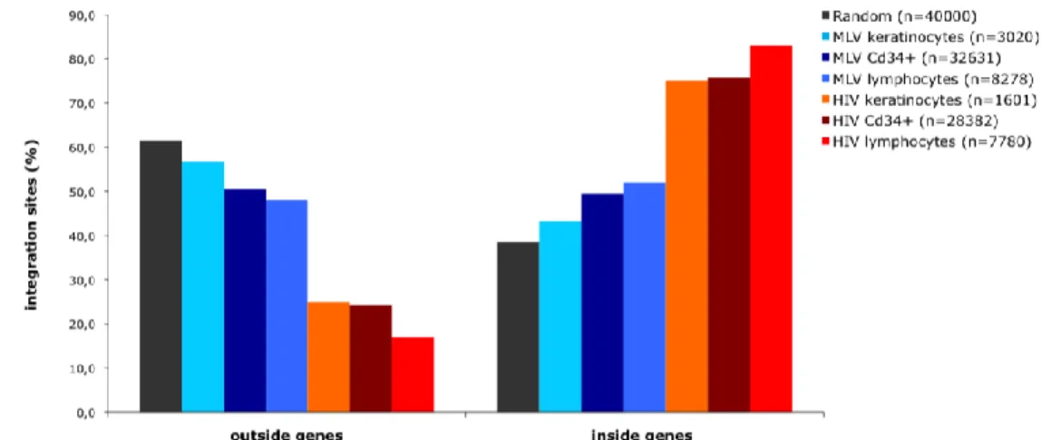
Viral and random integrations along the genome of the three cell-types, were classified as being inside genes or outside genes. Figure 7 represents the distribution as a % of the total number of integrations, showing that HIV has a high tendency to integrate inside genes 75% in keratinocytes and Cd34+HSC and a even higher preference in lymphocytes with 83% of integrations inside genes (p < 2.2e-16).
Figure 7. Distribution of MLV, HIV and random integration sites with respect to Known Genes (UCSC
definition) on primary human keratinocytes, lymphocytes and Cd34+HSC.
23
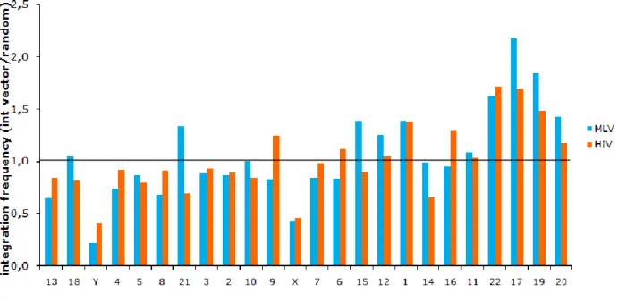
proportions with continuity correction, Rweb 1.03) preference to integrate inside genes than would be expected from a random distribution through the genome in all cell-types, although, with a significantly higher freuency in lymphocytes and Cd34+HSC in relation to keratinocytes with 52%, 49% and 43% of integrations inside genes, respectively (p 0,0001815). Random integrations were 38,5% inside genes and 61,5% outside, reflecting mostly the gene content of the human genome.Following the previous results, we hypothesized that chromosomes with an overrepresentation of viral integration would be the ones with higher gene density. To verify the proposed hypothesis, integration frequency along chromosomes of keratinocytes was analysed by comparison with the random distribution and then correlated with values of gene density for each human chromosome. In fact, we could confirm an association between viral integration and gene density, since the 4 more gene dense chromosomes (17, 19, 20, 22) (data on chromosome gene density taken from NCBI http://www.ncbi.nlm.nih.gov/genome/guide/human/) perfectly overlapped with the 4 more targeted ones (Figure 8), however, the variation observed in all the chromosomes could not be explained solely by the gene density parameter.
Figure 8. Frequency of integration of MLV (blue bars) and HIV (orange bars), in relation to the control,
according to the gene density of chromosomes in primary human keratinocytes.
MLV is attracted to TSS proximal regions while HIV is repelled
Single integrations were then classified into three groups (Figure 9), TSS proximal when located at a distance of +/-2,5 kb from the TSS of any gene (UCSC Known Gene track), intragenic when located inside a gene but not near a TSS and intergenic when outside any gene and at a distance higher then 2,5 kb from any TSS.
24
Figure 9. Distribution of MLV, HIV and random integration sites with respect to Known Genes (UCSC
definition) on primary human keratinocytes, lymphocytes and Cd34+HSC.
MLV showed a higher integration in TSS proximal regions in comparison to the control (p < 2.2e-16) in all cell-types. Lymphocytes showed the highest bias towards TSS regions accounting for 30,6% of all integrations, followed by Cd34+HSC with 22,3% and the lowest frequency for keratinocytes with 10,5% of integrations in this region. In contrast, HIV integrations were disfavoured by TSS proximal regions in keratinocytes with only 1,6% (p = 0.000291) of total integrations and had a similar distribution to the control in the other two cell-types, 3%.
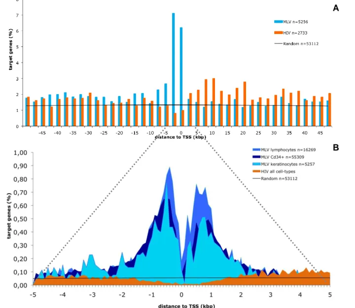
To further look at this particular feature, all integration events occurring within 50kb from the TSS of any Known Gene were plotted in 2,5 kb intervals in either direction of the TSS (Figure 10 A). MLV showed a high incidence of integrations within 5 kb from the TSS while HIV showed a moderate but statistically relevant higher incidence of integration downstream of the TSS confirming it’s favouritism for intragenic integrations. Mapping at a closer distance from the TSS at 5kb in both directions (Figure 10 B), revealed that MLV is attracted to TSS regions while HIV seems to be repelled by the same regions. Both MLV and HIV have a mirror plot with MLV increasing the number of integrations near the TSS while a decrease of integration is shown by HIV. In the region close to the TSS MLV shows a sudden drop of integration, in lymphocytes and Cd34+HSC this occurs in the 0bp to 100bp region while in keratinocytes the same drop is localized in the region 100bp downstream from the previous ones. A second drop is seen in the region from 300bp to 400bp from the TSS in lymphocytes and, once again, 100bp downstream in keratinocytes, but is not observable in Cd34+HSC. These drops in integration frequency are possibly due to occupancy from the basal transcription machinery. Both of these distinct characteristic patterns of integration represent a viral integration distribution non-dependent on cell specificities once it was observed in all the studied cell-types. Although, a significant higher frequency of integration by MLV was observed in lymphocytes, mainly at a distance from -1,7 to
25
1,7 kb from the TSS of these cells. HIV’s distribution showed no differences between all the studied cell-types. Random integrations were evenly distributed throughout the analysed region.Figure 10. Distribution of retroviral integrations around transcription start sites. The % of the total
number of targeted genes (n) is plotted on the Y axis. The black line indicates the distribution of control random sites. (A) Distribution of the distance of MLV (blue bars) and HIV (orange bars) integration sites in primary human keratinocytes from the transcription start site (TSS) of targeted genes at 50kp resolution. (B) Comparative analysis of distribution of retroviral integrations around transcription start sites on primary human keratinocytes, lymphocytes and Cd34+HSC at 5kbp resolution.
Retroviruses target mostly active genes
To understand if the genes targeted by retrovirus, in particular those with TSS proximal integrations, were active, an Affymetrix microarrays run (HG-U133 plus 2.0) was performed in triplicate to determine the expression profile of over 18,900 genes in keratinocytes activated in culture in the same conditions used for retroviral transduction. Affymetrix probe sets were re-annotated with custom CDF files according to the Bioconductor indications (Dai, Wang et al. 2005),
A
26
to obtain a single expression value for each gene. Expression levels were divided into four classes: absent (black portion of histogram bars), low (below the 25th percentile of the normalized distribution, yellow), intermediate (between the 25th and the 75th percentile, orange) and high (above the 75th percentile, red). The percentage distribution of the expression values of genes targeted by all integration/random sites (all ISs), TSS-proximal sites (TSS-proximal ISs) and intragenic sites (intragenic ISs) are shown by the left, middle or right group of bars, respectively.In keratinocytes, both MLV and HIV showed a significant preference for active genes, once 69% and 74% (p-value < 2.2e-16) respectively of targeted genes were active in all insertion sequences, compared to 58% of the random. A even bigger difference was revealed by MLV’s integration in TSS proximal regions with 85% (p-value < 2.2e-16) of active genes and 87% (p-value = 9.421e-10) active in intragenic integrations as opposed to 60% of activity both in TSS and intragenic regions with the random. HIV had no significant preference for TSS proximal active genes but showed an evident preference for active genes part of intragenic integrations (p-value < 2.2e-16).
Results previously obtained for Cd34+HSC (Cattoglio et al., Blood in press) revealed the same overall preference for active genes. In comparison to the actual data, we can suggest that there’s a even higher preference for HIV to integrate in active genes among the intragenic integrations in Cd34+HSC than in keratinocytes with 88,5% and 73% of targeted expressed genes, respectively.
Figure 11. Association between retroviral integration and gene activity in primary human keratinocytes. Histogram distribution of expression values from an Affymetrix microarray analysis of RNA
obtained from primary human keratinocytes. The number of genes belonging to each category is indicated in parenthesis under the correspondent bar. Genes targeted more than once are considered more than once for the analysis.
27
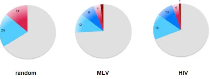
Moreover, with the Affymetrix data for gene expression in Cd34+HSC cells we analysed the proportion of genes hit (intronic and exonic integrations) by both viruses and the random dataset in comparison to the total number of genes in the genome. A significantly lower number of non-expressed genes were targeted by both MLV and HIV, corresponding to 6% and 5%, respectively, of the genome in comparison to 14% of the random distribution. No differences were seen in the proportion of expressed target genes by MLV and the random which corresponds to 20% of genes in the genome, however a significant difference was observed for HIV which targeted 26% of expressed genes (p-value < 2.2e-16). Globally MLV targeted less genes with a total of 24% in comparison to the random and HIV which targeted 34% and 31%, respectively.Regions highly targeted by retroviruses, defined as clusters of integration, were also analysed, comprising 10% of the genes in the genome in both viruses. Expressed genes hit by clusters accounted for 8% and 10% of the genes in Cd34+ cells for MLV and HIV, respectively, while non-expressed genes hit by clusters represented only 2% and 1% of the total number of genes in the genome.
Figure 12. Proportion of genes targeted by random, MLV and HIV distribution, in the genome of human Cd34+ cells. Blue represents targeted expressed genes, red represents targeted non-expressed
genes and grey non-targeted genes. Dark blue and red show the proportion of genes targeted in clusters.
MLV’s clusters are more packed while HIV’s expand through a wider chromatin region To analyse features present in highly targeted regions, clusters of viral integration in the genome were defined taking in consideration the size of the dataset and the distance from one integration to the consecutive one. A maximal distance between consecutive integrations corresponding to a false discovery rate of 0.01 was statistically established to define if an integration is inside or outside a cluster. For the keratinocytes dataset a distance of less or equal to 9,905 bp and 17,318 bp between two consecutive integrations was considered for MLV’s and HIV’s integrations, respectively. For the Cd34+HSC dataset we considered a window of three
28
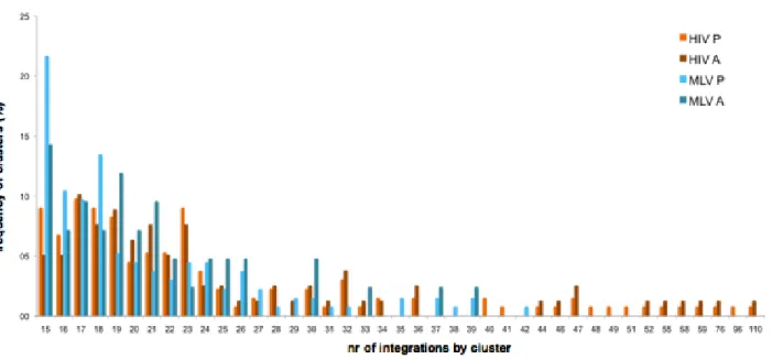
With this definition, 320 clusters for MLV and 102 for HIV in keratinocytes were identified, containing 23.6% (714) and 13.6% (218) of the total integration sites. For the Cd34+HSC dataset 3,497 MLV clusters were identified comprising 65.3% of all integrations and 2,446 HIV clusters with 50.6% of the total number of integrations.The size of clusters was very different between the two viruses, MLV’s clusters being more compact and HIV’s spreading through a larger region of the chromatin. Differences in clustering pattern were even more evident in the larger dataset for Cd34+HSC, where cluster size varied in length from 3,199 bp to 78,534 bp for MLV and from 7,271 bp to 200,508 bp for HIV’s clusters. Furthermore, HIV’s clusters have a maximum of 110 integrations by cluster while MLV’s maximum is less than half, with 42 integrations by cluster in Cd34+HSC.
Figure 13. Cd34+ cells cluster analysis. Graph shows the frequency of clusters in relation to the number of
integrations by clusters in both MLV (blue bars) and HIV (orange bars). Clusters were classified as having present expression (lighter bars) or absent expression (darker bars) according to its gene constitution. Clusters with both expressed and non-expressed genes were considered twice for analysis purposes.
MLV integration positively associates with epigenetic markers of promoters and enhancers Using UCSC track for epigenetic markers in keratinocytes, histone modifications present in clusters were analysed. MLV’s clusters were positively correlated with histone modifications associated with enhancers and promoters of active genes, namely H3K4me1, H3K4me2, H3K4me3, H3K9ac, H3K27ac and also with polymerase 2. Histone modifications associated with inactive genes and heterochromatic regions like H3K9me1 and H3K27me3, and H4K20me1 or to the body of actively transcribed genes like H3K36me3 were negatively correlated with MLV’s clusters in keratinocytes. For HIV’s clusters no obvious association with epigenetic markers was seen, therefore, we would have to perform a global analysis on all the integrations to have a better notion of its preferences.
29
Figure 14. MLV’s cluster of 5 integrations (marked green) associated with epigenetic markers for primary human keratinocytes available in the UCSC genome browser. Cluster’s position overlaps with picks relative to several epigenetic markers and polymerase 2.
Furthermore, possible associations with other genomic features annotated by UCSC were verified, namely CpG islands, phylogenetically conserved sequences and transcription factor binding sites that could be involved in the viruses choice to integrate.
The influence of CpG islands on viral integration was analysed by plotting integrations +/-10kb around the CpG islands’ midpoint. Mostly the same distribution observed before for TSS proximal regions, with a clearly higher incidence of MLV’s integrations and a negative correlation by HIV, was verified. However, as shown on previous works, most of the CpG islands chosen by MLV overlap with TSS proximal regions (Cattoglio, Facchini et al. 2007), therefore, this feature shouldn’t be considered as independent and instead as being correlated with the previous. That is, the higher integration near CpG islands should be a consequence of selection for TSS proximal regions. This feature showed the same pattern of integration in all studied cell-types (data not shown).
30
Figure 15. Distribution of retroviral integrations around CpG islands on keratinocytes. Distribution of
the distance of MLV (blue bars) and HIV (orange bars) integration sites from the midpoint of CpG islands at 100-bp resolution. The % of the total number of CpG islands (n) is plotted on the Y axis. The black line indicates the distribution of control random sites.
MLV has a superior integration in evolutionary conserved elements
Correlation of viral integration with phylogenetically conserved non-coding sequences (CNCs) among mammals was done using the database available on UCSC and the dataset on keratinocytes. Integration positions in the genome were plotted against the midpoint of conserved sequences along a +/-10kb distance (Figure 16). MLV revealed an enrichment of CNCs around integration sites. Looking at the -1kb to 1kb window around the midpoint of the CNC, 10,4% of MLV’s integrations locate in this region in comparison to 5,4% of the random distribution (p < 2.2e-16). HIV showed no significant difference in the distribution around this feature in comparison to the control. This particular pattern was similar between all analysed cell-types.
Figure 16. Distribution of retroviral integrations around conserved non-coding sequences on keratinocytes. Distribution of the distance of MLV (blue bars) and HIV (red bars) integration sites from
the midpoint of mammalian evolutionarily conserved non-coding sequences (CNCs) at 100bp resolution. The % of the CNCs (n) is plotted on the Y axis. The black line indicates the distribution of control random sites.
distance to mid point of CNC sequence (kbp)
C N C ( % )