Effects of Physical Activity on Brain Mitochondrial Function
100
0
0
Texto
(2) Balça, M. (2013). Effects of physical activity on brain mitochondrial function. Porto: Balça, M. Master thesis presented to the Faculty of Sport, University of Porto. KEY-WORDS: PHYSICAL ACTIVITY; EXERCISE; BRAIN; MITOCHONDRIAL FUNCTION.
(3) FUNDING The present work was supported by a research grant from the FCT (PTDC/DTP/DES/1246/2012 – FCOMP-01-0124-FEDER-028618) to António Ascensão; from Research Centre in Physical Activity, Health and Leisure (CIAFEL) I&D Unit (PEst-OE/SAU/UI0617/2011)..
(4)
(5) Dedicatória. “Aconteça o que acontecer, aprendo. Ganho sempre.” Marguerite Yourcenar. À Mana, Por fazeres da insegurança dos meus passos o caminho mais leve.
(6) ..
(7) AGRADECIMENTOS. Ao Professor Doutor José Magalhães, um muito obrigada pela disponibilidade e conhecimento científico, pela ajuda e maturidade, pela força e empurrão.. Ao Professor Doutor António Ascensão, um muito obrigada pela persistência e saber exigente, pela preocupação e otimismo, pela atenção cuidada todos os dias.. Ao CIAFEL, obrigada por me ter permitido todo este trabalho e, acima de tudo, por me ter encaminhado a uma melhor “descoberta científica”.. À Inês Aleixo, pela incondicional ajuda, disponibilidade e paciência. Pelas palavras acertadas nos momentos menos fáceis, pelo ombro amigo que me encheu de esperança tantas vezes. Obrigada pela coragem que me soubeste dar, por tornares este percurso possível e lhe dares sentido, Nocas!. À Estela, pela dedicação e conhecimento, pela partilha de inseguranças e preocupações. Obrigada pela amizade e companhia durante todo este tempo.. Ao Mariani e ao André, pelo companheirismo e cumplicidade dos bons tempos de Biotério, por fazerem das manhãs de trabalho um diário de alegria.. A todo o grupo, um muito obrigada por me terem recebido, ajudado e orientado desde sempre..
(8) À Mãe e à Mana, pelo amor verdadeiro e certo. Obrigada pela coragem e esperança, por estarem comigo dia após dia. Obrigada pela Vida.. Ao Pipe: pela telepatia, pela presença sem “porquês”, pelo lugar único e cúmplice. Obrigada por estares comigo.. Aos Avós, pela maturidade, sabedoria e perseverança, pelo papel fundamental na minha identidade, por serem uns verdadeiros Pais. Obrigada, Bó e Bu.. A toda a minha família, um muito obrigada por me acompanharem todos os dias e me serem uma certeza, independentemente do lugar.. Aos meus amigos: aos mais presentes e aos que deixaram de estar, obrigada por fazerem parte de mim, dos meus planos, das minhas recordações. Obrigada por me terem ensinado a crescer.. Ao desporto: um obrigado sentido e pensado por me ter traído a confiança..
(9) TABLE OF CONTENTS 1. Introduction .................................................................................................. 1 2. State of Art ..................................................................................................... 3 2.1. Neuro-adaptations to exercise ..................................................................... 3 2.1.1. Neuroplasticity .......................................................................................... 3 2.1.1.1. Cognitive function .................................................................................. 4 2.1.1.2 Structural and morphological alterations ................................................ 6 2.1.1.3. Neurochemical adaptations ................................................................... 8 2.2 Relevance of Mitochondria on Brain Function ............................................ 13 2.2.1 Brain mitochondrial response to physical exercise .................................. 19 2.2.1.1 Mitochondrial respiratory chain and antioxidant adaptations ................ 20 2.2.1.2. Mitochondrial permeability transition pore and apoptotic signaling ...... 22 2.2.1.3. Biogenesis and Dynamic ..................................................................... 24 2.3. Physical exercise: implications for brain health ......................................... 27 2.3.1. Aging and neurodegenerative diseases – role of mitochondria, oxidative damage and apoptotic cell death...................................................................... 27 2.3.2. Traumatic brain injury ............................................................................. 29 3. Aim............................................................................................................. 33 4. Materials and Methods............................................................................... 35 4.1 Reagents .................................................................................................... 35 4.2 Animals....................................................................................................... 35 4.3. Exercise protocols ..................................................................................... 36 4.3.1. Endurance training protocol.................................................................... 36 4.3.2. Voluntary physical activity ...................................................................... 37 4.4. Behavioral tests ......................................................................................... 37. ix.
(10) 4.4.1 Y-maze .................................................................................................... 37 4.4.2 Open field ................................................................................................ 37 4.5 Animal sacrifice, heart and soleus extraction ............................................. 38 4.6 Brain mitochondria isolation ....................................................................... 39 4.7 Mitochondrial respiratory activity ................................................................ 39 4.8 Mitochondrial electric transmembrane potential ......................................... 40 4.9 Mitochondrial calcium accumulation and mPTP induction .......................... 40 4.10 Mitochondrial oxidative damage ............................................................... 41 5. Results ....................................................................................................... 43 5.1 Characterization of animals and exercise protocols ................................... 43 5.2 Behavioral tests .......................................................................................... 45 5.2.1 Y-maze .................................................................................................... 45 5.2.2 Open Field ............................................................................................... 45 5.3 Brain mitochondrial oxygen consumption and transmembranar electric potencial ........................................................................................................... 47 5.4 Calcium induced mitochondrial permeability transition ............................... 48 5.5 Brain mitochondrial oxidative damage markers .......................................... 49 6. Discussion ................................................................................................. 51 6.1 Animals and exercise protocol.................................................................... 51 6.2. Exercise effects on cerebral, cardiac, body weights and citrate synthase activity .............................................................................................................. 52 6.3. Effects of physical exercise in behavioral tests ......................................... 54 6.4. Effects of physical exercise on brain mitochondrial respiratory activity and membrane potential .......................................................................................... 56 6.5. Effects of physical exercise on calcium-induced brain mitochondrial permeability transition ...................................................................................... 58 x.
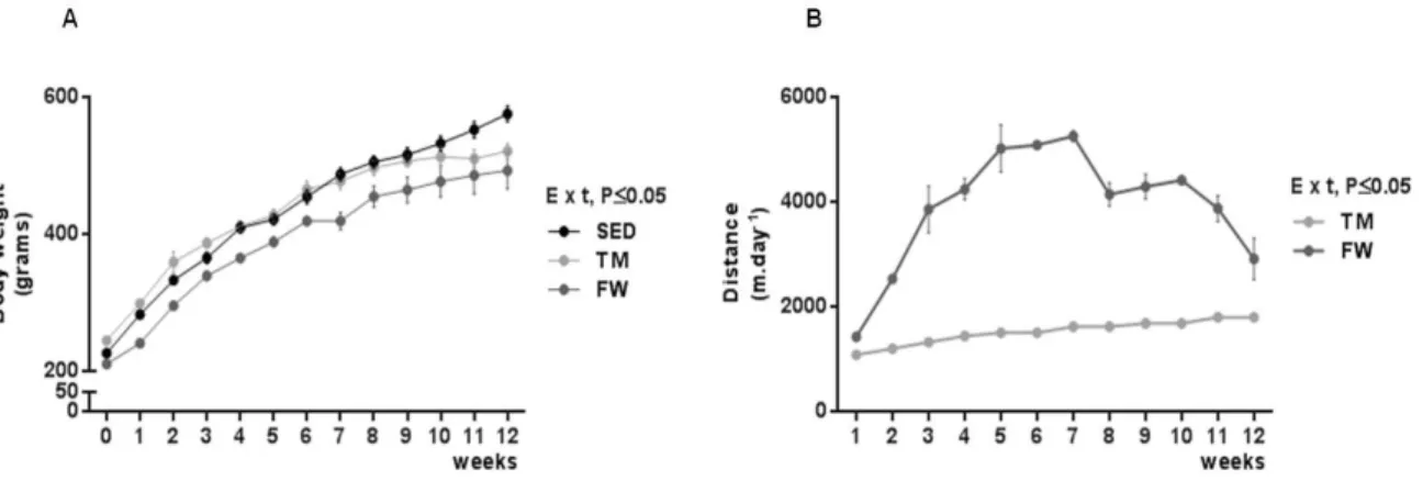
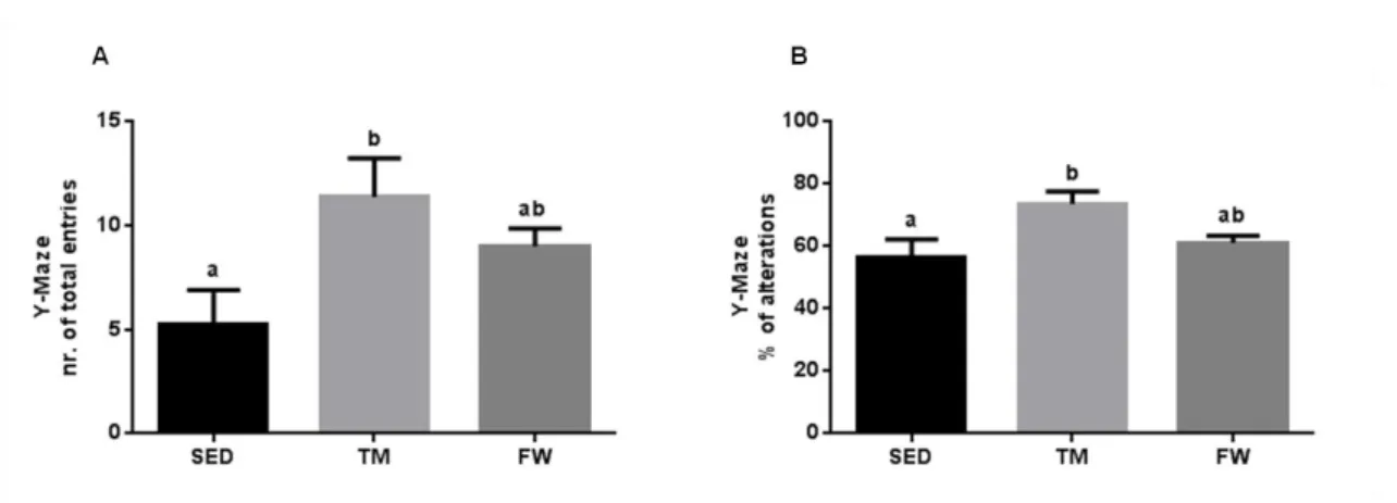
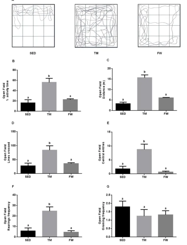
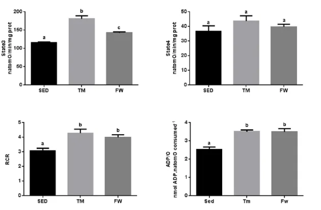
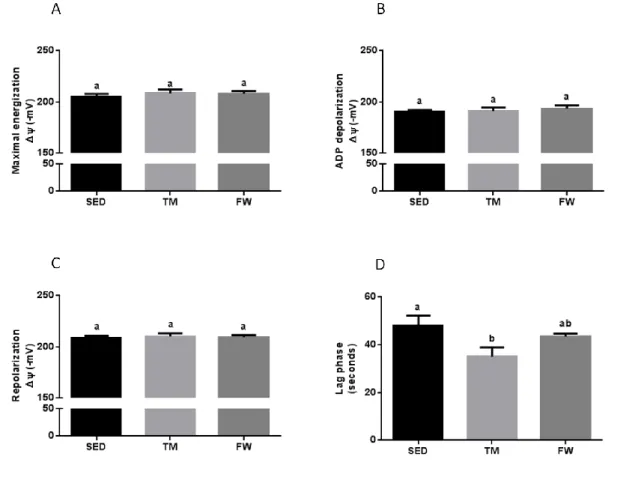
(11) 6.6. Effect of physical exercise on brain mitochondria oxidative stress markers ......................................................................................................................... 59 7. Conclusions ............................................................................................... 63 8. References ................................................................................................ 65. INDEX OF TABLES Table 1. Neurochemical alterations induced by exercise in healthy animals ... 10 Table 2. The Mitochondria Specialized Structures ........................................... 14 Table 3. Functions of the five complexes for ETC............................................ 17 Table 4. TM exercise protocol .......................................................................... 36 Table 5. Animal data and yield of mitochondrial protein isolation..................... 44. INDEX OF FIGURES Figure 1. Effect of physical activity on body mass alterations over time (A) and distance run per day by treadmill and free wheel groups during the 12 wks of protocol (B).. ..................................................................................................... 43 Figure 2. Effect of exercise on Y-maze behavior, number of total entries (A) and % of alterations (B).. ......................................................................................... 45 Figure 3. Effect of exercise on Open Field behavior; illustrative example of a SED, TM and FW animal travel pathway in 5 min exploration (A), % activity time (B), locomotive distance traveled (C), number of lines crossed (D), number of central entries (E), number of rearings and grooming performed (F and G).. ... 46 Figure 4. Effect of exercise on brain mitochondrial respiration.. ...................... 47 Figure 5. Effect of exercise on brain mitochondria ∆ψ fluctuations (maximal energization, ADP-induced depolarization, and repolarization) and ADP phosphorylation lag phase................................................................................ 48. xi.
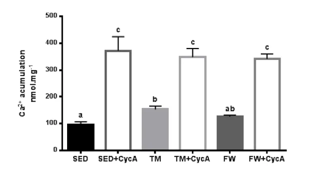
(12) Figure 6. Effects of chronic physical activity on brain mitochondria calciuminduced membrane depolarization.. ................................................................. 49 Figure 7. Effects of chronic physical activity on brain mitochondrial reduced sulfhydryl groups (A) and MDA contents (B).. .................................................. 50. xii.
(13)
(14) ABSTRACT The main purpose of the present work was to analyse the effect of physical activity and exercise training on brain mitochondrial function. Eighteen SpragueDawley male rats were randomly divided into three groups (n=6/group): sedentary, endurance treadmill training (TM, 5days/week with progressive increase of time and speed during 12-weeks), free wheel voluntary physical activity (FW, 24h/day with an unlimited access to running wheel). Behavioral tests were performed to measure spontaneous alternation behaviour and exploratory activity in the open field. In vitro brain mitochondrial oxygen consumption rates, transmembrane potential fluctuations, mitochondrial calcium accumulation until mitochondrial permeability transition pore (mPTP) opening and mitochondrial oxidative damage were evaluated. Both exercise groups increased spontaneous alternation and exploratory activity; however, on openfield, the evaluated behaviours were only increased in TM group. TM and FW exercise induced a significant increases in state 3, respiratory control and ADP/O ratios. In addition, only TM lowered the lag phase and accumulated significantly more Ca2+ before mPTP induction than the sedentary group. No significant differences was observed on brain mitochondrial oxidative damage (SH content and MDA levels). These data suggest that physical activity and more importantly forced exercise training results in improvements on the brain mitochondrial function.. KEYWORDS: PHYSICAL ACTIVITY; EXERCISE; BRAIN; BIOENERGETICS. xiii.
(15)
(16) RESUMO O presente trabalho teve como principal objetivo analisar o efeito do treino de endurance e da atividade física voluntária na função mitocondrial do cérebro. Foram divididos, aleatoriamente, dezoito machos Sprague-Dawley em três grupos (n = 6/grupo): sedentário, exercício em tapete rolante (TM, 5 dias/semana com aumento de tempo e velocidade, progressivamente, durante 12 semanas) e exercício na roda livre (FW, 24h / dia com acesso ilimitado à roda). Foram realizados testes para avaliar o comportamento alternado e espontâneo e, também, a atividade exploratória em espaço aberto. Foram avaliadas, in vitro, as taxas de consumo de oxigénio mitocondrial, as flutuações de potencial elétrico transmembranar, a quantidade de cálcio acumulada até a abertura do poro de permeabilidade transitória mitocondrial (mPTP) e o dano oxidativo mitocondrial. Os dois tipos de exercício induziram um aumento das alterações de comportamento e da atividade exploratória. No entanto, apenas o grupo TM apresentou um aumento significativo dos comportamentos avaliados em espaço aberto. O treino em tapete rolante e atividade voluntária induziram um aumento significativo do estado 3, do controlo respiratório e da razão ADP/O. Adicionalmente, no grupo TM foi observada uma redução da lag phase e uma maior capacidade de acumular Ca2. +. antes da indução mPTP. comparativamente ao grupo sedentário. Não foram observadas diferenças entre grupos nos marcadores de dano oxidativo mitocondrial (contéudo de grupos -SH e de MDA). Estes dados sugerem que a atividade física voluntária e, principalmente, o treino de endurance forçado promovem melhorias na função mitocondrial do cérebro.. PALAVRAS-CHAVE: ACTIVIDADE FÍSICA; EXERCÍCIO; BIOENERGÉTICA. xv.
(17)
(18) ABBREVIATIONS AND SYMBOLS. A.M.. Before Midday. Ach. Acetylcholine. ADP. Adenosine Diphosphate. ADP/O. Ratio between Phosphorylated ADP and Consumed Oxygen. AIF. Apoptosis Inducing Factor. AMP. Adenosine Monophosphate. AMPK. Adenosine Monophophate Kinase. ANT. Adenine Nucleotide Translocase. Apaf-1. Apoptosis Protease Activating. ATP. Adenosine Triphosphate. BDNF. Brain-Derived Neurotrophic Factor. BSA. Bovine Serum Albumin. Ca2+. Calcium Ion. CAT. Catalase. CS. Citrate Synthase. COX. Cytochrome c Oxidase. CO2. Carbon Dioxide. CsA. Cyclosporin A. Cyp D. Cyclophilin D. d. Day. Da. Dalton. DA. Dopamine. DNA. Deoxyribonucleic Acid. Drp1. Dynamin Related Protein 1. xvii.
(19) EndoG. Endonuclease G. ETC. Electron Transport Chain. FAD. Oxidied Flavin Adenine Dinucleotide. FADH2. Reduced Flavin Adenine Dinucleotide. Fis1. Fission 1. FW. Free Wheel. G. Glutamate. GABA. Gamma-Aminobutyric Acid. GPX. Glutatione Peroxidase. GSH. Reduced Glutathione. GSSG. Oxidied Glutathione. h. Hours. H2O2. Hydrogen Peroxide. HSP70. Heat Shock Protein of 70 kDa. KCl. Potassium Chloride. Kg. Kilogram. KH2PO4. Monopotassium Phosphate. m. Meters. M. Malate. MAPK. Mitogen-Activated Protein Kinase. Mfn 1. Mitofusin 1. Mfn 2. Mitofusin 2. mg. Milligrams. min. Minute. mL. Millliter. Ml. Microliter. μM. Micramolar. xviii.
(20) mM. Millimolar. mmol. Millimoles. MnSOD. Manganese Superoxide Dismutase. mPTP. Mitochondrial Permeability Transition Pore. mtDNA. Mitochondrial Deoxyribonucleic Acid. mtNOS. Mitochondrial Nitric Oxide Synthase. mRNA. Messenger Ribonucleic Acid. MΩ. Megaohm. NA. Noradrenaline. NaCl. Sodium Chloride. NAD. Oxidized Nicotinamide Adenine Dinucleotide. NADH. Reduced Nicotinamide Adenine Dinucleotide. NADPH. Reduced Nicotinamide Adenine Dinucleotide Phosphate. NGF. Nerve-Growth Factor. Nmol. Nanomol. nM. Nanomolar. NO. Nitric Oxide. NRF1. Nuclear Respiratory Factor 1. NRF2. Nuclear Respiratory Factor 2. NT-3. Neurotrophin-3. NT-4. Neurotrophin-4. O2. Oxygen. O2-. Superoxide Radical. ºC. Degrees Celsius. OH.. Hydroxyl Radical. Opa 1. Optic atrophy 1. PGC - 1α. Peroxisome Proliferator-Activated Receptor Gamma coactivato xix.
(21) RCR. Respiratory control ratio. RNA. Ribonucleic acid. ROS. Reactive oxygen species. rRNA. Ribosomal ribonucleic acid. s. Second. SED. Sedentary group. SEM. Standard Error of the Mean. SIRT 1. Silent information regulator T1. SOD. Superoxide dismutase. TM. Treadmill. TNF. Tumor necrosis factor. TPP+. Tetraphenylphosphonium. tRNA. Transfer ribonucleic acid. UCP. Uncoupling proteins. VDAC. Voltage-dependent Anion Channel. xg. Centrifugal Force. ΔμH+. Transmembrane Chemical Potential. Δψ. Transmembrane Electric Potential. xx.
(22) 1. INTRODUCTION Nowadays, it is well recognized that the adoption of a healthy life style is related to increased levels of physical activity. Evidences suggest that physical activity is able to induce overall benefits in skeletal muscle, cardiovascular system, liver and kidney metabolism and also in brain function (Cotman and Berchtold 2002, Klaus and Amrein 2012). At least in part, the improved mitochondrial function seems to be an important adaptation to physical activity and therefore an important target to counteract several chronic diseases associated with compromised mitochondrial viability (Bishop et al. 2010, Moreira et al. 2010). Epidemiological studies demonstrate that cognition is improved with physical activity in children, adult and aged persons (Hopkins et al. 2011, LeMoyne et al. 2012). Similarly, data from animal research suggests increases in behavioral tests performance with exercise training or voluntary physical activity (Fulk et al. 2004).. However,. besides. morphological,. vascular,. neurogenic. and. neurochemical adaptations induced by physical activity (Cotman and Berchtold 2002, Gomez-Pinilla et al. 2002, van Praag et al. 2005), brain mitochondrial machinery improvement also seems to have a crucial role in exercise-induced increased brain function (Ploughman 2008, Radak et al. 2001a). Physical activity is involved in a series of adaptations usually leading to the upregulation of brain tissue protective mechanisms (Ang and Gomez-Pinilla 2007, Dishman et al. 2006, Ma 2008). These adaptations include increased mitochondrial biogenesis and function, and improvement in mitochondrial antioxidant capacity, leading to a more effective control of free radical production and suggest that physical activity is an important mediator of increased brain function through mitochondrial-mediated mechanisms (Navarro and Boveris 2007, Steiner et al. 2011). However, research focused on brain mitochondrial response to exercise training in the brain is still scarce. Mitochondrial relevance to brain function is undoubted. Besides the classical oxidative. phosphorylation. and. inherent. adenosine. triphosphate. (ATP). production, these organelles are also critical to intracellular calcium (Ca2+) regulation as well as for redox and apoptotic signaling. Viable and fit 1.
(23) mitochondria are required to several ATP-depend process in neurons such as ion. transport,. receptors. function,. vesicle. release. and. recycling. of. neurotransmitters (Chan 2006, Hoppins et al. 2007, Steiner et al. 2011). Moreover, mitochondrial dynamics regulated by fusion and fission-related mechanisms are associated with the redistribution of mitochondria to distinct subcellular localizations, thus responding to high-energy requirements, and leading to biogenesis and mitochondrial deoxyribonucleic acid (mtDNA) mixing that is critical for the repair of defective mtDNA (Steiner et al. 2011). Therefore, we aimed to study the effects of 12 weeks of endurance exercise and voluntary physical exercise on brain cortex and cerebellum mitochondrial bioenergetics with special reference to oxidative stress and mitochondrial permeability transition.. 2.
(24) 2. STATE OF ART 2.1. Neuro-adaptations to exercise 2.1.1. Neuroplasticity Functional and structural alterations occur in the brain throughout lifespan. Neuroplasticity has replaced the formerly-held position that the brain is a physiological static organ. Actually, is known today that brain has the ability to change its structure and function throughout life (Pascual-Leone et al. 2005). Brain plasticity allows to acquire new information and learn skills which, in turn, enable an adequate response to environmental stimulation, and to recover from brain injuries (Gomes da Silva et al. 2012). Importantly, some studies demonstrated that brain plasticity decreases with age and is deregulated in patients with neurological disorders (Eisch et al. 2008, Lovden et al. 2013, Lynch et al. 2006). This lifelong brain capacity for behavioral flexibility is mainly driven by a mismatch between functional supply and environmental demand (Lovden et al. 2010). Physical activity is known to impact the entire body and its regular practice is associated with general health benefits. It is well accepted that physical activities are considered among the non-pharmacological strategies to reduce the risk of developing cardiovascular diseases and diabetes among other distinct pathologies (Klaus and Amrein 2012). However, several of these pathophysiological conditions, including hypertension and hyperlipidemia, glucose intolerance, diabetes mellitus and obesity also contribute to vascular dementia and to increased risk to develop neurodegenerative process (Ahlskog et al. 2011). Regular physical exercise has proven to accomplish significant outcomes against neurodegenerative-related mechanisms. The benefits of an active life style are associated with enhancing and protecting brain function. Several studies demonstrated that physical exercise is able to confer health protective. 3.
(25) benefits against distinct neurological diseases and ischemic stroke (for refs see Marques-Aleixo et al. 2012). Taking all together, behavioral and neurobiological manipulations, such as those related with exercise programs can have long-term and vigorous effects on brain function (Hopkins et al. 2011). In fact, the neuroplasticity appears to have a crucial association with environmental manipulations including physical exercise throughout the life. The main mechanisms that are involved in physical exercise-induced. brain. plasticity. include. structural,. functional. and. neurochemical alterations, which we will further address in detail in the following sections.. 2.1.1.1. Cognitive function An active lifestyle and increased levels of regular physical activity have been associated with improvements on cognitive functioning in children, adults and in elderly persons (LeMoyne et al. 2012). The level of physical activity seems to decrease in the course of lifetime. However, it has been reported that an active lifestyle can have a preventive role in cognitive health and in the aging-related decrease in cognitive function (Kaliman et al. 2011, Pluncevic 2012). Hopkins et al. (2011) demonstrated that the effects of regular exercise in anxiety, mood and cognitive function are significant. Similarly, Hillman et al. (2008) reviewed the benefits of exercise for the treatment of depression and some neurodegenerative disorders. In fact, intellectual appointment, social interaction and physical activity are associated with the maintenance of cognitive function and also with the protection against the onset of chronic diseases, including neuropathologies such as Alzheimer´s and Parkinson´s (for refs see Marques-Aleixo et al. 2012). Other authors described that children with low physical activity levels showed decreased neuroelectric activity and inferior cognitive performance when compared to physically fit children (Chaddock et al. 2010a). Moreover, it seems that they present better hippocampal plasticity and spatial memory in adulthood 4.
(26) and life (Gomes da Silva et al. 2012). Likewise, Chomitz et al. (2009) demonstrated that increased aerobic fitness and regular physical activity are associated with an enhanced academic performance in reading and mathematics. Chaddock et al. (2012) reported that children with higher physical activity levels may show better selective attention, inhibition of improper responses and maintenance of information in working memory. Similarly, another study conducted on children assessed the relationship between aerobic fitness and executive control. The conclusions suggest that fitness is associated with greater cognitive performance on an executive control task through enhanced cognitive control (Hillman et al. 2009). Cognitive abilities usually show a gradual decline after middle adulthood, which may lead to dementia in later life (Chaddock et al. 2012). Prakash et al. (2011) verified that cardiorespiratory fitness relates to cognition in old adults being higher levels of fitness associated with better behavioral performance. Moreover, Colcombe et al. (2004) established that highly fit humans appear to have an increased functioning of the attentional network on the brain and cognitive task when compared with low-fit persons. The same authors suggested that physical activity at an early phase of life is a beneficial influence on diverse cognitive functions for many years. Also, Scarmeas et al. (2009) suggested that physical activity can reduce or prevent functional decline associated with aging and decreases the probability of Alzheimer´s emergence. As mentioned previously, older adults with greater physical activity and aerobic training levels showed decreased of cognitive impairment and dementia, as well as enhanced function and brain structure (McAuley et al. 2011, Voss et al. 2011). A study conducted with older man and women showed that midlife physical activity helps to sustain cognitive function and may decrease or delay the possibility of dementia in late life (Chang et al. 2010). Further studies suggested that greater physical activity levels and aerobic fitness in adults over the age of 65 are associated with enhanced cognitive performance (Chaddock et al. 2012). Similarly, a study with adults over the age of 65, who participated in physical activity program more than three times per week for at least 15 min showed that this group of active seniors have 34% less risk to be diagnosed 5.
(27) with dementia compared with those who exercised less than three times per week (Larson et al. 2006). In general, both human and animal studies reported that physical activity and aerobic exercise training appears to have positive effects on non-pathological and pathological conditions on the aging brain (Kramer et al. 2006, Morie et al. 2010). Regular physical activity during life is associated with better school achievement and lower risk for cognitive decline, dementia and of risk for neurological disorders (Chaddock et al. 2012). Some of the mechanisms and targets by how exercise affects cognitive function will be addressed in the next sections.. 2.1.1.2 Structural and morphological alterations As previous referred, physical exercise is an important factor that positively modulates mental health throughout life (Hillman et al. 2008). Indeed, robust effects of physical activity have been reported in humans and rodents and have demonstrated that physical exercise is able to confer health protective benefits against several neurological diseases, seen at morphological level that could be translated into an increased brain function (Biedermann et al. 2012, van Praag et al. 1999). Erickson and colleagues (2011) reported that regular exercise increases hippocampus volume and induces plastic changes in specific brain structures. Exercise training seems to influence brain plasticity by hippocampus size, dendritic growth, cortical thickness and synapse formation, increased cell proliferation, increased hippocampal-dependent memory and learning and synaptic plasticity (Chaddock et al. 2010b, Pereira et al. 2007, Simpson and Kelly 2011, Voss et al. 2012). In fact, exercise has been described as a crucial factor in maintaining cerebrovascular integrity, increasing cerebral circulation, increasing dendritic connections and capillary growth and, finally, increasing the efficiency of the processing functions of the central nervous system (Radak et al. 2001a).. 6.
(28) Chaddock et al. (2012) showed that higher-fit children present more efficient neuroelectric activation, larger brain volumes in the hippocampus and basal ganglia than less fit children. Also, higher-fit children showed greater bilateral hippocampal volumes and superior relational memory task performance compared to lower-fit children. Additionally, bilateral hippocampal volume was found to mediate the relationship between fitness cardiorespiratory level (VO2 max) and relational memory (Voss et al. 2013, Voss et al. 2012). Similarly, it has been shown that young adults who did practice aerobic exercise for 3 months demonstrated a significantly increased hippocampal dentate gyrus blood volume over baseline (Ahlskog et al. 2011). Age-related alterations in brain structure interfere with the hippocampal. However, according to Erickson and colleagues (2011) physical activity in older adults increase hippocampal perfusion, cerebral blood volume and the size of the anterior hippocampus, leading to improvements in spatial memory. Verstynen and co-workers (2012). demonstrated that aerobic exercise. decreased the atrophy of several brain regions including the medial temporal lobe, basal ganglia and prefrontal cortex. This one year-long study with older adults, who participated in aerobic exercise tests and were submitted to magnetic. resonance. imaging. assessement,. concluded. that. higher. cardiorespiratory fitness levels were associated with increased gray matter volume in the dorsal striatum and better cognitive performance. At the same time, rodent models have demonstrated that voluntary aerobic exercise positively affects the hippocampus, increased cell proliferation, enhance hippocampal-dependent memory and learning processes, synaptic development, and angiogenesis (Chaddock et al. 2010b, Voss et al. 2012). Indeed, an adaptive response to functional and structural changes induced by chronic exercise training is angiogenesis (Roque et al. 2011). Angiogenesis is the neoformation of capillaries and a complex process requiring proliferation, migration and congregation of endothelial cells to form new vessels (Roudier et al. 2009). Huang et al. (2013) demonstrated that running exercise-induced increases of the capillaries in the female rat cortex might be one of the. 7.
(29) structural bases for the exercise-induced improvement in the spatial learning capacity of middle-aged female. The beneficial effects of physical exercise on brain functions suggest special attention of adult neurogenesis in cognitive and mental health (Yau et al. 2011). Thus, neurogenesis is the process by which neurons are generated from neural stem and progenitor cells. Hippocampal neurogenesis is known to be regulated by age stress, learning, seizures and exercise (Snyder et al. 2001). Yau et al. (2011) showed that exercise promotes dendritic plasticity and hippocampal neurogenesis. In elderly humans, imaging studies have shown changes in the hippocampus that may lead to cognitive decline. However, exercise training prevented. or. reversed. these. deleterious. morphological. and. behavior. consequences of aging (Snyder et al. 2001, van Praag et al. 2005). Moreover, research indicates that neurogenesis has a role in learning and memory. A study with wheel running in rodents resulted in a 3-4 fold or even larger enhance in the survival and formation of new neurons (van Praag 2009). In summary, physical exercise has unquestionable beneficial effects on brain health (Bernardi et al. 2013). Brain structure and function appears to be closely related to the practice of regular exercise which, in turn, seems to contribute to an increased aging brain function and to counteract neurodegeneration.. 2.1.1.3. Neurochemical adaptations Exercise-induced enhanced brain function could also be explained by neurochemical alterations. Neurotrophic factors (Cotman and Berchtold 2002), stress hormones (Schoenfeld and Gould 2012), and neurotransmitters (Meeusen and De Meirleir 1995) can be influenced by physical exercise and could have an important role in the development, plasticity and brain health. In fact, alteration in the concentration and activity of neurotransmitters, stress hormones and neurotrophic factors induced by exercise seem to modulate neurochemical alterations, including increased learning, cognition and behavior, higher adaptation to stress and anxiety situations, increased regulation of mood, 8.
(30) appetite and sleep, better response to inflammatory state and increased neural development (Cote et al. 2011, Goekint et al. 2012, Simpson and Kelly 2011). Additionally, some studies demonstrated that physical exercise modulates several physiological mechanisms that may increase brain resistance to aging and neurodegeneration (to refs see Marques-Aleixo et al. 2012). Table 1 summarizes some neurochemical alterations induced by physical exercise.. 9.
(31) Table 1. Neurochemical alterations induced by exercise in healthy animals Neurochemic factors. Main cognitive function. Role of physical exercise. References. Neurotransmitters and stress hormones ↑ or~. Goekint et al. (2012); Dey (1994); Ishikawa et al. (2013); Rethorst et al. (2012). ↑ or~. Foley et al. (2008); Goekint et al. (2012); Tsuchiya et al. (2012); Simpson et al. (2011). Increase heart rate and blood flow to skeletal muscle at the fight-or-flight response. ↑ or~. Goekint et al. (2012); Ishikawa et al. (2013); Madarame et al. (2013); Simpson et al. (2011). Acetylcholine (Ach). Memory; Learning; Lowers heart rate and force of cardiac contraction; Vasodilation. ↑. Parnow et al. (2012). Opioids. Analgesic effects, which decrease perception and reaction to pain; increase pain tolerance; Deficit respiratory; Sedation; Strong sense of euphoria. ↑ or~. Jonsdottir et al. (1997); Simpson et al. (2011). Serotonin (5HT). Memory; Learning ; Regulation of mood, appetite and sleep; Related with feeling well and happiness. Dopamine (DA). Cognition and behavior; Motivation; Working memory; Learning; Attention; Mood; Dreaming and sleep. Noradrenaline (NA). 10.
(32) GammaAminobutyric Acid (GABA). Regulation of mood, appetite and sleep; Related with decrease anxiety; Regulation of muscle tone. ↑. Urakawa et al. (2013); Ni et al. (2009); Hill et al. (2010). Glutamate. Learning; Memory; Increase Brain Function and Mental activity; Decrease fatigue. ↑. Mourzakis et al. (2002); Simpson et al. (2011). Cortisol. Adaptation to stress; Maintain glucose levels appropriate even in fasting; Catabolism of skeletal muscle and adipose tissue; increase vasoconstriction caused by adrenaline; Decrease the use of glucose, saving it to the brain. ↑,~or ↓. Zoladz et al. (2002); Dean, (2002); Kanaley et al. (2001). ↑ or~. Quirie et al. (2012); Cote et al. (2011); Boyce et al. (2007); Skup et al. (2000); Vivar et al. (2012); Goekint et al. (2012). Neurotrophic factors Brain-Derived Neurotrophic Factor (BDNF). Memory; Learning; Long-term memory; Neural development;. Nerve Growth Factor (NGF). Development; Maintenance of peripheral sympathetic and embryonic sensory; Maturation; Response to offending stimuli; Immune system and inflammatory cells. . ↑. Liu et al. (2011); Bonini et al. (2013); Vivar et al. (2012); Chae et al. (2012); Simpson et al. (2011). 11.
(33) Neurotrophin-3 (NT-3) Neurotrophin-4 (NT-4). Survival and differentiation of existing neurons; Growth of new neurons and synapses;. Learning; Memory; Long-term memory; Adult locomotory behaviour; Epidermis development. ↑. Sharma et al. (2010); Boyce et al. (2007); Skup et al. (2000); Cote et al. (2011). ↑. Skup et al. (2002); Funakoshi et al. (1995); Skup et al. (2000); Cote et al. (2011). ↑, significant increase; ↓, significant decrease; ~, no significant changes.. 12.
(34) Neurotransmitters can be catecholamines and tryptamines and are associated with emotionality, anxiety and behavior; stress hormones are necessary in “fight or flight” response and other individual stress situations and, finally, neurotrophic factors are proteins known to promote cell survival and functions particularly related to synaptic plasticity. Therefore those neurochemical parameters are intimately related with specific brain function and can be modulated by alterations induced by physical exercise.. 2.2 Relevance of Mitochondria on Brain Function As. previously. reported,. with. enhancing. life. expectancy,. age-related. neurodegenerative disorders resulting in structural and neurophysiological alterations in the brain are extremely complex. The literature shows that healthy mitochondria are crucial to the maintenance of the integrity of human systems, including brain and nervous system function (for refs see Marques-Aleixo et al. 2012). Several studies suggested a relationship between mitochondrial function and cognitive function in humans (Inoue et al. 2000). Mitochondrial respiration is fundamental. to. maintain. the. cortical. and. hippocampal. plasticity. for. establishment for spatial memory (Tanaka and Watanabe 2008). Therefore, it is increasingly important to find strategies to counteract the decline of mitochondrial function as this can be a decisive contributor to the aging process and acceleration of neuropathological conditions. Three-dimensional reconstruction indicates that mitochondria constitute a complex branched, forming a reticular network which alters in size, shape and complexity (Benard and Rossignol 2008b). The next table explains the main functions of the mitochondrial sub-structures (Table 2).. 13.
(35) Table 2. The Mitochondria Specialized Structures Structural. Main functions. components Outer membrane. - is needed for compartmentalization of the specific activity of mitochondria from the cytosol; - contains a great numbers of porins that permit free diffusion of small molecules;. Inner Membrane. - contains all the essential components of the electron transport chain; - compartmentalized proteins with five types of functions: 1) ATP synthase, 2) execute the redox reactions of oxidative phosphorylation, 3) specific transport proteins that control metabolite passage into and out of the mitochondria matrix, 4) protein import machinery and 5) mitochondria fusion and fission protein;. Inner Membrane Space. - space the protons (H+) that are pumped to create the proton motive force, which drive ATP production; - contains many proteins that may play important roles in cell death like the cytochrome c and the apoptosis inducing factor;. Cristae. - are the functional internal compartments formed by the many invaginations and folding of the inner membrane; - in this structure occurs the cellular respiration and grouped to protein complexes of the respiratory chain and the proteins. that. are. involved. in. the. iron/sulfer. group. biogenesis;. 14.
(36) Matrix. - is where enzymes catalyze the oxidation of pyruvate, the end product of glucose, which occurs in the cytosol, addicted to energy rich products that drive the electron transport chain; - in this structure are located the enzymes that carry out the ß-oxidation citric acid cycle and the mitochondrial DNA (mtDNA).. Mitochondria are the powerhouses of the cell able to generate large quantity of ATP; however, these important organelles also serve numerous other functions in addition to energy production, such as calcium regulation and cell signaling, thus determining cell survival or death. Cellular respiration, the process of breaking down food into energy, carbon dioxide and water, is related to several interconnected pathways and steps: glycolysis, tricarboxylic acid cycle (Kreb´s Cycle), beta-oxidation, electron transport chain (ETC) and oxidative phosphorilation. The ETC operates by a step-wise transfer of energy rich electrons from NADH and FADH2 donors to inferior energy acceptor molecules (complex I-IV, ubiquinone and cytochrome c). The NADH dehydrogenase (complex I) receives electrons from NADH and succinate desydrogenase (complex II) from FADH 2. These protein complexes are starting points of the ETC. The rest of the ETC operates despite of which complex (I or II) initiated the chain. The remaining carrier molecules (ubiquinone and cytochrome c) and protein complexes cytochrome bc1 (complex III) and cytochrome c oxidase (complex IV) continue the electron transfer until the reduction of molecular oxygen (O2) (Benard and Rossignol 2008b, Gilkerson et al. 2003a). Therefore, the pathways for the electrons transferency throughout the ETC can be described as follows:. 15.
(37) Table 3 summarizes the specific functionality of the diverse members of protein complexes of the ETC.. 16.
(38) Table 3. Functions of the five complexes for ETC Complexes. Function. Has the capacity to oxidize NADH. Proton pump to the intermembrane space of I – NADH Dehydrogenase mitochondria contributes to the (Coenzima Q redutase) electrochemical gradient of protons and can guide to the generation of reactive oxygen species (ROS). II - Succinate Desydrogenase Catalyze the oxidation of succinate to fumarate (Ubiquinone redutase) with the creation of ubiquinol from ubiquinone. Through ubiquinol, and simultaneously that III – Cytochrome bc1: ocurring explosion of a pair proton, an electron Cytochrome c Reductase is transferred to the Cytochrome c. IV – Cytochrome c Oxidase. O2 is reduced to water and energy used to pump protons produced.. Oxidative Phosporilation (OxPhos) is the electrochemical gradient (Δp) driven process of phosphorylating (adenine diphosphate). Three to four protons are transported down their (Δp) back to the matrix through the ATP synthase – complex V to produce 1 molecule of ATP (Boyer 1997). Another very important function related to mitochondrial network is calcium (Ca2+) regulation. Calcium is a signaling molecule and a second messenger that can regulate metabolism in the cell. In physiological conditions, the extracellular concentration of free ionic calcium is 1-2 mM and cellular range are between 50 and 100 nM being levels inside mitochondria in the scale of μM (Hansford and Zorov 1998). According to Gunter et al. (2000), three dehydrogenases associated to the Kreb´s Cycle as well F0F1ATPase and the ANT are activated by moderate concentrations of intramitochondrial calcium concentration. Mitochondria can modulate calcium signaling by exporting additional calcium into the intracellular cytosolic space (propagating) or by sequestering even. 17.
(39) insignificant enhances of calcium (inhibiting). The level of calcium loading into mitochondria is dependent the frequency and duration of stimuli. The capacity of mitochondria to regulate calcium with such an exact spatiotemporal control, underscores the important roles that calcium plays in the cell (Gunter et al. 2000). However, despite calcium regulates cell and even mitochondrial metabolism, large amounts of calcium influx into mitochondria may inhibit oxidative phosphorylation and directly affect the rate of ROS production. Unnecessary and excessive calcium loads can also generate assembly of the mitochondrial permeability transition pore (mPTP) (Kann and Kovacs 2007). The permeability transition pore complex is a channel that is assembled at the junctions between the inner membrane and the outer membrane and, despite some controversy is proposed to be composed and/or regulated by adenine nucleotide translocase (ANT), cyclophilin D (Cyp D), hexokinase, voltagedependent anion channels (VDAC) (Haworth and Hunter 1979). More recently, the ATP synthase as been purposed to have an important role in the regulation of the mPTP (Bonora et al. 2013). The formation of mPTP may have devastating cellular consequences as a result of the dissipation of mitochondrial proton gradients, liberation of cytochrome c and initiation of apoptosis (Haworth and Hunter 1979). It is also assumed that mitochondria play an important role on cell signaling, including cell survival and death pathways (Smaili et al. 2003). Cell death is an indispensable event that occurs by apoptosis and/or necrosis. Apoptosis, or planned cell death, is necessary for survival and plasticity, while necrosis is an unexpected death due to a lack of accessible ATP indicative of energy breakdown (Brown and Borutaite 2001). Cytochrome c and mitochondrial nitric oxide (mtNOS) are two crucially important molecules that play dual roles in cellular respiration and in these two cell survival and death signaling pathways. These molecules are directly or indirectly modulated by calcium levels. Cytochrome c is familiarly linked to mitochondrial respiration by transferring electrons between complex III and IV of the ETC. Ott et al. (2002) refer that apart from the cause or mechanism for cytochrome c extrusion, it is a potent incentive for initiation of apoptosis. Oxygen uptake is synchronized by mtNOS 18.
(40) that can reversibly inhibit cytochrome oxidase – complex IV – and the production of superoxide (O2-) from the electron transport chain. Under certain pathological conditions significant suppression of mitochondrial respiration and elevations of oxidative stress are observed (Smaili et al. 2003). Considering the importance of mitochondrial process in neuronal function, it has been suggested that mitochondria may possibly be a potential target for pharmacological and non-pharmacological. strategies. to. minimize. the. impact. of. several. neurodegenerative disorders (Marques-Aleixo et al. 2012). Calabrese et al. (2001) demonstrated that evidence for mitochondria susceptibility to damage in neurodegenerative disorders is, at least in part, based on decreases in respiratory chain complex activities. Such defects in respiratory complex activities, probably associated with oxidant/antioxidant balance perturbation, may have serious impact on energy metabolism contributing to cellular degeneration. The neurons are highly differentiated cells that need large amounts of ATP for preservation. of. ionic. gradient. across. the. cell. membranes. and. for. neurotransmission (Kann and Kovacs 2007). Since the majority of neuronal ATP is generated by oxidative metabolism, neurons depend on mitochondrial function and oxygen supply (Ames 2000). Although the brain is a noncontractile tissue, an increase on energy metabolism seems to indirectly influence neuronal function (Dishman et al. 2006). In addition, mitochondria are intracellular organelles containing their particular genomes (mtDNA) and playing an important role in ATP production through oxidative phosphorylation (Inoue et al. 2000). Mutations on mtDNAs and deficiencies in mitochondrial respiration are associated with an extensive diversity of disorders, such as mitochondrial and neurodegenerative diseases, diabetes and aging (Tanaka et al. 2008).. 2.2.1 Brain mitochondrial response to physical exercise As mentioned previously, physical activity and particularly the regular physical exercise has been considered a decisive strategy in the therapy of several pathologies.. Thus,. the. understanding. of. the. underlying. physiological 19.
(41) mechanisms that are associated with physical exercise-mediated benefits assume high relevance. Considering different mechanisms and targets, brain mitochondrial metabolism and function seems to be highly modulated by physical exercise. The study of some metabolic brain alterations induced by physical exercise has been based on the analysis of the content and/or activity of several enzymes, particularly those implicated in aerobic energy production pathways (Ding et al. 2006). In fact, an increase in ATP synthase content may be one of the adaptations by which exercise enhances energy production in brain mitochondria. Importantly, different exercise types such as free wheel voluntary running or forced running on a treadmill have distinct impacts on hippocampal mitochondrial proteins (for refs see Marques-Aleixo et al. 2012). In fact, several studies suggested that enhanced physical activity induced brain alterations in translational efficiency or protein constancy that may compensate upstream transcriptional responses (Marques-Aleixo et al. 2012). Physical exercise induces an increase of O2 consumption and energy utilization which, in turn, increases ATP synthesis. This suggests that exercise induces important adaptations in the mitochondrial activity of hippocampal neurons in order to sustain metabolic demands.. 2.2.1.1 Mitochondrial respiratory chain and antioxidant adaptations As previously mentioned, mitochondria play a decisive role in animal cells and are involved in aerobic energy production, converting oxygen and nutrients into ATP (Benard and Rossignol 2008a). The mitochondrial respiratory chain is the “locus” of a spatial series of redox reactions in which electrons are transferred from a donor molecule to an acceptor molecule. Oxidative phosporilation is an important cellular process that uses oxygen and electron derived from simple sugars, fatty acids and proteins to create ATP being five inner membrane located protein/enzymatic complexes (I-V) involved in this process (Gilkerson et al. 2003b). During oxidative phosphorylation, these complexes conduct. 20.
(42) chemical reactions that drive the production of ATP. In particular, they produce an irregular electron charge on either side of inner membrane and this disparity in electrical charge provides the proton-motive force for ATP production (Benard and Rossignol 2008a). Some studies analyzed brain mitochondrial adaptive responses to exercise and suggested that moderate running antagonize some age-related impairments in mitochondrial function in general and particularly, at ETC level (Boveris and Navarro 2008b, Navarro and Boveris 2007). Indeed, 24 weeks of moderate exercise improved ETC flux in brain mitochondria, by enhancing the activity of complexes I, III and IV, thus contributing to prevent the age-dependent mitochondrial function decline reported in sedentary rodents (Navarro et al. 2004). Also, moderate exercise reverted the progressive decrease in enzymatic activities of brain mitochondrial complexes I and IV (Navarro and Boveris 2007). Indeed, physical exercise seems to modulate mitochondrial respiratory chain, and it has been suggested that exercise may be a valuable strategy for neuroprotection by targeting these components of mitochondrial machinery (Boveris and Navarro 2008b). ROS (reactive oxygen species) can be produced from different enzymatic and non-enzymatic reactions and mitochondria is the major intracellular source of ROS (Chakrabarti et al. 2011). Radak et al. (2008) stated that the age-related accumulation of oxidative damage impairs brain function, and exercise due to the changes in redox homeostasis (increasing antioxidant/damage repair enzyme activity, decreasing oxidative damage and increasing resistance to oxidative stress), can attenuate the accumulation of damage, causing improved brain function. Navarro et al. (2004) described that moderate and regular exercise have an important role in the upregulation of antioxidant enzymes, the decrease in oxidative stress markers and in the increased of mitochondrial enzymatic activity. In fact, exercise appears to be elementary on brain mitochondria function through enhance antioxidant levels with consequent attenuation of ROS production (Navarro and Boveris 2007).. 21.
(43) In studies with older humans, either favorable brain redox adaptations or incapacity to revert the increased ROS that characterized aging process were found (Jolitha et al. 2006). Furthermore, exercise combined with antioxidant supplementation appears such as a possibility to reduce the age-dependent risk of oxidative modification of brain proteins and lipids (Chakrabarti et al. 2011). The same authors refer that this combination seems to substantially stimulate the endogenous antioxidant system. Indeed, the increased of mitochondrial and tissue antioxidant capacity induced by physical exercise is not exclusive of brain. The generation of ROS during exercise may play a crucial role as regulatory mediators and signaling molecules in adaptative response (Radak et al. 2001a).. 2.2.1.2. Mitochondrial permeability transition pore and apoptotic signaling In recent years the role of mitochondria in cell death has been a subject of particular attention by the scientific community. The increased of permeability of mitochondrial membranes is pivotal in cell death signaling (Tsujimoto and Shimizu 2007). In addition to the proposed mPTP components, including Cyp D, ANT and the VDAC, hexokinase, the phosphate carriers and more recently, ATP synthase might also be involved as potential components of mPTP (Bonora et al. 2013). Furthermore, some studies demonstrated that Cyp D seems to be the essential element of the mPTP opening (Halestrap and Brenner 2003, Kokoszka et al. 2004), but ANT and VDAC seem to be responsible for the interface with the apoptotic proteins (Crompton 2003). Additionally, it has been suggested that the mPTP state is regulated by proteins of the Bcl-2 family comprising pro-apoptotic fractions as Bax, Bak, Bok, and anti-apoptotic as Bcl-2, Bcl-XI, Bcl-w. The relative proportion of these proteins seems to have an important role in the apoptotic susceptibility (Bernardi et al. 2001, Tsujimoto 2003b).. 22.
(44) Several factors facilitate the assembly of these specific proteins leading to the opening of the mPTP. The regulation of mPTP appears to be modulated by the decreasing of transmembrane electrical potencial (Δψ), the increasing in inorganic phosphate concentration, the decreasing of the content of adenine nucleotides (ATP and ADP) and, finally, the increasing of oxidative stress. Importantly, it has been also suggested that mPTP is a process favored by dysregulation Ca2+ homeostasis (Tsujimoto and Shimizu 2007). Korshunov et al. (1997) showed that transient mPTP opening may contribute to the Δψ regulation and operate as a channel for fast release of Ca 2+. In fact, increasing Ca2+ concentration continues to be one of the main factors that lead to cellular apoptosis (Richter 1997). Cells have two essential apoptotic signaling pathways, the intrinsic and the extrinsic (Green and Evan 2002a). Regardless endoplasmic reticulum-mediated path, the commonly referred intrinsic pathway is activated by the release of different mitochondrial pro-apoptotic proteins to the cytosol (Green and Evan 2002b, Wang 2001), among which we can emphasize the cytochrome c. Once released into the cytoplasm, and in the presence of ATP, cytochrome c binds apoptosis activating factor protease (Apaf-1), enhances its oligomerization and the recruitment/activation of pro-caspase-9. The protein complex formed by cytochrome c, Apaf-1 and pro-caspase-9 is called apoptosoma (Desagher and Martinou 2000, Hill et al. 2003, Tsujimoto 2003a). Mitochondria contain other pro-apoptotic proteins in the intermembrane space such as Smac/DIABLO, HtrA2/Omi, apoptosis inducing factor (AIF), and endonuclease G (EndoG) (Green and Evan 2002b, Wang 2001). Together, Smac/DIABLO and HtrA2/Omi are able to prevent inhibitory apoptosis proteins (IAPs) to facilitate the activation of caspases. Moreover, AIF and EndoG translocase are related to an apoptotic stimulus that appear to be responsible for chromatin condensation and DNA fragmentation (Ravagnan et al. 2002). Programmed cell death is essential in the development, morphogenesis, tissue alteration and immune regulation also being connected to several pathologies such as neurodegenerative disorders (for refs see Marques-Aleixo et al. 2012). Apoptosis is a mechanism characterized by cell contraction, chromatin 23.
(45) condensation, DNA disintegration, protrusions of the plasma membrane and apoptotic bodies formation (Fiers et al. 1999). Skulachev et al. (2000) suggested that apoptotic process appears to be preceded by changes in mitochondrial membranes and mediated by ROS. Thus, as previously mentioned, the antioxidants have the capacity to protect and prevent the negative consequences caused oxidative stress. Additionally, increasing evidences provide support that oxidative stress and apoptosis are closely linked physiological phenomena and are implicated in the pathophysiology of some of chronic diseases, including Alzheimer’s and Parkinson’s as well as ischemia of heart and brain (Calabrese et al. 2000). In. fact,. inappropriate. apoptotic. responses. are. implicated. in. several. neurodegenerative conditions, and there are some reports showing that physical exercise affords protection against deleterious stimuli and/or pathophysiological conditions (Sim et al. 2004). A study with trained animals during 21 days showed that endurance training prevented increase in Bax levels caused by chronic stress on cortical mitochondria, suggesting that exercise inhibit Bax translocation to mitochondria with resultant decrease in its proapoptotic activity (Haack et al. 2008). On the other hand, Um et al. (2008) reported a decrease of several proapoptotic proteins, including caspases 9 and 3 as well as an increased Bcl-2 protein after an exercise training program. Therefore, and according to Murer et al. (2001), physical exercise can possibly alter the mitochondrial efficiency and apoptotic signaling with preventive and/or therapeutic implications in some pathological conditions.. 2.2.1.3. Biogenesis and Dynamic There is increasing evidence suggesting a crucial role of mitochondrial dysfunction in the aging process and in a large number of brain diseases (Chaturvedi and Flint Beal 2013). Deleterious alterations in mitochondrial biogenesis and dynamics could be negatively associated with mitochondrial function, contributing to an impaired brain function. However, stimulation of. 24.
(46) mitochondrial biogenesis could be a compensatory adaptation against excessive fission and degradation mechanisms that characterize most of neurodegenerative pathologies (Knott et al. 2008). It is well established that exercise training induces not only an increase in muscle mitochondria, but also in brain mitochondria (Holloszy 1967). However, little research has focused on the brain mitochondrial response to physical activity and/or exercise training. Given the important role of mitochondrial dysfunction in some pathological disorders and neurodegenerative conditions, the implications for exercise induced increase in brain mitochondrial biogenesis is potentially large (Steiner et al. 2011). Mitochondrial biogenesis can be promoted via an increase in transcriptional coactivators of Silent Information Regulator T1 (SIRT1), which interacts with deacetylates and activates the peroxisome proliferator-activated receptor- coactivator 1-alpha (PGC-1α) (Cotman et al. 2007). PGC-1α is considered the “master regulator” of mitochondrial biogenesis, antioxidant defense and was also associated with neuroprotection (Puigserver and Spiegelman 2003, Wareski et al. 2009). Increasing in rodent brains PGC-1α seem to be related to increases in exercise tolerance (Steiner et al. 2011). Same authors verified that brain adaptations to endurance training induced mitochondrial biogenesis through PGC-1α and SIRT1 mRNA overexpression and, simultaneously, increased mtDNA. Wareski et al. (2009) referred that PGC-1α overexpression in neuronal cells of the cortex, midbrain and cerebellum was related to increased mitochondrial density. In addition to mitochondrial biogenesis, mitochondrial dynamics is equally important in brain structure and function. Neurons are also particularly sensitive to changes in mitochondrial movement and distribution (for refs see MarquesAleixo et al. 2012). Mitochondrial dynamics is regulated by fission and fusion mechanisms, which are regulated by fission and fusion-related proteins, respectively. Fission-related proteins include dynamin-related protein 1 (Drp1) and fission 1 (Fis1), while mitofusins (Mfn1/2) and optic atrophy type 1 (Opa1) operate as fusion proteins. These fission/fusion-associated proteins are present in the neurons and are able to promote fast alterations in mitochondrial dynamics that can be significant in context of mitochondrial metabolism 25.
(47) (Nakamura et al. 2010). Additionally, the unbalance of mitochondrial dynamics in neurons could also contribute to the interference to Ca2+ homeostasis (Knott and. Bossy-Wetzel. 2008),. mitochondria. depolarization,. translocation. of. proapoptotic mediators to mitochondria and to the release of cytochrome c leading to apoptosis (Yuan et al. 2007). Also, brain mitochondrial dysfunction can result from negative alterations in the fission-fusion machinery concomitant with neurodegenerative disorders (Chan 2006). Chen et al. (2007) reported that excessive fragmented mitochondria have been associated with inhibition of cell growth, a decrease in mitochondrial respiration and loss of mitochondrial membrane potential. Twig et al. (2008) verified that increased fusion and decreased levels of fission inhibit. mitophagy,. consequently. establishing. a. relationship. between. mitochondrial dynamics and mitophagy. Mitophagy is a selective autophagy of mitochondria (Lemasters 2005) and could be triggered by the alteration of K+/H+ activity and failure of cation homeostasis, the damage of oxidative phosphorylation and enhanced ROS system (Twig et al. 2008). Finally, dysfunction in mitochondrial dynamics, negative changes in ROS production and antioxidant capacity, disruption of Ca2+ homeostasis and mtDNA mutations are interrelated and can have deleterious consequences in the aging process or neurodegenerative disorders (Chan 2006, Knott and Bossy-Wetzel 2008). Despite the relevance of mitochondrial dynamic in brain mitochondrial health and on the pathogenesis of some neurodegenerative disorders, and the previous described role of physical exercise as a possible strategy for brain mitochondria protection, to our knowledge, only one study observed an increased expression of Mfn1 and Mfn2 in trained rats after 6-weeks of training (Liu and Zhou 2012).. 26.
(48) 2.3. Physical exercise: implications for brain health 2.3.1. Aging and neurodegenerative diseases – role of mitochondria, oxidative damage and apoptotic cell death The aging process refers to a series of time-dependent changes at molecular and cellular levels leading to several characteristic features that compromise the functional fitness of the organism (Chakrabarti et al. 2011). Aging is associated with a general decline of physiological functions with a more marked effect on those that depend on central nervous system, such as behavior and cognitive performances (Navarro et al. 2004). Oxidative stress, mitochondrial dysfunction, inflammatory response, changed cell signaling and gene expressions are some of the causes of brain aging that lead to morphological ant structural alterations in the brain along with metabolic deficits and cognitive decline (Chakrabarti et al. 2011). In fact, morphological dysfunction seems to be closely related to aging and to several neurodegenerative diseases (Moreira et al. 2010). Bishop et al. (2010) reported that the decrease of mitochondrial function is related to shorter lifespan and may contribute to brain aging and, enhancing neuronal vulnerability to age-related pathologies. The central nervous system is very susceptible to aging, since it is extremely metabolically active, uses a great amount of oxygen, has an elevated content of free fatty acids that can be simply damaged by free radicals, and has limited regenerative properties (Toescu and Verkhratsky 2003). The overproduction of mitochondrial ROS. could. be. a. decisive. contributor. to. brain. senescence. and. neurodegeneration (Boveris and Navarro 2008a). In fact, progressive accumulation of ROS could compromise brain cell structure and its components, particularly mitochondria structure and function, and trigger apoptotic pathways that can result in neuronal death. The “free-radical theory of aging” sustains that critical cellular components are under regular attack by free radicals, contributing to age-related functional declines seen in normal aging, as well as in degenerative diseases (Harman 1956). However, this is a controversial topic, with some studies reporting higher activities of the. 27.
(49) antioxidant enzymes, like superoxide dismutase (SOD) and catalase (CAT) in older animals, and others a decreased in SOD activity in older animals (Ochoa et al. 2011) and no differences in CAT activity between older and young rats (Meng et al. 2007). Accordingly, Ochoa et al. (2011) suggest that compensatory mechanisms of the redox systems in brain mitochondria might occur in order to counteract the enhanced oxidative stress associated with aging. When the production of ROS is significantly high, the endogenous antioxidant capacity became deficient increasing brain susceptibility (for refs see Marques-Aleixo et al. 2012). In fact, signs of oxidative damage seems to be enhanced in neurodegenerative disorders, and are normally associated by decreased levels of antioxidants (Gilmer et al. 2010a). Mitochondrial DNA (mtDNA) is particularly vulnerable to high levels of oxidative stress suggesting that the accumulation of mtDNA mutations may play an important role in aging and in neurodegenerative diseases (Yang et al. 2008). As previously referred, damaged ETC components could contribute to mitochondrial bioenergetic deficiency resulting in elevated levels of ROS production (Ames et al. 1993). Additionally, Correia et al. (2010) suggested that mitochondrial oxidative stress and mtDNA injure also contribute to neuronal cell degradation and death through the improved susceptibility mPTP. Regular physical exercise seems to retard the accumulation of cell damage and physiological dysfunction that is characteristic of the aging process and neurodegeneration (Navarro et al. 2004). Some studies suggested that implementing continuous exercise programs for individuals in the early stages of Parkinson´s disease has resulted in improved daily activity, motor performance and overall functional independency (Comella et al. 1994, Lau et al. 2011). Lau et al. (2011) examined the effects of treadmill exercise on movement and balance coordination, changes in dopamine neuron biomarkers, mitochondrial functions, and neurotrophic factor activities in mouse model of Parkinson´s disease with moderate neurodegeneration. These authors concluded that exercise produces neuronal and mitochondrial protection, suggesting that increased exercise activity would be a non-pharmachological neuroprotective approach for mitigating neurodegenerative diseases such as 28.
(50) Parkinson. Therefore, lesions on Substantia Nigra pars compacta found in mouse model of Parkinson´s disease were decreased when these animals were submitted to a chronic treadmill exercise (Lau et al. 2011). Additionally, evidences suggest that voluntary physical exercise decreases both cortical and hippocampal ß-amyloid peptide levels in a mouse model of Alzheimer´s disease (Um et al. 2008).. 2.3.2. Traumatic brain injury As previously mentioned, there is a large amount of experimental evidence that established an age-related decline in mitochondrial function (Navarro and Boveris 2004, Navarro et al. 2002). Mitochondria appear to have an increased susceptibility to perturbation with age, suggesting that the increased mitochondrial dysfunction observed following brain injury may impede recovery in aged animals (Gilmer et al. 2010b). Traumatic brain injury (TBI) is defined by the Center for Disease Control as a blow, bump or penetrating injury to the head that disrupts the function of the brain. Experimental focal TBI results in a fast and important loss of neurons in the tissue instantly underneath the site of the impact. TBI consists of a primary insult resulting from the biomechanical forces directly damaging neuronal tissue and the depth of the insult is directly proportional to extent of neuronal tissue loss (Gilmer et al. 2009). Several of these injuries happened in young adulthood and result in significant injury on cognitive, motor, emotional functioning, main and persisting deficits to executive functioning, attention, memory, and speed of processing compromise psychosocial functioning and quality of life (Hawthorne et al. 2009). Imaging studies have also shown evidence of TBI-induced deterioration, including decreased cerebral blood flow, decreases in whole brain volume, lesion expansion, atrophy of discrete gray and white matter structures including the hippocampus, and reduced white matter integrity (Frasca et al. 2013). Nevertheless, extensive tissue loss is partially due to the disruption of mitochondrial respiration occurring fast after the insult (Xiong et al. 1997). Associated with this insult there is a large influx of cytosolic calcium that 29.
(51) overloads mitochondria, which results in metabolic dysfunction and increased in oxidative damage (Gilmer et al. 2010b). Same authors referred that neuronal survival is intimately related to mitochondrial homeostasis, owing to the fact that mitochondria supply the centre nervous system with a majority of ATP and regulate Ca2+ within the cell. As results of these two functions, mitochondrial usually operate close to their physiological peaks and are very susceptible to cellular perturbations (Fineman et al. 1993). Several therapeutic interventions that aim stabilizing mitochondria have shown promising results by reducing overall neuronal tissue damage such as enhancing neurological outcome following TBI (Xiong et al. 1997, 2005). Finsterer et al. (2008) reviewed many drugs that might be utilized to improve aspects of mitochondrial bioenergetics in several respiratory chain diseases, some of which may be effective in TBI as well. Significant early respiration changes in mitochondrial bioenergetics may represent part of the primary injury difficult to mitigate through to pharmacologic intervention (Gilmer et al. 2010b). Gilmer et al. (2009) performed a detailed analysis of mitochondrial bioenergetics following mild, moderate and severe injuries. These authors reported that damage to mitochondria could occur at several locations in the molecular machinery, which could be guilty for declines in respiration. Some of the possible location of damage are transporter proteins responsible for importing substrates inside mitochondria; enzymes needed to initiated the Kreb´s cycle such as pyruvate dehydrogenase; several complexes of the ETC used for production of proton motive force; or the ANT that exchanges ADP for ATP across the inner mitochondrial membrane (Gilmer et al. 2009). In fact, functional, biochemical and. structural changes that occur within. the. mitochondria following TBI, which suggest that therapeutic intervention aimed at assisting mitochondria, need to be initiated early for possible neuroprotection (Gilmer et al. 2010b). As a large majority of people with TBI are young and likely to survive into older age, it seems to be important find many possible ways to counteract the TBI insults that are characterized by set of biochemical cascades. To our. 30.
(52) knowledge, there are no reports targeting mitochondrial adaptations to physical exercise as a possible strategy against TBI injuries. However, it is important to highlight that as both exercise and TBI interfere with brain mitochondrial function by different mechanisms, it is reasonable to hypothesize that these organelles may be central to explain the physiological outcome of the combination. Indeed, people with TBI frequently present a sedentary lifestyle, require endurance and have a decline in peak aerobic capacity compared to health sedentary persons (Bhambhani et al. 2003). Increased physical activity and exercise training improves cardiorespiratory fitness in some populations with physical and cognitive impairments. Consequently, increasing the endurance and cardiorespiratory fitness of people with TBI would seem to have important preventing and treatment-related implications (Mossberg et al. 2010). The same authors reported that assessing endurance capacity and cardiorespiratory fitness early in the TBI rehabilitation is fundamental and also providing effective and available training modalities appear to be an imperative consideration for persons with TBI. On the other hand, while the relationship of physical exercise and fatigue following a TBI has not been extensively studied, fatigue is frequently reported by persons who have incurred a TBI. Given the degree of cognitive, motor and behavioral impairments that affect TBI population, enhancing aerobic fitness may be demanding. It seems to be positive improving physical endurance and/or metabolic capacity in patients with a TBI (Bhambhani et al. 2003).. 31.
(53)
(54) 3. AIM The aim of the present study was to analyze the effect of treadmill endurance training and free wheel voluntary physical activity on animal behavioral and brain mitochondrial bioenergetics, giving particular emphasis to the permeability transition pore opening susceptibility and to oxidative damage markers.. We can define as specific purposes of this work, to study the effects of two chronic exercise (TM and FW) models:. -. On animal behavioral Y-Maze and Open field tests;. -. On end-points associated with brain mitochondrial oxygen consumption;. -. On the fluctuations of brain mitochondrial transmembrane electrical potential;. -. On the susceptibility to calcium-induced brain mitochondrial permeability transition pore opening;. -. On brain mitochondrial oxidative damage markers.. 33.
(55)
(56) 4. MATERIALS AND METHODS. 4.1 Reagents Deionized water (18.7 MΩ) from an arium®611VF system (Sartorius, Göttingen, Deutschland) was used. All other chemicals were purchased from Sigma Aldrich (Sintra, Portugal).. 4.2 Animals All experiments involving animals were conducted in accordance with the European Convention for the Protection of Vertebrate Animal Used for Experimental and Other Scientific Purposes (CETS no. 123 of 18 march 1986 and 2005 revision) and the Commission Recommendation of 18 June 2007 on guidelines for the accommodation and care of animals used for experimental and other scientific purposes (C (2007) 2525). The authors are accredited by the Federation of Laboratory Animal Science Associations (FELASA) for animal experimentation. Eighteen Sprague-Dawley male rats (aged 3 weeks) were randomly divided into three groups (n = 6 per group): sedentary (SED), treadmill endurance training (TM), free wheel voluntary physical activity (FW). Only male rats were used at this point to avoid hormone-dependent alterations in drug mitochondrial function/toxicity. During the experimental protocol, animals were housed in collective cages (two rats per cage) and were maintained in a room at normal atmosphere (21–22◦C; 50–60% humidity), receiving food (Scientific Animal Food and Engineering, A04) and water ad libitum in 12-h light/dark cycles.. 35.
Imagem

+6

Documentos relacionados
We also see that at the 20-year mark, the percentage contributions of the different components do not change much with respect to education levels, human capital accounts for two
É nesta mudança, abruptamente solicitada e muitas das vezes legislada, que nos vão impondo, neste contexto de sociedades sem emprego; a ordem para a flexibilização como
We then applied a Kruskal-Wallis (H) test to the time spent searching for food, maximum distance ventured and transport velocity variables and a Mann–Whitney (U) test to the
Maputo: Universidade de Maputo – Faculdade de Ciências de Educação Física e Desporto, Universidade do Porto – Faculdade de Ciências do Desporto e de Educação Física, Actas do
Primeiro, implica ser o discurso um modo de ação, uma forma em que as pessoas podem agir sobre o mundo e especialmente sobre os outros, como também um modo de
Do estudo efetuado e respetiva análise, e dando resposta à pergunta de partida “Qual a perceção dos consumidores hoteleiros portugueses acerca da ISO 9001 em hotelaria?”,
O presente trabalho realizou um comparativo da qualidade entre a produção de sementes, salvas por produtores rurais e comerciais de feijão, e
Desta forma, para a individualização em mapa dos diferentes ambientes deposicionais representativos da Planície Costeira presentes na área desta bacia, adotou-se a proposição
