Universidade de Aveiro 2010
Departamento de Biologia
Liliana de Jesus
Vieira da Silva
Efeitos centrais da insulina e IGF1 na neuropatia
diabética
Central effects of insulin and IGF1 in diabetic
neuropathy
Universidade de Aveiro 2010
Departamento de Biologia
Liliana de Jesus
Vieira da Silva
Efeitos centrais da insulina e IGF1 na neuropatia
diabética
Central effects of insulin and IGF1 in diabetic
neuropathy
Dissertação apresentada à Universidade de Aveiro para cumprimento dos requisitos necessários à obtenção do grau de Mestre em Biologia Molecular e Celular, realizada sob a orientação científica da Professora Doutora Isaura Tavares, Professora Associada com Agregação do Instituto de Histologia e Embriologia da Faculdade de Medicina do Porto, e co-orientação da Professora Doutora Maria de Lourdes Pereira, Professora Associada com Agregação do Departamento de Biologia da Universidade de Aveiro.
O júri / The jury
Presidente / president Professora Doutora Helena Abreu Silva
Professora Auxiliar do Departamento de Biologia da Universidade de Aveiro
Vogais / examiners committee Professora Doutora Isaura Ferreira Tavares
Professora Associada com Agregação do Instituto de Histologia e Embriologia da Faculdade de Medicina da Universidade do Porto (Orientadora)
Professora Doutora Maria de Lourdes Gomes Pereira
Professora Associada com Agregação do Departamento de Biologia da Universidade de Aveiro (Co-Orientadora)
Professor Doutor Armando Alberto da Nova Pinto de Almeida
Agradecimentos / Acknowledgments
À Professora Doutora Isaura Tavares pela sua orientação, motivação, apoio e proeminente visão crítica ao longo das diversas fases da minha tese.
À professora Doutora Maria de Lourdes por ter assumido a co-orientação da minha tese, após o triste e precoce desaparecimento do Professor Doutor Edgar Cruz.
À licenciada Carla Morgado pela convivência diária, apoio e ideias prestadas. Aos meus amigos de sempre, Abílio, Cândida, Magda, Patrícia, Sónia e Joana, por estarem sempre presentes em todos os momentos da minha vida pessoal e profissional.
Aos meus colegas e membros do grupo de investigação, especialmente à Patrícia Terra, pela sua amizade incondicional e por todo o apoio prestado no trabalho laboratorial, sempre que precisei.
A todas as pessoas que trabalham no Departamento de Histologia, cuja simpatia é absolutamente inquestionável.
Palavras-chave Diabetes tipo 1, Neuropatia Diabética, vias de modulação da dor, insulina,
factor de crescimento derivado de insulina, serotonina, noradrenalina.
Resumo
Este estudo avaliou se o tratamento de ratos diabéticos induzidos com estreptozotocina, com insulina ou factor de crescimento derivado da insulina (IGF1), em doses que não revertem a hiperglicemia, afectam os sinais comportamentais da neuropatia diabética e a ativação neuronal na medula espinhal. Foi também avaliada a participação de algumas das principais áreas do tronco cerebral (VLPAG), envolvidas na modulação descendente da dor. Uma semana após a indução da diabetes, foi iniciado o tratamento dos animais, 3 vezes por semana durante 3 semanas, com soro fisiológico, insulina (2 IU) ou IGF1 (2,5 mg / Kg). O tratamento com insulina ou IGF1 preveniu sinais comportamentais de neuropatia diabética, denominada alodínia mecânica. A avaliação comportamental dos animais através do teste de formol evidenciou que, quer a insulina quer o IGF1, previnem a elevada frequência de espasmos observados nos ratos diabéticos, para valores semelhantes aos dos controlos. Quanto à activação nociceptiva da expressão de c-fos no corno dorsal da medula espinal, esta foi inibida por ambos os tratamentos. A melhoria das acções comportamentais e da activação nociceptiva dos neurónios espinhais mediada pelo tratamento com IGF1 é susceptível de ser devida aos efeitos na modulação dolorosa descendente proveniente do tronco cerebral, mediada pela serotonina e noradrenalina. Ratos diabéticos apresentaram elevados números de neurónios imunorreactivos para TpH (marcador de neurónios serotoninérgicos) no RVM, ou para TH (marcador de neurónios noradrenérgicos) no núcleo celular noradrenérgico A5 (pontine). Estes números foram normalizados para níveis controlo, mas apenas quando tratados com IGF1, uma vez que a insulina não afecta estes parâmetros. Observou-se que os níveis de serotonina e noradrenalina na medula espinhal estavam aumentados, bem como os ratos tratados com insulina. Estes resultados evidenciaram que a insulina e o IGF1 possuem diferentes efeitos no sistema nervoso, sendo que os efeitos centrais devem-se essencialmente ao IGF1.
Keywords Type 1 Diabetes, Neuropathic Pain, pain modulation pathways, insulin, insulin-like growth factor, serotonin, noradrenaline.
Abstract This study evaluates if the treatment of Streptozotocin-induced diabetic rats with insulin or insulin growth factor 1 (IGF1), in doses that do not reverse hyperglicemia, affect behavioural signs of diabetic neuropathy and neuronal activation at the spinal cord. The participation of main brainstem areas involved in descending modulation of pain was also evaluated (VLPAG). One week after diabetes induction, the animals were injected, 3 times per week, with saline, insulin (2 IU) or IGF1 (2.5 mg/Kg) during 3 weeks. Treatment with insulin or IGF1 prevented behavioural signs of diabetic neuropathy, namely mechanical allodynia. Behavioural evaluation of the animals by the formalin test showed that insulin and IGF1 strongly prevented the higher frequency of flinching behavior to values similar to controls. Nociceptive activation of c-fos expression induced by formalin at the spinal dorsal horn was inhibited by both treatments. The improvement of behavioural actions and nociceptive activation of spinal neurons mediated by IGF1 treatment are likely to be due to effects in descending pain modulation from the brainstem, mediated by serotonin and noradrenaline. Diabetic rats presented higher numbers of neurons immunoreactive for TpH (marker of serotoninergic neurons) at the rostroventromedial medulla or for TH (marker of noradrenergic neurons) at the pontine A5 noradrenergic cell group. These numbers were normalized to control levels only after IGF1 treatment, but not after insulin. The levels of serotonin and noradrenaline at the spinal cord were increased in non treated-diabetic rats and insulin treated-rats. These results show that insulin and IGF1 appear to have different effects on the nervous system, with more central effects being ascribed to IGF1.
MSc Thesis Central effects of insulin and IGF1 in diabetic neuropathy
Table of Contents
Index of abbreviations ... vi 1. INTRODUCTION ... 1 1.1. Pain 1.1.1. General considerations ... 11.1.2. Ascending sensory pathways ... 5
1.1.3. Descending nociceptive pathways ... 7
1.1.3.1. PAG-RVM system ... 8
1.1.3.2. Noradrenergic system ... 9
a) Noradrenaline ... 10
b) Spinal and supraspinal mechanisms of pain modulation ... 12
1.1.3.3. Serotoninergic system ... 13
a) Serotonin ... 14
b) Spinal and supraspinal mechanisms of pain modulation ... 16
1.2. Type 1 diabetes 1.2.1. General considerations ... 17
1.2.2. Diabetic neuropathy ... 17
1.2.2.1. Treatment ... 19
1.2.3 Neurochemistry and pharmacology ... 21
1.2.3.1. The insulin/IGF system ... 21
1.2.3.2. Noradrenaline and serotonin ... 23
2. AIMS ………..………24
3. MATERIAL AND METHODS ... 25
3.1. Drugs ... 25 3.2. Animals ... 25 3.3. Induction of diabetes ... 25 3.4. Treatment ... 25 3.5. Behavioral tests ... 27 3.5.1 Randall-Selitto test ... 27 3.5.2 Formalin-evoked flinching ... 27
MSc Thesis Central effects of insulin and IGF1 in diabetic neuropathy
3.6. Harvest of biological material ... 28
3.7. Immunocytochemistry ... 29
3.7.1 Processing of spinal cord and PAG sections for c-fos Evaluation ... 29
3.7.2 Processing of brainstem for Tyrosine Hidroxilase (TH) and Tryptofan Hydroxylase (TpH) Evaluation ... 30
3.8. ELISA ... 31
3.9. Analysis of data ... 31
4. RESULTS ………..…32
4.1. Glycemia and Body Weights (BW) ... 32
4.2. Behavioral tests ... 32
4.2.1 Randall-Selitto test ... 32
4.2.1 Formalin-evoked flinching ... 33
4.3. Spinal and PAG evaluation of c-fos expression ... 34
4.4 Tyrosine Hydroxilase (TH) evaluation ... 37
4.5 Tryptofan Hydroxylase (TpH) evaluation ... 39
5. DISCUSSION ………..41
6. FINAL CONCLUSION AND FUTURE PERSPECTIVES ... 44
7. PUBLICATIONS WITH DATA FROM THIS MASTER THESIS ... 45
MSc Thesis Central effects of insulin and IGF1 in diabetic neuropathy
Index of Figures
Figure 1: Diagram illustrating the factors that influence pain perception……… ... 1
Figure 2: Nociceptive pain………. 2
Figure 3: Inflammatory pain……… .... 3
Figure 4: Dysfunctional pain……… . 4
Figure 5: Neuropathic pain………..… . 4
Figure 6: Ascending sensory pathway that transmits nondiscriminative touch, pain, and temperature sensations from the body……… ... 7
Figure 7: Descending pain modulatory system……… ... 8
Figure 8: Location of noradrenergic cell groups A5, A6 and A7 in the pons………..…10
Figure 9: Structure and synthesis of noradrenaline……… 11
Figure 10: Summary of the neuronal pathways from the PAG to noradrenergic neurons in the A5, A6 and A7 cell groups……… ... 13
Figure 11: Structure and synthesis of serotonin………15
Figure 12: General principles of pharmacotherapy for DN……… 20
Figure 13: Structural homology between insulin and IGF1 receptors……… ... 22
Figure 14: Interactions among ligands and receptors of the insulin family………..…… 23
Figure 15: Schematic overview of the experimental protocol……….…26
Figure 16: Photograph of a rat in the Randall-Selitto test………..…27
Figure 17: Photograph of three rats in Perspex boxes during the formalin test……….…28
Figure 18: Sequential pictures that represent some of the the steps performed during the transcardiac perfusion……… ... 29
Figure 19: Effects of insulin and IGF1 on body weight………..…32
Figure 20: Time-course changes of mechanical response thresholds in control and treated rats….……… ... 33
MSc Thesis Central effects of insulin and IGF1 in diabetic neuropathy Figure 21: Sum of flinches measured during 60 minutes, after injection of 0.2% formalin into the hindpaw of control rats, non-treated STZ rats, STZ treated with insulin and STZ rats treated with IGF1………... 34 Figure 22: Mean numbers of fos-IR neurons counted bilaterally in laminae I-V of the L4-L5 spinal segments (A) and in VLPAG (B)……… ... 35 Figure 23: Photomicrographs of Fos labeled neurons in L4-L5 spinal segments of control rats (A), STZ+saline (B), STZ+Ins (C) and STZ+IGF1 (D) treated rats……… 35 Figure 24: Photomicrographs of Fos labeled neurons in VLPAG of control rats (A), STZ+saline (B), STZ+Ins (C) and STZ+IGF1 (D) treated rats……… ... 36 Figure 25: A - Number of TH-IR in A5 and A7 noradrenergic cell groups, and labeling density in the A6 noradrenergic cell group.……… 37 Figure 25: B - Noradrenaline content at lumbar spinal cord……….…37 Figure 26: Representative example of noradrenergic neurons of the A5 cell group that were characterized by the presence of tyrosine-hydroxylase immunoreactivity of the neuron………...……… ... 38 Figure 27: A - Number of serotorinergic neurons of the RVM by their immunoreactivity to tryptofan hydroxylase (TpH-IR).……… 39 Figure 27: B - Serotonin content at lumbar spinal cord……… ... 39 Figure 28: Representative example of serotorinergic neurons in the RVM that were characterized by the presence of tryptofan-hydroxylase immunoreactivity of the neurons……… ... 40
MSc Thesis Central effects of insulin and IGF1 in diabetic neuropathy
Index of Tables
Table 1: General description of the anatomical and functional aspects to the ascending sensory pathways……… . 5 Table 2: Range of treatments available for neuropathic pain……….…… 19
MSc Thesis Central effects of insulin and IGF1 in diabetic neuropathy
Index of Abbreviations
The abbreviations are listed in alphabetical order. Each abbreviation is followed by the structure name. The nomenclature and abbreviation used to designate brain nuclei and fiber tracts are in accordance with those used by Paxinos and Watson (2005) or result from a simplification of it.
Abbreviation
Definition
A1-A7 Noradrenergic cell groups 5-HT
5-HTP
5-hydroxytryptamine 5-hydroxytryptophan AADC Aromatic acid decarboxylase
ABC Avidin-biotin complex ACC Anterior cingulated cortex
ALS Anterolateral system ATP Adenosine -5´-triphosphate CNS
CPC
Central nervous system Chronic pain syndrome CVLM Caudal ventrolateral medulla DLPAG Dorsal lateral periaqueductal gray DMPAG Dorsal medial periaqueductal gray
DN Diabetic neuropathy DOPA DCML DLPT DAB Dihidroxyphenylalanine Dorsal column-medial lemniscal Dorsolateral pontine tegmentum 3,3´-diaminobenzidine
DPN Diabetic polyneuropathy DR Dorsal raphe nucleus DRG Dorsal root ganglion
DRt Dorsal reticular nucleus F.T. Formalin Test
GABA γ-aminobutyric acid GH Growth hormone
IASP International Association for the Study of Pain IGF1
IGF2
Insulin growth factor 1 Insulin growth factor 2
IGF1R Insulin growth factor 1 receptor IGF2R Insulin growth factor 2 receptor IGFBPs IGF-binding proteins
IGFs Insulin growth factors IR Insulin receptor
MSc Thesis Central effects of insulin and IGF1 in diabetic neuropathy LC
LPAG
Locus coeruleus
Lateral periaqueductal gray MAO Monoamine oxidase
NA Noradrenaline NMDA NGF NT-3 NCF NAD NSS NHS NRM N-methyl-D-aspartate Nerve growth factor Neurotrophin-3 Nucleus cuneiformis Neuroaxonal dystrophy Normal swine serum Normal horse serum Nucleus raphe magnus PAH Phenylalanine
PB Parabrachial nucleus PBS Saline phosphate buffer
PBST 0.1 M saline phosphate buffer containing 0.3% Triton X-100 PNS Peripheral nervous system
R.S. Randall-Selitto RO Raphe obscurus
RVLM Rostral ventrolateral medulla RVM Rostroventromedial medulla
SG Substantia gelatinosa STT Spinothalamic tract STZ Streptozotocin
SNRI Serotonin noradrenaline reuptake inhibitor TH Tyrosine hydroxylase
TH-IR Immunoreactivity to tyrosine hydroxilase TpH Tryptophan hydroxylase
TpH1 Tryptophan hydroxylase 1 TpH2 Tryptophan hydroxylase 2
TpH-IR Immunoreactivity to tryptophan hydroxylase VLM
VLPAG
Ventrolateral medulla
MSc Thesis Central effects of insulin and IGF1 in diabetic neuropathy
Chapter 1: Introduction
1.1 Pain
1.1.1 General Considerations
The International Association for the Study of Pain (IASP) defines pain as “an unpleasant sensory and emotional experience associated with actual or potential tissue damage, or described in terms of such damage”. Pain progression is characterized by “sensory-cognitive-affective-illness behavior” (Pearce, 2005). From the moment that a nociceptive stimulus is applied to the periphery until pain perception occurs, multiple factors are recruited, such as cognitive and affective conditions (Fig. 1). Althought pain protects the body against potentially damaging stimuli and motivates patients to seek health care, if it persists longer than the reasonable expected healing time, it should be considered chronic pain. Chronic pain syndrome (CPS) is often a pejorative descriptor and it has been defined as chronic anxiety and depression, anger, and changed lifestyle, all with a variable, but significant level, of neurologically based pain. Chronic pain is associated with chronic pathological processes and its pathophysiology is multifactorial, complex and poorly understood (Almeida et al., 2004).
From Tracey and Mantyh, 2007 Fig. 1: Diagram illustrating the factors that influence pain perception.
MSc Thesis Central effects of insulin and IGF1 in diabetic neuropathy Depending on the type of nociceptive stimulus and the pathways involved, several pain syndromes can be defined: nociceptive, inflammatory, dysfunctional and neuropathic pain.
Nociceptive pain (Fig. 2) occurs in response to noxious stimuli and continues only in its presence. It alerts to intense thermal or mechanical stimuli, as well as exogenous and endogenous chemical mediators (Dhaka et al., 2006). It is mediated by high-threshold unmyelinated C or thinly myelinated Aδ primary sensory neurons that feed into nociceptive pathways of the central nervous system (CNS) (Woolf and Ma, 2007). Certain diseases may generate recurrent or ongoing noxious stimuli to produce chronic nociceptive pain.
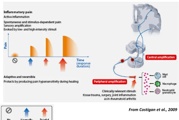
Inflammatory pain (Fig. 3) is caused by tissue damage and by the subsequent inflammatory response. The first lesion and the inflammatory process cause alterations in the activities of Aδ and C fibers. These fibers are responsible for sensitization, recruitment of nociceptors normally silent and activation of ionic channels and membrane receptors. Tipically, inflammatory pain disappears after resolution of the initial tissue injury. However, in some situations, chronic pain may persist after inflammation disappears, due to permanent changes in the nerve fibers (Michaud et al., 2007; Pace et al., 2006).
MSc Thesis Central effects of insulin and IGF1 in diabetic neuropathy
Fig. 3: Inflammatory pain.
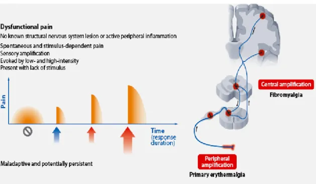
Dysfunctional pain (Fig. 4) is a group of pain syndromes caused by malfunction of the somatosensory system. This malfunction can be considered a disease in its own right (Costigan et al., 2009). Dysfunctional pain occurs in situations in which there is no identifiable noxious stimulus nor any detectable inflammation or damage to the nervous system. It is unclear in most cases what causes the manifestation or persistence of dysfunctional pain. This type of pain appears to result from an autonomous amplification of nociceptive signals inside the CNS (Nielsen et al., 2008) with a disturbed balance of excitation and inhibition in central circuits (Julien et al., 2005).
Neuropathic pain (Fig. 5), the pain state that will be addressed in this Master thesis, is defined by the IASP as “pain initiated or caused by a primary lesion or dysfunction in the nervous system” (Merskey and Bogduk, 1994). Neuropathic pain is a subcategory of the larger group of pain syndromes and can be divided into peripheral and central neuropathic pain. The former results from lesions to the peripheral nervous system (PNS) caused by metabolic diseases, mechanical trauma, neurotoxins, infection, or tumor invasion (Dworkin et al., 2003; Woolf and Mannion, 1999). Central neuropathic pain most commonly results
MSc Thesis Central effects of insulin and IGF1 in diabetic neuropathy from spinal cord injury, stroke, or multiple sclerosis (Ducreux et al., 2006). In both cases, the primary disease (such diabetes mellitus) and the neural damage it causes are only the initiators of a cascade of changes that induce and sustain neuropathic pain.
Fig. 4: Dysfunctional pain.
Fig. 5: Neuropathic pain.
MSc Thesis Central effects of insulin and IGF1 in diabetic neuropathy
1.1.2 Ascending sensory pathways
The ascending sensory pathways are the main avenues by which information concerning the body’s interaction with the external environment, its internal condition, and the position and movement of its parts, reach the brain.
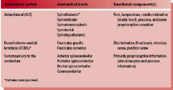
Anatomically, the ascending sensory systems consist of three distinct pathways (Table 1): the anterolateral system (ALS), the dorsal column-medial lemniscal (DCML) pathway, and the somatosensory pathways to the cerebellum. The anterolateral system, which includes the spinothalamic, spinoreticular, spinomesencephalic, spinotectal, and spinohypothalamic tracts, conveys predominantly pain and temperature sensation, as well as nondiscriminative touch, pressure, and some proprioceptive sensation. The dorsal column medial lemniscal pathway (which includes the fasciculus gracilis, fasciculus cuneatus, and medial lemniscus) relays discriminative (fine) tactile sense, vibratory sense, and position sense. The somatosensory pathways to the cerebellum, which include the anterior, posterior, and rostral spinocerebellar, as well as the cuneocerebellar tracts, relay primarily proprioceptive (but also some pain and pressure) information (Patestas and Gartner, 2006).
Table 1: General description of anatomical and functional aspects of the ascending sensory pathways.
MSc Thesis Central effects of insulin and IGF1 in diabetic neuropathy The output from the dorsal horn to higher centres in the brain is carried by projection neurons located at the spinal cord along ascending pathways. These ascending pathways carries primarily sensory information and so provides the sensory component of the pain experience (Mello and Dickenson, 2008).
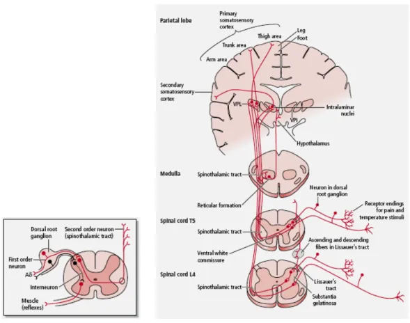
The anterolateral system (ALS) is the main ascending nociceptive pathway and transmits nociceptive, thermal and nondiscriminatory touch information to higher brain centers, generally by a sequence of three neurons and interneurons (Fig. 6). The sequence of transmission consists of a first order neuron whose cell body is located in the dorsal root ganglion and transmits sensory information from peripheral structures to the dorsal horn of the spinal cord. These neurons have two main types of fibers: the fast-conducting myelinated Aδ fibers and the slow-conducting unmyelinated C fibers. The second order neuron has its cell body within the dorsal horn of the spinal cord. Their axons usually decussates and ascends in the direct pathway of the ALS (spinothalamic tract) to synapse in the contralateral thalamus, and sending some collaterals to the reticular formation (as it ascends through the brainstem); in the indirect pathway of the ALS (spinoreticular tract) to synapse in the reticular formation, and sending some collaterals to the thalamus; or as spinomesencephalic, spinotectal, or spinohypothalamic fibers to synapse in several brainstem nuclei. The third order neuron has its cell body in the thalamus, and their axon ascends ipsilaterally to terminate in the somatosensory cortex (Patestas and Gartner, 2006).
Approximately 85% of the nociceptive fibers from the spinal cord ascending in the ALS, terminate in the brainstem reticular formation. From there, the information eventually reaches the thalamus via multiple additional synapses that occur in the brainstem. The reticular formation sends fibers transmitting nociceptive input not only to the thalamus but also to the hypothalamus, which is associated with the autonomic and reflex responses to nociception, and the limbic system, which mediates the emotional component of nociception.
MSc Thesis Central effects of insulin and IGF1 in diabetic neuropathy
Adapted from Patestas and Gartner, 2006
Fig. 6: Ascending sensory pathway that transmits nondiscriminative touch, pain, and temperature sensations from the body.
1.1.3 Descending nociceptive pathways
Studies that link anatomy and pharmacology of dorsal horn neurons and descending facilitatory pathways provided a better understanding of the neuronal plasticity associated with a peripheral insult. The supraspinal structures receive and integrate nociceptive information to regulate behaviour, and are potential sources of descending influence on nociceptive processes.
Supraspinal control of nociception at the spinal cord originates from many brain regions and plays a critical role in determining the experience of both acute and chronic pain (Heinricher et al., 2009). This descending control can be inhibitory or facilitatory and originate from a number of supraspinal sites like periaqueductal gray matter (PAG), parabrachial nucleus, nucleus tractus solitarius, nucleus raphe magnus, rostroventromedial medulla (RVM), noradrenergic cell groups (A5, A6 and A7), dorsal reticular nucleus (DRt) and ventrolateral medulla (VLM) (Yoshimura and Furue, 2006) (Fig. 7). Under normal conditions,
MSc Thesis Central effects of insulin and IGF1 in diabetic neuropathy a “balance” is maintained between inhibitory and facilitatory pathways. When the equilibrium is disturbed, for example, after a disease like diabetic neuropathy, the excitation dominates and, consequently, pain is potentiated in intensity or persistence (Porreca et al., 2002; Suzuki et al., 2004).
From Tracey and Mantyh, 2007
Fig. 7: Descending pain modulatory system. ACC - anterior cingulated cortex; DLPT - dorsolateral pontine tegmentum; NCF - nucleus cuneiformis; PAG - periaqueductal gray matter; +/- indicates both pro- and anti- nociceptive influences, respectively.
1.1.3.1 PAG-RVM system
The best studied descending pain control is the PAG-RVM system. The periaqueductal gray (PAG) area is subdivided in dorsal medial PAG (DMPAG), dorsal lateral PAG (DLPAG), lateral PAG (LPAG) and ventrolateral PAG (VLPAG), according to Paxinos and Watson (Paxinos and Watson, 2005). The PAG has a key role in descending mechanisms that modulate spinal nociceptive activity and is interconnected with the hypothalamus and limbic forebrain structures including the amygdala and also receives direct spinomesencephalic input (Fields, 2000). The VLPAG plays a crucial role in pain control (Behbehani, 1995). The
MSc Thesis Central effects of insulin and IGF1 in diabetic neuropathy PAG projects to the RVM, which, in turn, sends output to spinal cord laminae important in nociceptive function. The PAG control of dorsal horn responses is highly selective for noxious inputs. Understanding the PAG-RVM system is of considerable importance from both a behavioral and therapeutic point of view.
As previously stated, the PAG does not project directly to the spinal cord. Instead, its principle descending projection is to the RVM, which can be considered the output of the midline pain-modulation system. The RVM includes the serotonin-rich nucleus raphe magnus and adjacent reticular formation, and projects diffusely to dorsal horn laminae important in nociceptive processing, including the superficial and deep dorsal laminae. It also receives dense input of noradrenergic nerve fibers arising from the A1, A2, A5, A6 and A7 cells groups (Herbert and Saper, 1992; Kwiat and Basbaum, 1990).
Fields and Colleagues (1983) have characterized cells in the RVM that may constitute the physiological basis for generation of biphasic modulation of spinal nociceptive transmission. They have operationally defined three classes of neurons, named “ON-cells”, “OFF-cells” and “NEUTRAL-cells”. “ON-cells” are the facilitating outputs from the RVM whereas “OFF-cells” function as the antinociceptive output (inhibits nociceptive transmission in the spinal dorsal horn). Neutral-cells had no consistent correlated response with behavior reactions and could represent a subtype of ON- or OFF-cells (Ellrich et al., 2000).
It is thought that neutral-cells can be recruited to become ON- or OFF-cells during the development of chronic pain states. Manipulations that increase nociceptive responsiveness, thus indicating facilitation, also increase ON-cell activity, whereas the opposite occurs with OFF-cells which are inhibited by morphine antagonists. These experimental outcomes suggest that supraspinal sites can contribute to development or maintenance of exaggerated pain behaviors produced by noxious (e.g. hyperalgesic) and non-noxious (e.g. allodynic) peripheral stimuli, even in the absence of an obvious pathology (Urban and Gebhart, 1999).
1.1.3.2 Noradrenergic system
Catecholamines, such as adrenaline and noradrenaline (NA), modulate noxious transmission at the spinal dorsal horn. Noradrenergic cell groups involved in pain modulation encompasse mainly the A1 to A7 cell groups and are located in the brainstem. Although the origin of noradrenaline at the spinal cord is supraspinal, a few spinal neurons, mostly near the central canal, also possess NA (Willis and Coggeshall, 2004).
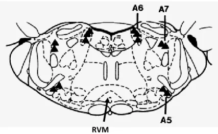
MSc Thesis Central effects of insulin and IGF1 in diabetic neuropathy The A1 cell group is located ventrolaterally at the medulla oblongata at the level of the area postrema, A2 is distributed throughout the dorsal vagal complex, A3 is in the medullary reticular formation, and A4 surrounds the fourth ventricle. The A5 cell group is in the ventrolateral pons, A6 (locus coeruleus and nucleus subcoeruleus) is dorsally in the pons and A7 is in the lateral part of the pons, close to the lateral lemniscus (Fig. 8). The A5, A6 and A7 noradrenergic cell groups have significant descending noradrenergic projections to the spinal dorsal horn and are designated as the descending noradrenergic pain inhibitory pathway (Fig. 8). Evidence for a relevant role of the A5, A6 and A7 in descending pain modulation comes from studies showns that stimulation of those areas induces analgesia, which can be blocked by intrathecal administration of α2-adrenoceptors antagonists (Pertovaara, 2006).
At the periphery, the sympathetic system is the main neuronal source of noradrenaline (Pertovaara, 2006). Several studies, such as does done by McLahlan et al. (1993) and Perl (1999), have demonstrated that NA released from sympathetic nerve terminals produces hyperalgesia by acting on the dorsal root ganglion cells and the peripheral nerves under pathological conditions such as nerve injury.
From Pertovaara, 2006 Fig. 8: Location of noradrenergic A5, A6 and A7 cell groups in the pons.
a)
Noradrenaline
Noradrenaline has been implicated in the modulation of endogenous analgesic mechanisms via the descending inhibitory pain pathways in the brain and spinal cord. An imbalance in these inhibitory mechanisms may contribute to central sensitization and hyperexcitability of the spinal and supraspinal pain transmitting pathways leading to
MSc Thesis Central effects of insulin and IGF1 in diabetic neuropathy persistent pain (Basbaum and Fields, 1984; Clark and Proudfit, 1993; Fields and Basbaum, 1999; Fields et al., 1991).
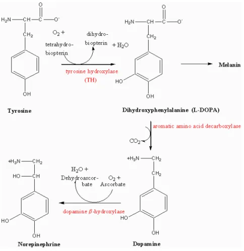
Noradrenaline, as other catecholamines dopamine and epinephrine, possesses two hydroxyl groups and one amine group bound to a benzene ring. It is biosynthesized from tyrosine that is first converted to dihidroxyphenylalaline (DOPA) by tyrosine hydroxylase (TH). After that, DOPA is converted to dopamine by aromatic amino acid decarboxylase. In noradrenergic cells dopamine is further converted to noradrenaline by dopamine-beta-hydroxylase (Fig. 9) (Pertovaara, 2006).
From www.rpi.edu/dept/bcbp/molbiochem/BiochSci/sbello/tyr_norep.gif Fig. 9: Structure and synthesis of noradrenaline.
Blockade of peripheral noradrenergic receptors by antidepressants may contribute to a peripheral analgesic action because, as already mentioned, peripheral release of noradrenaline and serotonin is known to be hyperalgesic (Mico et al., 2006).
MSc Thesis Central effects of insulin and IGF1 in diabetic neuropathy
b) Spinal and supraspinal mechanisms of pain modulation
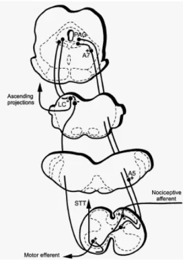
The source of spinal noradrenaline is descending axons originating in the noradrenergic neuronal cell groups of the brainstem (Fig. 10) (Jones, 1991; Proudfit, 1988), particularly the locus coeruleus, but also the A5 and A7 noradrenergic cell groups (Kwiat and Basbaum, 1992). Stimulation of these noradrenergic cell groups produces spinal release of noradrenaline and are connected with other pain control centers that receive projections from the PAG (Bajic and Proudfit, 1999). Curiously, the noradrenergic innervation of the spinal cord differs according to the rat strain, which was shown to affect descending pain modulation (West et al., 1993; Clark and Proudfit, 1992).
The distribution of various adrenoceptor types varies with descending pain modulation structure (Day et al., 1997; Nicholas et al., 1993; Scheinin et al., 1994; Wang et al., 1996) and has a role in pain control.
The spinal dorsal horn is a critical link for all ascending pain pathways and the spinal cord receives strong innervation from descending noradrenergic pathways.
As will be mentioned below, noradrenaline receptors are classically divided into two main categories: alpha- and beta-adrenoceptors. Alpha-adrenoceptors are classified into subtypes alpha- 1A, 1B, 1D, 2A, 2B and 2C, and beta-adrenoceptors into subtypes beta- 1, 2 and 3 (Aantaa et al., 1995; Ruffolo and Hieble, 1994). Like 5-HT, NA can variably influence nociceptive processing, depending on the mix and subtype of adrenoceptors activated, as well as on the presence, duration, and nature of pain.
It has been demonstrated that electrical stimulation of the noradrenergic A5, A6 and A7 cell groups produces antinociceptive effects (Burnett and Gebhart, 1991; Yeomans et al., 1992; West et al., 1993; Tsuruoka et al., 2004). These antinociceptive actions involve not only direct membrane hyperpolarization and enhancement of inhibitory transmitter release, but also reduce the excitatory transmitter release. These presynaptic actions of NA are tested in SG that receives inputs predominantly from Aδ and C afferent fibers, which is known to carry the nociceptive information (Yoshimura and Furue, 2006). It has subsequently been shown that activation of α2-adrenoceptors within the noradrenergic cell groups of the brainstem may produce pronociceptive effects in some contitions (Ossipov and Gebhart, 1986; Pertovaara et al., 1994), contributing to maintenance of chronic pain and
MSc Thesis Central effects of insulin and IGF1 in diabetic neuropathy hyperalgesia (Baron, 2000). Spinal and supraspinal effects of noradrenaline are, therefore, likely to be opposite.
Ventrolateral pathways have a major role in mediating descending antinociceptive influences from the noradrenergic cell groups. This was shown by the finding that the antinociceptive effect induced by locus coeruleus stimulation is blocked by a lesion of the ventrolateral part of the spinal cord, but not of the dorsolateral funiculus (Mokha et al., 1986; Tsuruoka et al., 2004).
Fig. 10: Summary of the neuronal pathways from the PAG to noradrenergic neurons in the A5, A6 and A7 cell groups. STT - spinothalamic tract.
1.1.3.3 Serotoninergic system
The serotoninergic system has been recognized as one of the main neurotransmitter systems participating in pain transmission, processing and controlling. Both pro-nociceptive and anti-nociceptive effects have been attributed to serotonin (5-HT) depending on the site and the receptor subtype it acts on (Roberts, 1989; Eide and Hole, 1993; Kayser et al., 2010).
Similar to a noradrenergic system, the descending serotonin containing fibers arise from the nucleus raphe magnus (NRM), a nucleus located at the RVM, the main source of
MSc Thesis Central effects of insulin and IGF1 in diabetic neuropathy serotoninergic fibers in the spinal cord. These fibers innervate almost all brain areas, through its ascending and descending projections. Other serotoninergic nuclei, the raphe pallidus (RP) and the raphe obscurus (RO), are also within the vicinity of RVM. However, the contribution of the latter two nuclei is thought to be small (Millan, 2002; Basbaum et al., 1984; Ruda et al., 1986 and Ho et al., 1991).
Like the noradrenergic system, the serotoninergic fibers also descend through the dorsolateral funiculus and bifurcate their axon collaterals to innervate dorsal horn neurons, in particular lamina I and II, central canal, intermediolateral nucleus, and ventral horn (Millan, 2002; Basbaum et al., 1984). Traditionally, actions of 5-HT in the dorsal horn have been considered as dedicated to the suppression of nociceptive transmission (Basbaum and Fields, 1984; Eide and Hole, 1993), but the role of 5-HT depends on the spinal receptors (see bellow).
a)
Serotonin
Serotonin (5-hydroxytryptamine, 5-HT), a metabolite of the essential amino acid tryptophan, has been the subject of intense biological research since the early 1950s. With increasing knowledge, it has emerged as a mediator of several functions in the human body, including mood, appetite, regulation of gastrointestinal motility and pain modulation (Keszthelyi et al., 2009). At the periphery, the action of serotonin is mainly seen as an inflammatory mediator that is released from platelets and mast cells after tissue injury, and exerts direct actions (excitatory) on C-fibers (Eide and Hole, 1993; Dray, 1995).
Serotonin is synthesized from tryptophan through hydroxylation and decarboxylation. These processes are catalysed by the tryptophan hydroxylase (TpH) and the aromatic acid decarboxylase (AADC), respectively (Gershon and Tack, 2007; Lesurtel et al., 2008) (Fig. 11). Tryptophan hydroxylase (tryptophan 5- monooxygenase) belongs to a superfamily of aromatic amino acid hydroxylases, together with phenylalanine (PAH) and tyrosine hydroxylases (Fitzpatrick, 1999). Its expression is limited to a few specific cells: brainstem neurons, pinealocytes, mast cells, mononuclear leukocytes, intestinal enterochromaffin cells and bronchopulmonary neuroendocrine epithelial cells. There are two known isoforms of TpH. Tryptophan hydroxylase 1 (TpH1) is localized in enterochromaffin cells and the pineal gland and tryptophan hydroxylase 2 (TpH2) is present in neurons (Walther et al., 2003). Both isoforms employ molecular oxygen and the cofactor tetrahydrobiopterin to convert tryptophan to 5-hydroxytryptophan (5-HTP), which is then converted by AADC to serotonin.
MSc Thesis Central effects of insulin and IGF1 in diabetic neuropathy 5-HT is released from the synaptic vesicles to the synaptic space by a Ca2+-dependent process, while its reuptake from synaptic space to 5-HT neurons is carried out by the membrane-bound 5-HT transporter occurring in axons, bodies and/or dendrites of 5-HT neurons. 5-HT is catabolized by mitochondrial type A monoamine oxidase (MAO) (Filip and Bader, 2009).
Fig. 11: Structure and synthesis of serotonin.
5-HT can produce anti-nociceptive and pro-nociceptive effects depending on the targetted receptor. More than 15 receptor subtypes have been identified for 5-HT, providing it with a diverse repertoire in pain and other responses. It is noteworthy that pain, particularly chronic pain, can be accompanied by fear, anxiety, depression, aversion and sleep disturbances, which are all underpinned by monoamine-mediated neurotransmission. However, this complexity has caused problems in deducing the role of 5-HT in nociception, and earlier studies often presented conflicting views on the pro-nociceptive and anti-nociceptive actions of this transmitter (Millan, 2002).
Althought the physiological function of 5-HT containing cells in pain modulation has been questioned (Gao and Mason, 2000), at least a proportion of “on” cells of the RVM are likely to contain 5-HT. Tonic activation of 5-HT-mediated and non-5-HT-mediated brainstem facilitatory influences is one contributor to the development and maintenance of central sensitization in sustained pain states (Urban and Gebhart, 1999; Porreca et al., 2002).
From Gwaltney-Brant et al., 2000
(TpH)
MSc Thesis Central effects of insulin and IGF1 in diabetic neuropathy
b) Spinal and supraspinal mechanisms of pain modulation
Based on structural (amino acid sequence), biochemical (postreceptor mechanisms of signal transduction) and pharmacological differences, 5-HT receptors were classified into 7 families (5-HT1-5-HT7) and at least 14 different subtypes (Millan, 2002; Ruda et al., 1986;
Filip and Bader, 2009). All these receptors are expressed in the brain, but some subclasses are also expressed within the spinal cord. Among the 14 5-HT receptor families identified so far, much of the pain research has focused on 5-HT1, 5-HT2 and 5-HT3 receptors, but the role
played by other 5-HT receptors in nociception has been poorly or not thoroughly investigated (Colpaert, 2006; Eide and Hole, 1993; Kayser et al., 2010). Neurochemical findings indicate that stimulation of 5-HT1A, 5-HT1B and 5-HT1D receptors inhibits 5-HT release
in the brain, and stimulation of 5-HT4 and 5-HT6 receptors cause an increase in the levels of
this neurotransmitter. On the other hand, stimulation on 5-HT1A facilitates NA release in the
brain, and stimulation on 5-HT2A and 5-HT2C inhibits the release in this neurotransmitter
(reviewed by Filip and Bader, 2009).
Autoradiographic studies show that among these 14 subtypes, the 5-HT1A, 5-HT1B,
5-HT1D, and 5-HT3 binding sites are abundant in the spinal level, suggesting the presence of the
receptors on peripheral sensory nerves.
At the periphery, 5-HT7 receptors have been found in the superficial laminae of the
dorsal horn, postsynaptically in local interneurons and presynaptically in peptidergic fibers and in astrocytes. However, it may be also present in supraspinal areas. Available data suggest a pro-nociceptive role of 5-HT7 receptors when activation occurs at the periphery
(Meuser et al., 2002; Rocha-González et al., 2005). Activation of 5-HT2A and 5-HT3 receptor
subtypes present on C-fibers was already shown to underlie such a peripheral pro-nociceptive effect on 5-HT (Obata et al., 2000; Sommer, 2004).
Electrophysiological studies have disclosed the antinociceptive mechanisms of descending 5-HT systems in the spinal cord level. 5-HT directly hyperpolarize SG neurons, inhibit the glutamate release from the Aδ and C afferent fibers, and increase GABA and glycine release from interneurons. Although some excitatory effects have been reported, the overall effect of 5-HT tends to be anti-nociceptive, in a manner similar to what occurs with NA (Yoshimura and Furue, 2006).
MSc Thesis Central effects of insulin and IGF1 in diabetic neuropathy
1.2 Type I diabetes
1.2.1 General Considerations
Type 1 diabetes is one of the most widespread chronic illness in the world and, in general, is considered primarily a T-cell mediated disease arising through a complex interaction of immune, genetic and environmental factors, results from autoimmune destruction of insulin-producing ß-cells. An interplay between genetic susceptibility and environmental factors is thought to provide the fundamental element for disease and provides potential targets for both prediction and prevention of disease (Van den Driessche, 2009; Devendra, 2004). This disease occurs most commonly in people of European descendent and affects 2 million people in Europe and North America (Gillespie, 2006).
Type 1 diabetes has two types of complications: microvascular and macrovascular. Macrovascular complications include myocardial infarction, stroke and large vessel peripheral vascular disease. One of the most frequently-occuring microvascular complications is diabetic neuropathy (DN), that will addressed below.
“STZ-rats” is a common model of type I diabetes. Streptozotocin (STZ) selectively destroys pancreatic islet beta-cells, similar to what occurs in type I diabetes in humans. STZ-rats develop hyperglycemia and several complications typical of diabetes, such as diabetic neuropathy and retinopathy (Bhuyan et al., 1974; McCall, 1992).
1.2.2 Diabetic Neuropathy
Neuropathy is on of the most common complications associated with diabetes. It is characterized by sensory, autonomic and motor dysfunctions that may lead to debilitating clinical complications. Autonomic neuropathy is characterized by symptons ranging widely from minor papillary and sweating problems to significant disturbances in cardiovascular reflexes, alimentary, and genitourinary function (Schmidt et al, 1999). Sensory neuropathy is characterized by inability to perceive stimuli, such as pain and heat. Motor neuropathy is characterized by atrophy and weakness in muscles. Conduction velocity generally is reduced in sensory, motor and probably sympathetic nerves and synaptic transmission can be abnormal as well (Schiller and Rahamimoff, 1989). This decreased neuronal activity is
MSc Thesis Central effects of insulin and IGF1 in diabetic neuropathy consistent with the reduction in brain weight and neocortical volume, which is associated with a reduction of the number of cortical neurons (Jakobsen et al., 1987).
The predominant form of DN is a symmetric polyneuropathy that is primarily sensory-motor and often includes the autonomic system. Patients with this disease usually present a reduction or loss of small fibre-mediated sensation results in loss of pain sensation (heat pain, pin-prick) and temperature perception to cold (Aδ) and warm (C) stimuli. Evoked pain such as allodynia (pain due to a stimulus that does not normally cause pain) and hyperalgesia (severe pain due to a stimulus that normally causes slight pain), with a distal and symmetrical distribution, may be present (Otto et al., 2003; Ziegler, 2008). Typically, DN is more severe at night, and often prevents sleep causing a constant state of tiredness (Quattrini et al., 2003).
The prevalence of these neuropathies is higher in males and generally progress with age and duration of disease. At the time of diagnosis, neuropathy is present in 10% of diabetic patients but increases to 50% in patients with a 25-year history of the disease (Feldman et al., 1999).
In light of current knowledge, there is little understanding about the pathophysiology of painful diabetic neuropathy, especially as regards to its effects on the CNS. Hyperglycemia is usually cause of diabetic neuropathy. It is thought to envolve the following pathways: activation of the polyol pathway, non-enzymatic glycosylation (glycation) which leads to abnormal cross-linking of proteins and altered protein function along with an excessive production of reactive oxygen species (Oates, 2002; Obrosova, 2002; Ryle and Donaghy, 1995). However, other theories suggest that some mechanisms leading to neuropathy may be independent of the glycemic state. Although systemic or local hyperglycemia in non-diabetic animals can induce mechanical hyperalgesia, impaired sensory and motor nerve conduction velocity and nerve blood flow, recent studies shows that there is a strong possibility that lack of insulin itself, resistance to its actions, or decresead availability of its related molecules (the insulin growth factors-IGFs), contributes to diabetic neuropathy. This may be explaned by the fact that both insulin and IGF1 act as neuronal growth factors essential for proper neuronal activity (Ishii, 1995; White, 2003). Therefore, the severity of diabetic neuropathy may be dependent on the combined loss of insulin and IGF1 activities, beyond the effects of hyperglycemia (Ishii, 1995). These ligands are also involved in cell survival, synaptogenesis, neurite (axon and dendrite) outgrowth and nerve regeneration of neurons. To strengthen this theory, it was shown that systemic IGF1 levels in diabetic patients are slightly lower than those of nondiabetic individuals, and serum
MSc Thesis Central effects of insulin and IGF1 in diabetic neuropathy IGF1 levels in diabetes patients with DN are lower than those in diabetic patients without DN (Guo et al., 1999; Migdalis et al., 1995). In addition, the study by Schmidt et al. (2003), demonstrated that type 2 diabetic rats do not develop sympathetic neuroaxonal dystrophy (NAD), whereas rat models of type 1 diabetes suffer from NAD. This is due to the fact that type 1 diabetic rats do not possess insulin, whereas type 2 diabetic rats have some insulin.
A decline in insulin and IGF activity, as well as others neurotrophic molecules such neuronal growth factor (NGF) and neurotrophin 3 (NT-3) in diabetes, may lead to impaired production and axonal transport of tubulins and neurofilaments, diminished microtubule and neurofilament contents, and a dwindling of axonal diameters. Diminished axonal transport and/or receptor tyrosine kinase activity may contribute to the slower conduction velocity seen early in diabetes prior to the detection of structural alterations in axons (Ishii, 1995).
Intensive insulin therapy has been shown to reduce the incidence of neuropathy in a population of type 1 diabetic patients (DCCT Research Group, 1993). The effects of IGF1 in the treatment of diabetic neuropathy in humans remain unknown.
1.2.2.1 Treatment
One common comorbid psychiatric diagnosis encountered in patients with dysfunctional and neuropathic pain is depression. Several drugs appear to have different efficacy on the various symptoms and signs of diabetic neuropathic pain (Table 2).
Table 2: Range of treatments available for neuropathic pain.
MSc Thesis Central effects of insulin and IGF1 in diabetic neuropathy The drugs that are used for alleviating DN can be broadly divided into two groups: conventional analgesics, such as tramadol and oxycodone, and adjunctive agents, such as antidepressants and anticonvulsants (Chong and Hester, 2007). Several authors consider the tricyclic antidepressants (TCA) to be the drug treatment of choice for neuropathic pain. The TCAs have numerous mechanisms of action including inhibition of noradrenaline and 5-HT uptake from the synaptic cleft (Finnerup et al., 2005; Finnerup and Jensen, 2006; Jensen et al., 2006). However, their use is limited by high rates of adverse events and several contraindications. The light of current knowledge, antidepressants with dual selective inhibition of serotonin and noradrenaline (serotonin noradrenaline reuptake inhibitors-SNRIs), such as duloxetine and vanlafaxine, proved to be the most effective for relieving the pain of diabetic neuropathy (Goldstein et al., 2005). These antidepressants relieve pain by increasing synaptic availability of noradrenaline and 5-HT in the spinal cord, which inhibit locally nociceptive transmission and reduce pain perception (Fig. 12) (Jensen et al., 2006).
According to Park HJ. et al, 2010, TCAs, gabapentin and pregabalin are recommended to be the first-line treatment options for patients with neuropathic pain, whereas opioid analgesics should be reserved as second or third-line options in most cases.
Fig. 12: General principles of pharmacotherapy for DN.
MSc Thesis Central effects of insulin and IGF1 in diabetic neuropathy
1.2.3 Neurochemistry and pharmacology
1.2.3.1
The Insulin/IGF system
Insulin, insulin-like growth factor 1 (IGF1) and insulin-like growth factor 2 (IGF2) are three closely polypeptide hormones with amino acid sequences considerably homologous that mediate a variety of metabolic and mitogenic effects by binding to their specific receptor tyrosine kinases present on the surface of target tissues and cells (Joan and Michael, 1981; Ursula et al., 2009). Insulin is synthesized predominantly in pancreatic beta cells from which its release is regulated by nutrient stimuli. IGF1 is synthetized by the liver upon stimulation by pituitary growth hormone (GH). It is also synthesized locally in many tissues, including the brain (Bondy and Cheng, 2004).
Because of the considerable structural homology of insulin and IGF1 receptors, a distinct overlap exists in binding affinity between insulin and IGFs (Fig. 13). Although some studies have indicated that the IGF-binding sites in some tissues exhibit a certain degree of affinity for insulin, and the high affinity insulin receptor specifically interacts with IGF1 and IGF2 (Zapf, 1978), at physiological concentrations, insulin and IGF1 exclusively bind to their cognate receptors (Ursula et al., 2009). Differences in kinetics of ligand binding and receptor activation by the insulin and IGF1 receptors may be one of the factors determining their specificity (Shymko et al., 1997).
There are three separate receptors that can bind insulin and the IGFs: insulin receptor (IR) (Ullrich et al, 1985; Ebina et al, 1985), IGF1 receptor (IGF1R) (Ullrich et al, 1986) and IGF2 receptor (IGF2R) (Morgan et al, 1987). A fourth, orphan member of the family is insulin receptor-related receptor (IRR), that it is thought to mediate the action of a ligand(s) identical or very similar to insulin, IGF1 or IGF2 (Fig. 14) (Shier and Watt, 1989). In addition to IGF1R and IGF2R, the IGF system includes 6 IGF-binding proteins (IGFBP1 to IGFBP6), which in addition to their main role as carriers of IGF1 and IGF2 in the circulation, also regulate the biological actions of IGFs either by inhibiting or potentiating their binding to cell surface receptors (Baserga et al., 1999).
Insulin and type I IGF receptors are structurally similar, both being comprised of α2ß2
heterotetramers. Like the insulin receptor, the IGF1 receptor is a membrane glycoprotein of Mr 300000-350000, consisting of two α-subunits (Mr~135000), that contains the extracellular ligand binding domain, and two ß-subunits (Mr~90000) that are connected by disulfide bonds to form the functional ß-α-α-ß heterotetrameric receptor complex
MSc Thesis Central effects of insulin and IGF1 in diabetic neuropathy (Massague and Czech, 1982; Kull et al., 1983). The intracellular domain of the membrane-spanning ß-subunits contains a tyrosine kinase consensus sequence that can be activated by ligand and binding. The type II IGF receptor is comprised of a single polypeptide that does not have a tyrosine kinase domain (Ishii, 1995). Ligand binding triggers a conformational change that enables the receptors to bind ATP. Autophosphorylation increases their kinase activity and catalyses substrate phosphorylation to engender growth or metabolic responses.
Fig. 13: Structural homology between insulin and IGF1 receptors.
Despite the functional and structural similarities, these two hormones are thought to play different biological roles during mammalian development and mature life. Whereas insulin plays a key role in regulation of a variety of metabolic processes, the IGFs appear to be more potent in regulation of cellular growth and differentiation. This is consistent with the fact that both IGF1 and IGF2 are expressed in various embryonic and adult tissues (Ullrich et al., 1986; Shin et al., 1994).
Autoradiographic studies reveal the widespread distribution of insulin, type I IGF and type II IGF receptors throughout the brain, suggesting a role in the CNS (Bondy et al., 1992).
MSc Thesis Central effects of insulin and IGF1 in diabetic neuropathy
From Kitamura et al, 2003 Fig. 14: Interactions among ligands and receptors of the insulin family.
The decline in the levels of growth factors necessary for peripheral nerve function has been postulated as one of the metabolic alterations that originate nerve dysfunction and subsequently diabetic neuropathy (Kamiya et al., 2006). In particular, decreased IGF1 levels have been postulated as a possible cause of neurological disorders in diabetes (Guo et al., 1999).
1.2.3.2
Noradrenaline and serotonin
Studies with STZ-rats showed that type I diabetes induces changes in the neurotransmitter content of brainstem, but the results are not consistent. In the study made by Padayatti and Paulose (1999), the STZ rats was shown an increase in 5-HT content in brainstem of diabetic rats, that was associated with a reduced affinity for serotonergic receptors. The insulin treatment with doses that do not reverse hyperglycemia reversed that parameter. On the other hand, in the same study, the NA content in diabetic brainstem was not affected. This last result is not, however, concensual since other study has shown that there is a reduction in noradrenergic neuronal activity in diabetic neuropathy, with a reduction in noradrenaline synthesis, associated with a difficulty to release this neurotransmitter from synaptic terminal. In the study led by Gallego et al (2003), diabetes altered NA levels in the sympathetic nervous system but not in large regions of the brain (medulla, pons, midbrain and striatum) of diabetic rats.
MSc Thesis Central effects of insulin and IGF1 in diabetic neuropathy
Chapter 2: Aims
The effects of diabetic neuropathy have been widely studied at the periphery but its central causes remain understudied. Since several studies support the hypothesis that deficiencies in insulin and/or IGF1 signaling may play an important role in the development of diabetic neuropathy in diabetes type I, independently of glycemic condition, we designed an experimental protocol to evaluate the effects of insulin and IGF1 treatment of a rat model of type I diabetes (the STZ-diabetic rat) in a paradigm that does not revert the hyperglycemic state of the animals. The main objectives of this study were to evaluate the effects of low doses of insulin and IGF1 in:
1) Pain behavioral responses. The pain behavioral responses will be assessed using the Randall-Selitto test, that measures mechanical hyperalgesia, and the formalin test, that is a widely employed mean to measure responses to a persistent noxious stimulus.
2) Neuronal activity at spinal cord and PAG, using c-fos expression to monitor neuronal activation. Fos is a nuclear protein encoded by the proto-oncogene c-fos, which is visualised by immunocytochemical techniques. Fos protein levels increase in the brain and spinal cord, in response to various types of noxious stimuli. c-fos expression is currently used to study nociception and has been employed as a functional marker of nociceptive activation of large neuronal populations (Harris, 1998).
3) The expression of NA and 5-HT in brainstem areas that project directly to the spinal cord and are involved in pain modulation. The numbers of noradrenergic neurons in the A5, A6 and A7 cell groups of the brainstem were counted after immunostaining for TH. As to 5-HT, the numbers of serotoninergic neurons were quantified at the RVM after immunostaining for TpH. The spinal levels of NA and 5-HT were quantified using appropriated ELISA kits.
MSc Thesis Central effects of insulin and IGF1 in diabetic neuropathy
Chapter 3: Material and Methods
1. Drugs
Streptozotocin was purchased from Sigma–Aldrich (St. Louis, USA); insulin was
purchased from Sanofi Aventis and IGF1 was offered by Ipsen Portugal.
2. Animals
A total of 40 male Wistar rats (Charles River Laboratories, Barcelona, Spain), weighing 300–350 g at the beginning of the experiments, were used. The experiments were performed in accordance with the ethical guidelines of the European Community Council Directive 86/609/EEC and the ethical guidelines for the study of pain in conscious animals (Zimmermann, 1983).
Animals were housed two per cage, in a room with a constant temperature (22 ± 2 ◦C) and humidity (55 ± 5%) and with a 12-h light/dark cycle, and received food and water ad libitum.
3. Induction of diabetes
Experiments started 1 week after the rats were fully adapted to their environment. Diabetes was induced by an intraperitoneal (i.p.) injection of STZ (60 mg/kg body weight) dissolved in citrate buffer (pH = 4.5) (STZ group; n = 30). Controls received equal volumes of citrate buffer (pH = 4.5) (control group; n = 10). Three days after the injection, glucose concentration was measured in tail vein blood samples using a glucose oxidase impregnated test strip (Accu Chek Sensor Comfort, Roche Diagnostics, Germany). Only rats with glucose concentration higher than 270 mg/dl were considered diabetic and included in the study. All animals were monitored daily and weighed regularly during the study period.
4. Treatment
MSc Thesis Central effects of insulin and IGF1 in diabetic neuropathy
Fig. 15: Schematic overview of the experimental protocol. R.S. - Randall-Selitto; F.T. - Formalin Test.
Diabetes was induced in day 0 and afterwards, the diabetic rats were divided into three subgroups: treated with insulin (STZ + INS; n = 10), treated with IGF1 (STZ + IGF1; n = 10), and injected withvehicle (STZ + saline; n = 10).
Mechanical nociception was assessed for the first time on day 0. On day 7, treatment of various STZ groups started. Diabetic rats were subcutaneously injected 3 times per week during 3 weeks with 2 IU of insulin, 2,5 mg/Kg of IGF1, and 300 µl of saline. Control rats received an equivalent volume of vehicle. The injections were performed always at the same time of the day.
All rats were weekly tested for evaluation of mechanical
nociception during the 4 weeks of the study, using the Randall Selitto test (see “Behavioral tests”). Blood glucose concentration was also assessed, immediately before the injection of insulin, the IGF1 or saline to confirm, if the hyperglycemia was maintained in diabetic rats, in spite of the treatment protocols.
MSc Thesis Central effects of insulin and IGF1 in diabetic neuropathy
5. Behavioral tests
After i.p injection, all animals were evaluated once a week to confirm the existence of behavioral signs of diabetic neuropathy in diabetic rats, using the Randall-Selitto test (Fig. 14).
All animals were habituated to handling and testing environment before the behavioral evaluation was started.
5.1 Randall-Selitto test:
Mechanical nociceptive thresholds were quantified by means of an Analgesymeter (Ugo-Basile, Comerio, Italy). All animals were subjected to a time-course behavioural evaluation of mechanical nociception, at 1, 2, 3 and 4 weeks after STZ or saline injection. An increasing pressure was applied on the dorsal area of the hindpaw with a cone-shaped plunger. The nociceptive threshold was defined as the force, in grams, at which the rat attempted to withdraw the paw, vocalized or struggled. The mechanical threshold of each animal was the average of three consecutive measurements, taken at 5-min intervals.
Fig. 16: Photograph of a rat in the Randall-Selitto test.
5.2 Formalin-evoked flinching:
Four weeks after of diabetes induction, all groups of animals (n = 5 per group) were singly housed in Perspex boxes (Fig. 17) and given 60 min to habituate to the testing environment. Animals were then injected sub-dermally, into the right hindpaw dorsum, with
MSc Thesis Central effects of insulin and IGF1 in diabetic neuropathy 50 µl of 0.2 % formalin. Flinching behaviors were counted in successive periods of 5-min during 60 min (Zhao et al., 2006; Ceseña and Calcutt, 1999).
Photo kindly provided by José Lopes Fig. 17: Photograph of three rats in Perspex boxes during the formalin test.
6. Harvesting of biological material
Two hours after formalin injection, animals from each experimental group were sacrificed by transcardiac perfusion to perform the immunohistochemistry for c-fos expression at the spinal cord and PAG and for TH (noradrenergic marker) and TpH (serotoninergic marker) in the brainstem. The perfusions were performed after anaesthesia with an i.p. injection of 35% chloral hydrate (1ml/kg body weight). Vascular perfusion was performed with 200 ml of phosphate-buffered saline (PBS), followed by 1000 ml of 4% paraformaldehyde in 0.1 M phosphate buffer (PB), pH 7.4 (Fig. 18 A-C).
Spinal segments L4-L5 (which receive input from the injected paw; Grant and Robertson, 2004), and the brainstem were removed, post-fixed for 2–4 h in the same fixative, and cryoprotected overnight in 30% sucrose in 0.1 M PBS, at 4ºC (Fig. 18 D-E). Coronal sections, 40 µm thick, were obtained using a freezing microtome, and every fourth section was collected in 0.1 M PBS.
One additional set of animals (n=5 per group) were sacrificed by decapitation and the lumbar spinal segments (L4-L5) were removed immediately. Then, the dissected tissue samples were stored at -80ºC until analysis with ELISA kits, for noradrenaline and 5-HT quantification.
MSc Thesis Central effects of insulin and IGF1 in diabetic neuropathy
Photos kindly provided by Diana Nascimento
Fig. 18: Sequential pictures that represent some of the steps performed during the transcardiac perfusion. A - The anesthetized rat were placed in a cork board where they underwent the perfusion; B - With the aid of forceps and scissors, cut up the chest by the diaphragm; C - The excess blood was removed by passing a washing solution (Tyrode) in the cardiovascular tissue of the rat. D - Dissection of the brain is done carefully with the help of a gouge suitable for this purpose; E and F - The ganglia and the spinal cord were removed from the remaining tissue of the animal.
7. Immunocytochemistry
7.1 Processing of spinal cord and PAG section for c-fos Evaluation
Fos protein was immunohistochemically detected by the avidin - biotin - peroxidase method, using the compound 3,3´-diaminobenzidine (DAB) as chromogen, as described previously (Morgado and Tavares, 2007; Morgado et al., 2009). Briefly, the sections were treated with 1% hydrogen peroxidase for 10 min to inhibit endogenous peroxidase activity.
In order to decrease background staining, incubation in primary antibody was preceeded by immersion the sections in a blocking solution of 10% normal swine serum (NSS, serum produced in the animal species from which the respective secondary antibody was obtained) in 0.3% Triton X 100 in PBS (PBST) with 0.1 M glycine for 2 h.