https://doi.org/10.1177/2048872617716387
European Heart Journal: Acute Cardiovascular Care 1 –6
© The European Society of Cardiology 2017 Reprints and permissions:
sagepub.co.uk/journalsPermissions.nav DOI: 10.1177/2048872617716387 journals.sagepub.com/home/acc EUROPEAN SOCIETY OF CARDIOLOGY®
Background
In the context of acute coronary syndrome (ACS), the inci-dence of high-degree atrioventricular (AV) block is 3–14%, complete AV block being the more severe manifestation of AV conduction disturbance. The presence of heart block has been associated with worse short-term prognosis regardless
Complete atrioventricular block
in acute coronary syndrome:
prevalence, characterisation and
implication on outcome
Silvia Aguiar Rosa, Ana Teresa Timóteo, Lurdes Ferreira,
Ramiro Carvalho, Mario Oliveira, Pedro Cunha,
André Viveiros Monteiro, Guilherme Portugal,
Luis Almeida Morais, Pedro Daniel and Rui Cruz Ferreira
Abstract
Purpose: The aim was to characterise acute coronary syndrome patients with complete atrioventricular block and to
assess the effect on outcome.
Methods: Patients admitted with acute coronary syndrome were divided according to the presence of complete
atrioventricular block: group 1, with complete atrioventricular block; group 2, without complete atrioventricular block. Clinical, electrocardiographic and echocardiographic characteristics and prognosis during one year follow-up were compared between the groups.
Results: Among 4799 acute coronary syndrome patients admitted during the study period, 91 (1.9%) presented with
complete atrioventricular block. At presentation, group 1 patients presented with lower systolic blood pressure, higher Killip class and incidence of syncope. In group 1, 86.8% presented with ST-segment elevation myocardial infarction (STEMI), and inferior STEMI was verified in 79.1% of patients in group 1 compared with 21.9% in group 2 (P<0.001). Right ventricular myocardial infarction was more frequent in group 1 (3.3% vs. 0.2%; P<0.001). Among patients who underwent fibrinolysis complete atrioventricular block was observed in 7.3% in contrast to 2.5% in patients submitted to primary percutaneous coronary intervention (P<0.001). During hospitalisation group 1 had worse outcomes, with a higher incidence of cardiogenic shock (33.0% vs. 4.5%; P<0.001), ventricular arrhythmias (17.6% vs. 3.6%; P<0.001) and the need for invasive mechanical ventilation (25.3% vs. 5.1%; P<0.001). After a propensity score analysis, in a multivariate regression model, complete atrioventricular block was an independent predictor of hospital mortality (odds ratio 3.671;
P=0.045). There was no significant difference in mortality at one-year follow-up between the study groups.
Conclusion: Complete atrioventricular block conferred a worse outcome during hospitalisation, including a higher
incidence of cardiogenic shock, ventricular arrhythmias and death.
Keywords
Acute coronary syndrome, complete atrioventricular block
Date received: 15 August 2016; accepted: 31 May 2017
Cardiology Department, Santa Marta Hospital, Portugal
Corresponding author:
Sílvia Aguiar Rosa, Santa Marta Hospital, Avenida Oscar Monteiro Torres, n. 51, 3 esquerdo, 1000-217 Lisbon, Portugal,
Email: [email protected]
of the location of myocardial infarction. The impact on long-term prognosis remains uncertain but it seems to be more relevant in anterior myocardial infarction.1–9 However,
the majority of previous studies dedicated to this complica-tion were performed before the introduccomplica-tion of primary per-cutaneous coronary intervention (PCI). The aim of the present study was to assess the incidence of complete AV block in patients admitted with ACS, predominantly treated with primary PCI, to characterisze this population and eval-uate the prognosis including short and long-term survival.
Methods
A prospective analysis of patients admitted with ACS was carried out at a tertiary centre from 2005 to 2015.
At admission patients presented with symptoms thought to be due to acute coronary artery disease and electrocar-diographic changes consistent with and/or elevated bio-markers of myocardial necrosis. This study included patients with ST-segment elevation myocardial infarction (STEMI), non-ST-segment elevation myocardial infarction (NSTEMI) and unstable angina (UA). STEMI was defined as a ST-segment elevation for more than 30 minutes and the remaining cases were considered non-ST-segment ACS, NSTEMI if troponin raised the upper reference limit and UA if there were no changes in biomarkers.
Patients were divided according to the presence of com-plete AV block: group 1 (G1), with comcom-plete AV block; group 2 (G2), without complete AV block.
In each patient, baseline clinical characteristics including demographic characteristics and comorbidities were col-lected. Laboratory data on admission, electrocardiographic and echocardiographic parameters were also analysed.
The outcome variables studied were heart failure, car-diogenic shock, mechanical complication, ventricular arrhythmias, the need for invasive mechanical ventilation, stroke, major bleeding and acute kidney disease. All-cause mortality was evaluated during hospitalisation, at 30 days after discharge and at one-year follow-up.
Follow-up evaluation by telephonic contact was per-formed to check the occurrence of cardiac events and death, at 30 days and one year after discharge.
The study protocol is according to the Declaration of Helsinki.
Statistical analysis
Statistical analysis was performed using dedicated software SPSS Statistics (v.19; IBM SPSS, Chicago, IL, USA). Continuous variables were expressed as mean ± standard deviation and categorical variabled were expressed as a percentage. Study groups were compared using Student’s t test or Wilcoxon–Mann–Whitney’s test for continuous var-iables, and Pearson’s chi-square or Fisher’s exact test for categorical measures, as appropriate.
Propensity score analysis was used to compare both groups adjusting for confounding factors, using one to one match between patients with and without complete AV block. The analysis was performed using the R open source statistical programming language and the package MatchIt. From the data of propensity score analysis a multivariate regression model was built to identify the predictors of hospital mortality, using clinical character-istics at presentation, including gender, age, anterior STEMI, inferior STEMI, previous ACS, diabetes, smok-ing, chronic kidney disease, Killip class greater than I, left ventricular ejection fraction (LVEF) less than 35% and complete AV block.
95% Confidence intervals (CIs) were used and a P value less than 0.05 was considered statistically significant.
Results
During study period 4799 ACS patients were admitted with a mean age of 63.7±13.1 years, 69.8% were men. STEMI was present in 2673 (55.7%) patients, of whom 2255 (84.4%) underwent primary PCI and 205 (7.7%) thrombo-lytic therapy.
Among all the population, 91 (1.9%) patients presented with complete AV block and were included in G1.
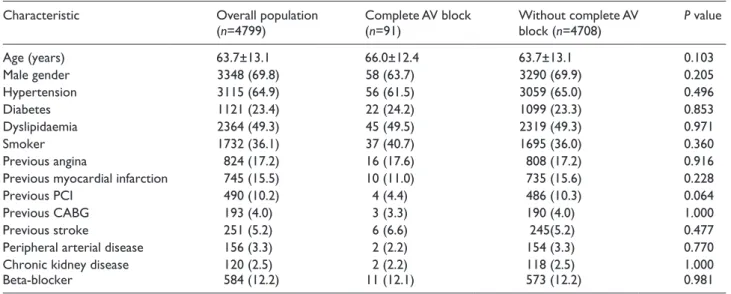
Baseline characteristics of the two groups are presented in Table 1. Heart block patients were slightly older (66.0±12.4 vs. 63.7±13.1 years; P=0.108) but there was not difference in gender (69.9% men in all the population). Cardiovascular risk factors were similar between patients with and without complete AV block, as well as comor-bidities (peripheral arterial disease and chronic kidney dis-ease). No significant differences were observed between the groups regarding the prevalence of coronary artery dis-ease, including previous myocardial infarction, PCI and coronary artery bypass graft surgery (CABG). About 12% of patients took beta-blockers in both groups.
Although angina was the most frequent symptom in both groups at admission, the main symptom was syncope in 13.6% of G1 patients in contrast with 1.5% in G2. The frequency of other manifestations, such as dyspnoea, was not significantly different between the groups. Complete AV block patients presented with lower systolic blood pres-sure (107.8±27.8 vs. 136.9±28.6 mmHg; P<0.001) and heart rate (59.7±23.4 vs. 79.2±21.3 beats per minute;
P<0.001) and higher Killip class (Killip >I in G1 25.0% vs.
G2 14.9%; P=0.013). On electrocardiogram, 86.8% of G1 patients presented with STEMI, inferior STEMI being the most frequent diagnosis in this group (79.1%) in contrast with only 21.9% of patients in G2. There was no significant difference in the presence of left and right bundle branch block between the groups (Table 2).
Among patients who underwent fibrinolysis, complete AV block was observed in 7.3% in contrast to 2.5% in patients submitted to primary PCI (P<0.001).
The presence of multivessel disease was 54% in both groups; however, LVEF was significantly lower in G1, in which 48.1% of patients had LVEF less than 50% com-pared with 37.2% in G2.
Reflecting the higher incidence of inferior STEMI, right ventricular myocardial infarction was significantly more frequent in G1 (3.3% vs. 0.2%; P<0.001).
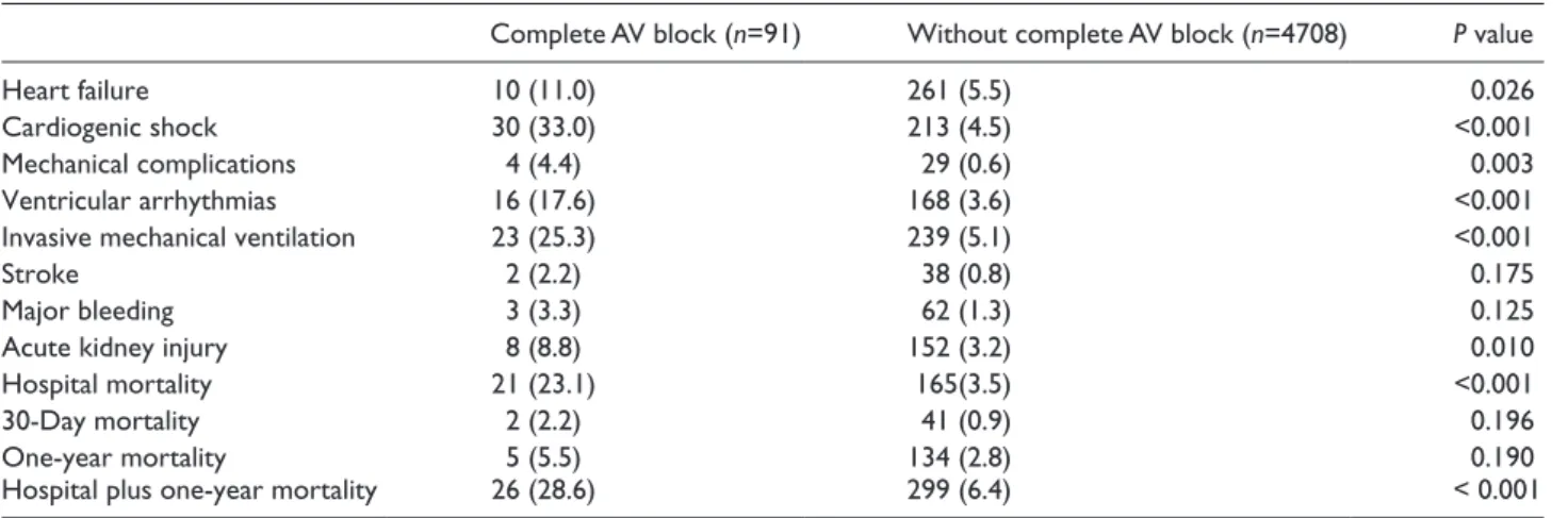
Complications during hospitalisation are shown in Table 3. With regard to hospital outcomes, patients with complete AV block presented with a worse prognosis. In G1, the incidence of heart failure was 11%, double that in G2, and cardiogenic shock was about seven times more fre-quent (33.0 vs. 4.5%; P<0.001). Ventricular arrhythmias and mechanical complications were also more frequent in
Table 1. Baseline clinical characteristics.
Characteristic Overall population
(n=4799) Complete AV block (n=91) Without complete AV block (n=4708) P value
Age (years) 63.7±13.1 66.0±12.4 63.7±13.1 0.103 Male gender 3348 (69.8) 58 (63.7) 3290 (69.9) 0.205 Hypertension 3115 (64.9) 56 (61.5) 3059 (65.0) 0.496 Diabetes 1121 (23.4) 22 (24.2) 1099 (23.3) 0.853 Dyslipidaemia 2364 (49.3) 45 (49.5) 2319 (49.3) 0.971 Smoker 1732 (36.1) 37 (40.7) 1695 (36.0) 0.360 Previous angina 824 (17.2) 16 (17.6) 808 (17.2) 0.916
Previous myocardial infarction 745 (15.5) 10 (11.0) 735 (15.6) 0.228
Previous PCI 490 (10.2) 4 (4.4) 486 (10.3) 0.064
Previous CABG 193 (4.0) 3 (3.3) 190 (4.0) 1.000
Previous stroke 251 (5.2) 6 (6.6) 245(5.2) 0.477
Peripheral arterial disease 156 (3.3) 2 (2.2) 154 (3.3) 0.770
Chronic kidney disease 120 (2.5) 2 (2.2) 118 (2.5) 1.000
Beta-blocker 584 (12.2) 11 (12.1) 573 (12.2) 0.981
Chronic kidney disease – creatinine >2.0 mg/dl; haemodialysis or renal transplantation.
AV: atrioventricular; PCI: percutaneous coronary intervention; CABG: coronary artery bypass graft.
Table 2. Clinical presentation, electrocardiographic and echocardiographic parameters at admission.
Complete AV block
(n=91) Without complete AV block (n=4708) P value
Main sympton at presentation <0.001
Angina 63 (77.8) 3508 (87.2)
Dyspnoea 2 (2.5) 89 (2.2)
Syncope 11 (13.6) 61 (1.5)
Killip class >I 20 (25.0) 641 (14.9) 0.013
I 60 (75.0) 3650 (85.1)
II 5 (6.3) 400 (9.3)
III 4 (5.0) 147 (3.4)
IV 11 (13.8) 94 (2.2)
Heart rate 59.7±23.4 79.2±21.3 <0.001
Systolic blood pressure 107.8±27.8 136.9±28.6 <0.001
Left bundle branch block 4 (4.4) 181 (3.8) 0.780
Right bundle branch block 2 (2.2) 219 (4.7) 0.443
STEMI 79 (86.8) 2594 (55.1) <0.001
Anterior STEMI 14 (15.4) 1016 (21.6) 0.154
Inferior STEMI 72 (79.1) 1030 (21.9) <0.001
Right ventricular myocardial
infarction 3 (3.3) 8 (0.2) 0.001
Multivessel disease 41 (54.7) 1429 (54.6) 0.994
Left ventricular ejection fraction 0.013
LVEF >50% 41 (51.9) 2443 (62.8)
LVEF 35–50% 25 (31.6) 1138 (29.3)
LVEF <35% 13 (16.5) 307 (7.9)
complete AV patients, as well as acute kidney injury and the need for invasive mechanical ventilation. Major bleeding and stroke were similar in both groups.
Among patients who developed complete AV block, eight (8.8%) underwent definitive pacemaker implantation. These patients were older, with a mean age of 72.0±12.6 years, 75% were men and six (75%) patients had STEMI, of those four had inferior STEMI.
Importantly, hospital mortality was almost eightfold higher in patients with complete AV block (23.1% vs. 3.5%;
P<0.001). In heart block patients, the mortality was 18.1%
in inferior STEMI patients and 50% in anterior STEMI patients. However, there was no significant difference in mortality after discharge evaluated at 30 days (2.2% vs. 0.9%; P=0.183) and at one-year follow-up (5.5% vs. 2.8%;
P=0.136).
A propensity score analysis between study groups was performed before determination of the predictors of inhos-pital mortality (Table 4).
After a propensity score analysis, in a multivariate regression model the predictors of inhospital mortality were complete AV block (odds ratio (OR) 3.671; 95% CI
1.032–13.076; P=0.045), LVEF less than 35% (OR 3.994; 95% CI 1.012–15.765; P=0.048), anterior STEMI (OR 6.401; 95% CI 1.597–25.655; P=0.009), Killip class greater than I (OR 3.674; 95% OR 1.042–12.951; P=0.043) and age (OR 1.072; 95% CI 1.013–1.134; P=0.016).
Discussion
In the present study, the presence of complete AV block was verified in 1.9% of ACS patients and was associated with a worse hospital outcome in the context of ACS.
We found an incidence lower than previously described.1–5
This could be due to the fact that our data are more recent and were collected in the primary PCI era, with higher repercussion rates. Furthermore, in our study NSTEMI patients were included, conferring less risk of complete AV block. Finally, we just included complete AV block, in con-trast with previous studies that comprised high-degree AV block (second and third-degree AV block).
In contrast with previous data, we did not find an asso-ciation with gender and cardiovascular risk factors and the presence of complete AV block.2,10 Also, there was no
Table 3. Complications during hospitalisation, short and long-term mortality.
Complete AV block (n=91) Without complete AV block (n=4708) P value
Heart failure 10 (11.0) 261 (5.5) 0.026
Cardiogenic shock 30 (33.0) 213 (4.5) <0.001
Mechanical complications 4 (4.4) 29 (0.6) 0.003
Ventricular arrhythmias 16 (17.6) 168 (3.6) <0.001
Invasive mechanical ventilation 23 (25.3) 239 (5.1) <0.001
Stroke 2 (2.2) 38 (0.8) 0.175
Major bleeding 3 (3.3) 62 (1.3) 0.125
Acute kidney injury 8 (8.8) 152 (3.2) 0.010
Hospital mortality 21 (23.1) 165(3.5) <0.001
30-Day mortality 2 (2.2) 41 (0.9) 0.196
One-year mortality 5 (5.5) 134 (2.8) 0.190
Hospital plus one-year mortality 26 (28.6) 299 (6.4) < 0.001
AV: atrioventricular.
Table 4. Comparison of patients with and without complete AV block after match.
Complete AV block Without complete AV block P value
Age (years) 66.27±12.38 66.1±13.3 0.935
Male gender 41 40 0.861
Smoker 25 30 0.382
Diabetes 15 15 1.000
Chronic kidney disease 1 1 1.000
Previous ACS 7 7 1.000
Anterior STEMI 10 12 0.641
Inferior STEMI 55 56 0.825
Killip Kimball class 18 15 0.548
LVEF <35% 10 9 0.805
significant difference in beta-blocker medication before ACS between the two study groups.
As expected, complete AV block was more frequent in inferior STEMI, in relation to hypoperfusion of the AV nodal artery that normally arises from the right coronary artery and also secondary to an increase of parasympathetic tonus. AV nodal presents with two mechanisms of protec-tion against ischaemia. On the one hand, this structure receives collateral supply from septal perforators of the left anterior descending artery and adjacent venous sinusoids.11
On the other hand, it has a high intracellular content of gly-cogen.12 This fact explains the transition nature of complete
AV block in ACS and the resolution of this conduction dis-turbance after revascularisation.13–15 In our study only 8.8%
of patients with complete AV block underwent a definitive pacemaker implantation, this ratio being in line with previ-ous data.16
The incidence of AV block was about 2–13% before the introduction of thrombolytic therapy. Since the beginning of this therapy the incidence decreased but mortality con-tinued to be high.1,2 With the primary PCI approach the
fre-quency of complete AV block in the context of ACS decreased to 3%.2,13 Our study is in agreement with these
data, as we found that among patients who underwent fibrinolysis complete AV block was observed in 7.3% in contrast with 2.5% in primary PCI patients. It is likely that patients who underwent primary PCI had a higher reperfu-sion rate and consequently developed less severe conduc-tion disorders secondary to prolonged ischaemia and irreversible necrosis.
The association between complete AV block and worse short-term outcomes has been shown in several papers.10,13
In accordance with the published literature, in our regis-try patients who presented with complete AV block had higher hospital mortality. In our population, complete AV block patients had a sevenfold higher incidence of hospital mortality. After a propensity score analysis, in a multivariate regression model, we found that patients with complete AV block presented with about a fourfold increased risk of hos-pital death, in line with previous studies.1–5,16,17 Among the
heart block group, the mortality was substantially higher in anterior STEMI compared with inferior STEMI (50.0% vs. 18.1%). This is line with the study by Kim and coworkers that demonstrated that the negative prognostic impact of high-degree AV block differs according to the location of infarction, being more prominent in anterior STEMI.18
Complete AV block was an independent predictor of hos-pital mortality, with an OR of 3.671. In the same multivari-ate analysis, inferior STEMI was not a predictor of mortality, reflecting the fact that more than association with inferior location, complete AV block could be a surrogate of exten-sive myocardial infarction, with consequent haemody-namic and electrical impairment. In our population, during hospitalisation AV block patients had a higher incidence of heart failure, cardiogenic shock, ventricular arrhythmias,
mechanical complications, acute kidney injury and the need for invasive mechanical ventilation. The other independent predictors of hospital mortality were age, anterior STEMI, LVEF less than 35% and Killip class greater than I.
In Kim’s work the univariate analysis showed that the incidence of 30-day major adverse cardiac events, includ-ing all causes of death, non-fatal myocardial infarction and revascularisation, for patients with heart block was approx-imately two times higher than for those without heart block; however, this association was not significant in multivari-ate analysis.10 In the present study, we did not find an
asso-ciation between complete AV block and poor prognosis at 30-day and one-year follow-up. Before discharge the majority of G1 patients recovered AV conduction without the need for a pacemaker, consequently they presented with a similar prognosis after discharge compared with G2 patients. This similar prognosis was also demonstrated by the study of Singh et al., in which patients with heart AV block who survived the initial hospitalisation had similar long-term survival when compared with patients without conduction disorders.4
Study limitations
This was a single-centre retrospective study based on an ACS registry. All registries have the risk of underreporting, which could lead to underestimation of the incidence of AV block and right ventricular infarction. Furthermore, our registry does not specify the timing of the development of complete AV block during hospitalisation.
Conclusion
Despite the decreased incidence of complete AV block in the context of ACS in the primary PCI era, this conduction disorder continues to be associated with a worse short-term outcome. In the present study, complete AV block conferred a higher risk of heart failure, cardiogenic shock, ventricular arrhythmias, mechanical complications and death during hospitalisation. After discharge the survival was similar between patients with and without heart block.
Conflict of interest
The authors declare that there is no conflict of interest. All authors take responsibility for all aspects of the reliability and freedom from bias of the data presented and their discussed interpretation.
Funding
This research received no specific grant from any funding agency in the public, commercial, or not-for-profit sectors.
References
1. Harpaz D, Behar S, Gottlieb S, et al. Complete atrioventricular block complicating acute myocardial infarction in the throm-bolytic era. SPRINT Study Group and the Israeli Thromthrom-bolytic
Survey Group. Secondary Prevention Reinfarction Israeli Nifedipine Trial. J Am Coll Cardiol 1999; 34: 1721–1728. 2. Nguyen HL, Lessard D, Spencer FA, et al. Thirty-year trends
(1975–2005) in the magnitude and hospital death rates asso-ciated with complete heart block in patients with acute myo-cardial infarction: a population-based perspective. Am Heart
J 2008; 156: 227–233.
3. Spencer FA, Jabbour S, Lessard D, et al. Two dedicate long trends (1975–1997) in the incidence, hospitalization, and long term death rates associated with complete heart block complicating acute myocardial infarction: a community wide perspective. Am Heart J 2003; 145: 500–507.
4. Singh SM, FitzGerald G, Yan AT, et al. High-grade atrioven-tricular block in acute coronary syndromes: insights from the Global Registry of Acute Coronary Events. Eur Heart J 2015; 36: 976–983.
5. Aplin M, Engstrøm T, Vejlstrup NG, et al.; TRACE Study Group. Prognostic importance of complete atrioventricu-lar block complicating acute myocardial infarction. Am J
Cardiol 2003; 92: 853–856.
6. Clemmensen P, Bates ER, Califf RM, et al. Complete atrio-ventricular block complicating inferior wall acute myocar-dial infarction treated with reperfusion therapy. TAMI study group. Am J Cardiol 1991; 67: 225–230.
7. Berger PB, Ruocco NA, Ryan TJ, et al. Incidence and prog-nostic implications of heart block complicating inferior myo-cardial infarction treated with thrombolytic therapy: results from TIMI II. J Am Coll Cardiol 1992; 20: 533–540. 8. Pokorney SD, Radder C, Schulte PJ, et al. High-degree
atrio-ventricular block, asystole, and electro-mechanical disso-ciation complicating non-ST-segment elevation myocardial infarction. Am Heart J 2016; 171: 25–32.
9. Goldberg RJ, Zevallos JC, Yarzebski J, et al. Prognosis of acute myocardial infarction complicated by complete heart block (the Worcester Heart Attack Study). Am J Cardiol 1992; 69: 1135–1141.
10. Kim HL, Kim SH, Seo JB, et al.; Other Korea Acute Myocardial Infarction Registry, Korea Working Group on
Myocardial Infarction Investigators. Influence of second- and third-degree heart block on 30-day outcome following acute myocardial infarction in the drug-eluting stent era. Am
J Cardiol 2014; 114: 1658–1662.
11. Kennel AJ and Titus JL. The vasculature of the human atrio-ventricular conduction system. Mayo Clin Proc 1972; 47: 562–566.
12. Jedeikin LA. Regional distribution of glycogen and phos-phorylase in the ventricles of the heart. Circ Res 1964; 14: 202–211.
13. Shacham Y, Leshem-Rubinow E, Steinvil A, et al. High degree atrioventricular block complicating acute myocardial infarction treated with primary percutaneous coronary inter-vention: incidence, predictors and outcomes. Isr Med Assoc
J 2015; 17: 298–301.
14. Simons GR, Sgarbossa E, Wagner G, et al. Atrioventricular and intraventricular conduction disorders in acute myocar-dial infarction: a reappraisal in the thrombolytic era. Pacing
Clin Electrophysiol 1998; 21: 2651–2653.
15. Wei JY, Markis JE, Malagold M, et al. Cardiovascular reflexes stimulated by reperfusion of ischemic myocar-dium in acute myocardial infarction. Circulation 1983; 67: 796–801.
16. Gang UJ, Hvelplund A, Pedersen S, et al. High-degree atrioventricular block complicating ST-segment elevation myocardial infarction in the era of primary percutaneous coronary intervention. Europace 2012; 14: 1639–1645. 17. Meine TJ, Al-Khatib SM, Alexander JH, et al. Incidence,
predictors, and outcomes of high-degree atrioventricular block complicating acute myocardial infarction treated with thrombolytic therapy. Am Heart J 2005; 149: 670–674. 18. Kim KH, Jeong MH, Ahn Y, et al.; Other Korea Acute
Myocardial Infarction Registry Investigators. Differential Clinical implications of high-degree atrioventricular block complicating ST-segment elevation myocardial infarction according to the location of infarction in the era of primary percutaneous coronary intervention. Korean Circ J 2016; 46: 315–323.