MESTRADO EM ONCOLOGIA
ESPECIALIZAÇÃO EM ONCOLOGIA LABORATORIAL
Differential expression of cadherins in
bladder cancer: role in
epithelial-mesenchymal transition
Cláudia Martins Lima
M
2019Clá
udia
Ma
rtins
Lima
D
iffe
re
nt
ial
ex
pr
es
sio
n o
f c
ad
he
rin
s in
b
lad
de
r c
an
ce
r:
ro
le
in
e
pit
he
lia
l-m
es
en
ch
ym
al t
ra
ns
itio
n
M
.IC
B
A
S
201
9
D
iffe
re
nt
ial
ex
pr
es
sio
n o
f c
ad
he
rin
s in
b
lad
de
r c
an
ce
r:
ro
le
in
e
pit
he
lia
l-m
es
en
ch
ym
al t
ra
ns
itio
n
Clá
udia
Ma
rtins
de
Lima
IN STI TU T O D E CIÊ N C IA S B IO MÉ DI CA S A BE L S A LA Z A RCláudia Martins de Lima
Differential expression of cadherins in bladder cancer: role in
epithelial-mesenchymal transition
Dissertação de Candidatura ao grau de Mestre em Oncologia –
Especialização em Oncologia Laboratorial submetida ao Instituto de Ciências
Biomédicas de Abel Salazar da Universidade do Porto
Orientadora: Professora Doutora Carmen de Lurdes Fonseca Jerónimo
Professora Associada Convidada com Agregação
Departamento de Patologia e Imunologia Molecular
Instituto de Ciências Biomédicas Abel Salazar - Universidade do Porto
Investigadora Auxiliar e Coordenadora do Grupo de Epigenética e Biologia do
Cancro
Centro de Investigação
Instituto Português de Oncologia do Porto Francisco Gentil, E.P.E
Coorientador: Professor Doutor Rui Manuel Ferreira Henrique
Professor Catedrático Convidado
Departamento de Patologia e Imunologia Molecular
Instituto de Ciências Biomédicas Abel Salazar - Universidade do Porto
Assistente Graduado de Anatomia Patológica
Investigador Sénior do Grupo de Epigenética e Biologia do Cancro
Centro de Investigação
Instituto Português de Oncologia do Porto Francisco Gentil, E.P.E
“All truths are easy to understand once they are discovered; the point is to discover them” Galileo Galilei
This study was funded by a grant of the Research Centre of
Portuguese Oncology Institute of Porto
vii
AGRADECIMENTOS
Gostaria de agradecer a todas as pessoas que diretamente e indiretamente contribuíram para a realização desta dissertação.
Em primeiro lugar gostaria de agradecer à Professora Doutora Carmen Jerónimo por ser orientadora deste trabalho e por me ter dado a oportunidade de realizar a tese de mestrado no Grupo de Epigenética e Biologia do Cancro. Foi aqui que adquiri uma nova visão de “ciência” e um enorme enriquecimento profissional.
Em segundo lugar, ao Professor Doutor Rui Henrique, coorientador desta tese, agradeço a contribuição enquanto patologista, a disponibilidade no esclarecimento de dúvidas, assim como, as sugestões para melhorar este trabalho.
Ainda, quero agradecer ao Professor Doutor Manuel Teixeira, Diretor do Centro de Investigação do IPO do Porto, por ter possibilitado e permitido a realização deste trabalho nestas instalações.
Agradeço também ao Engenheiro Luís Antunes pela contribuição na análise estatística.
Também quero deixar um agradecimento ao Doutor Bruno Costa da Universidade de Braga pela colaboração na análise in silico.
Um enorme agradecimento à Sara Reis, uma das pessoas fundamentais na concretização deste projeto. Obrigada pelos ensinamentos, tanto laboratoriais como pessoais, pelo incentivo e por me dizer sempre para acreditar no meu trabalho. Sem dúvida, das melhores pessoas que me podia ter acompanhado nesta experiência.
Ao João Lobo, um enorme agradecimento pela grande disponibilidade e paciência, já que mesmo estando na Holanda conseguiu avaliar todas as lâminas de imunohistoquímica.
À Catarina Teixeira, quero deixar um especial agradecimento, visto que esteve sempre a meu lado, por me fazer rir todos os dias, por ser tão o meu oposto, pela boa disposição e pelas histórias diárias. Sem dúvida, uma das melhores com quem podia ter dividido esta experiência.
À Ana Lameirinhas gostaria de agradecer pelos inúmeros conselhos, pelo enorme sentido estético, pelas ideias e enorme disponibilidade em ajudar-me sempre que necessitava, tornando-se numa ajuda fundamental na realização deste trabalho.
Gostaria também de agradecer à Daniela Barros pela enorme ajuda na análise estatística, assim como na organização da base de dados.
Ao José Pedro, agradeço por me ter ajudado quando mais precisava, especialmente naquelas semanas em agosto, por ter aturado as minhas aleatoriedades e por me fazer sempre rir.
viii
Também, à Sandra Nunes, quero agradecer pela ajuda que me deu na parte da metilação e por ter tido paciência para me explicar todo o processo de desenhar primers.
À Rita Oliveira agradeço com muito carinho por me ter acompanhado nestes dois anos de mestrado, assim como, pelos bons momentos passados no laboratório, especialmente quando passávamos dias inteiros a fazer extração de DNA e imunohistoquímica. Ainda, pelo enorme sentido de humor e sobretudo por nunca ter reclamado comigo por invadir a secretária dela com lâminas e blocos.
À Mariana, a minha companheira de escrita e de entrega de tese, quero agradecer pelos desabafos e conselhos, pela tranquilidade que sempre transmitiu e pelos muitos momentos de diversão. Ainda, quero agradecer pela partilha diária de memes e por ser sempre a minha companheira nos jantares.
Também, gostaria de agradecer à Vera Constâncio pela troca de ideias sempre que necessitava e ainda, pelas inúmeras conversas de culinária de pratos criados por ela.
Ao Gonçalo, o colega de bioquímica que virou parceiro de memes, quero agradecer por todo o apoio e por me fazer rir, especialmente nas alturas que mais precisava.
Igualmente, quero agradecer à Carina Maia por ter tornado os meus dias no processamento muito mais animados e por ser uma pessoa tão positiva.
Ainda, gostaria de agradecer à Sofia Salta por estar sempre disponível para esclarecer as minhas dúvidas, principalmente quando me sentia completamente perdida.
Gostaria também de agradecer à Catarina Macedo, à Helena, à Nair, à Vânia Camilo e à Vera Gonçalves e por todos os conselhos e pela imensa partilha de conhecimento. Ao Leonardo e às meninas mais novas, Ana Rita, Filipa Silva (a minha nova companheira de secretária) e Luísa Pinheiro espero que partilhem das mesmas boas experiências durante esta vossa nova etapa.
Quero ainda agradecer às minhas melhores amigas, Beatriz Bessa e Rita Osório, por serem as minhas companheiras de sempre e para sempre. Por ouvirem sempre os meus desabafos, por me incentivarem e por estarmos sempre juntas nas nossas pequenas conquistas. Também, a todos os jantares, saídas, tardes a vermos filmes da Fox Life e horas e horas de gargalhadas.
Às minhas amigas de licenciatura, Anastasyia Hryshkina, Catarina Marques, Rafaela Abrantes e Telma Costa, agradeço todos os momentos inesquecíveis vividos nestes últimos 5 anos, a começar nas aulas de bioquímica e a acabar em todos os convívios.
Por fim, gostaria de agradecer aos meus pais e ao meu irmão por tudo o que fizeram por mim, por todo o apoio e carinho, assim como por todas as oportunidades que me proporcionaram. Sem dúvida que se estou a concluir mais uma etapa é graças a vocês.
ix
RESUMO
Introdução:
Metade dos casos de cancro da bexiga não-músculo-invasivos (CBNMI)exibem elevadas taxas de recorrência, sendo que 5-25% dos CBNMI apresentam progressão para cancro de bexiga músculo-invasivo (CBMI). A transição epitélio-mesenquima (TEM) tem sido associada à progressão de não-músculo-invasivo para músculo-invasivo e, consequentemente, com a metastização, já que as células epiteliais perdem a polaridade e as estruturas de adesão célula-célula, adquirindo características mesenquimais. O switch das caderinas (SC) é um hallmark de TEM, em que a expressão normal das caderinas é substituída por um padrão anormal de expressão das mesmas. Apesar de o switch das caderinas entre E-caderina e N- e/ou P-caderina ter sido sugerido no cancro de bexiga, mais estudos são necessários realizar para clarificar o SC. Além disso, o papel da R-caderina ainda não foi abordado neste modelo tumoral. Consequentemente, o padrão de expressão das 4 caderinas foi avaliado em cancro de bexiga e, simultaneamente, os respetivos mecanismos de regulação que podem ajudar a entender a progressão tumoral.
Métodos: Primeiramente, a análise in silico foi realizada através da base de dados The
Cancer Genome Atlas (TCGA). Além disso, a expressão de E-, N-, P- e R-caderina foi
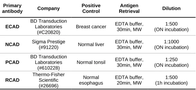
avaliada por imunohistoquímica em 124 pacientes com tumor primário de bexiga e em 40 casos de bexiga normal, sendo estes últimos utilizados como controlos. Para além disso, os mecanismos de regulação das caderinas foram investigados através da indução de
transforming growth factor-β1 (TGF-β1) nas linhas celulares SV-HUC1, RT-112 e 5637.
Simultaneamente, a metilação de CDH1, CDH2 e CDH3 foi avaliada em todas as linhas celulares de bexiga. Ainda, a metilação do promotor de CDH1, foi estudada no tecido normal e tumoral de bexiga.
Resultados:
A análise in silico dos dados relativos aos níveis de transcrito de CDH1 eCDH3 constantes na base do TCGA revelou que as amostras tumorais de bexiga (n=408)
são significativamente maiores do que em amostras de tecido normal (n=19). Contudo os níveis de transcrito de CDH4 foram significativamente menores em amostras de tecido normal. Uma elevada proporção significativa de CBMI demonstrou positividade para P-caderina membranar comparativamente com CBNMI. Para além disso, amostras tumorais demonstraram uma maior proporção de casos positivos para R-caderina membranar do que para amostras de bexiga normal, apesar de CBMI apresentar menor expressão do que CBNMI. A exposição de TGF-β1 in vitro não apresentou associação significativa com as
x
alterações da expressão das caderinas, no entanto alterações morfológicas foram observadas em quase todas as linhas celulares de bexiga. Ainda, os níveis de metilação dos promotores das caderinas correlacionaram-se negativamente com a respetiva expressão de proteína.
Especificamente, foram encontradas fortes correlações negativas entre CDH1/E-caderina e CDH3/P-caderina, enquanto uma moderada correlação negativa foi observada entre
CDH2 e N-caderina. Em pacientes, a metilação de CDH1 foi significativamente maior em
tecidos tumorais do que em tecidos normais. No entanto, não foram observadas associações inversas entre a metilação de CDH1 e expressão de E-caderina.
Conclusões: A desregulação na expressão das caderinas foi associada com tumorigénese
na bexiga. De facto, na base de dados TCGA, os níveis de transcrito de CDH1, CDH3 e
CDH4 encontravam-se significativamente alterados no tecido bexiga tumoral
comparativamente com os tecidos normais, enquanto na cohort do IPO do Porto apenas E-caderina e R-caderina demonstraram diferenças nestes dois tipos de tecido. Particularmente, E-caderina e R-caderina demonstraram uma diminuição significativa em CBMI comparativamente com CBNMI, enquanto a expressão de P-caderina foi significativamente maior nos tumores invasivos. Apesar de TGF-β1 parecer não ter influência na regulação da expressão das caderinas, a metilação dos promotores parece ser um importante mecanismo de regulação nesta neoplasia.
xi
ABSTRACT
Background: About half of non-muscle invasive bladder cancers (NMIBCs) display high
recurrence rates, from which 5-25% show progression to muscle invasive bladder cancer (MIBC). Epithelial-mesenchymal transition (EMT) has been associated with non-muscle to muscle invasive progression and, consequently, with metastases formation, since epithelial cells lose their polarity and cell-cell adhesion structures, acquiring mesenchymal features. Cadherin switch (CS) is a hallmark of EMT, where normal cadherins’ expression is replaced by an abnormal pattern of cadherins expression. Although, cadherin switch between E-cadherin and N- and/or P-E-cadherin has been suggested for bladder cancer, more studies are required to clarify CS. Moreover, the involvement of R-cadherin has not been yet addressed in this cancer type. Hence, the expression patterns of the 4 cadherins were assessed in bladder cancer and, simultaneously, the respective regulatory mechanisms that might help to understand the tumoral progression were also tackled.
Methods:
Firstly,
in silico analysis was performed using The Cancer Genome Atlas(TCGA) dataset. Moreover
,
E-, N-, P- and R-cadherin’s expression was assessed by immunohistochemistry in a cohort of 124 primary bladder tumor patients and 40 normal bladder tissues which were used for control purposes. Additionally, cadherins regulatory mechanisms were evaluated by performing transforming growth factor-β1 (TGF-β1) treatment in SV-HUC1, RT-112 and 5637 cell lines. Simultaneously, CDH1, CDH2 andCDH3 methylation status was evaluated in bladder cell lines. Moreover, CDH1 promoter
methylation was also determined in normal and bladder tumor tissues.
Results: In TCGA dataset CDH1 and CDH3 transcript levels were significantly higher in
invasive bladder tumors (n=408) than in normal tissues (n=19). On the other hand, CDH4 transcript levels were significantly lower in normal samples. A significantly higher proportion of MIBCs showed positivity for membrane P-cadherin compared with NMIBCs. Furthermore, tumor samples presented a higher proportion of cases positive for membrane R-cadherin compared with bladder normal tissues, although MIBCs showed a lower expression than NMIBCs. In vitro exposure of TGF-β1 did not significantly associate to cadherins’ expression alterations, whereas morphological alterations were apparent in almost all bladder cell lines. Additionally, cadherins’ promoter methylation levels, negatively correlated with their respective protein expression. Specifically, a strong inverse correlation was found for CDH1/E-cadherin and CDH3/P-cadherin, whereas CDH2/N-cadherin displayed a moderate inverse correlation. In patients, CDH1 methylation levels were
xii
significantly higher in cancer than in normal tissues. Nonetheless, no inverse association was found between CDH1 methylation and E-cadherin expression in bladder tissues.
Conclusions: Cadherins’ expression deregulation is associated with bladder
tumorigenesis. Indeed, in TCGA dataset, CDH1, CDH3 and CDH4 transcript levels were significantly changed in tumoral bladder tissues compared to normal tissues, whereas in IPO Porto´s cohort only E-cadherin and R-cadherin immunoexpression differed between these two tissues types. Particularly, E-cadherin and R-cadherin showed a significant decreased in MIBCs comparing with NMIBCs, while P-cadherin presented a higher expression in the invasive tumors. Moreover, although TGF-β1 seems to not play a major role in cadherin’s expression regulation, CDH1, CDH2 and CDH3 promoter methylation might constitute an important regulatory mechanism in this malignancy.
xiii
TABLE OF CONTENTS
INTRODUCTION ... 1
EPIDEMIOLOGY ... 3
UROTHELIAL CELL CARCINOMA ... 4
DIAGNOSIS ... 6
CURRENT TREATMENTS ... 7
UROTHELIAL CARCINOGENESIS ... 7
EPITHELIAL-MESENCHYMAL TRANSITION (EMT) ... 9
CADHERINS ...10
CADHERINS REGULATORY MECHANISMS ...13
TGF-β signaling pathways ...13
Epigenetics ...15
Cadherins promoter methylation ...16
AIMS ...19
MATERIAL AND METHODS ...23
IN SILICO ANALYSIS ...25
CLINICAL SAMPLES ...25
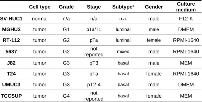
CELL LINES CULTURE ...25
TGF-Β1 TREATMENT ...26
DNA EXTRACTION ...27
CELL LINES ...27
PARAFFIN-EMBEDDED TISSUES ...27
SODIUM BISULFITE TREATMENT OF DNA ...28
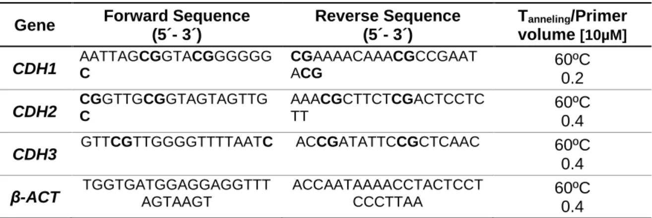
QUANTITATIVE METHYLATION-SPECIFIC PCR (QMSP) ...28
IMUNOFLUORESCENCE ...31
PROTEIN EXTRACTION AND QUANTIFICATION ...31
WESTERN BLOT ...32
STATISCAL ANALYSES ...32
RESULTS ...35
IN SILICO ANALYSIS ...37
CLINICAL AND PATHOLOGICAL DATA ...38
CADHERINS EXPRESSION IN TUMORS ...39
CADHERINS EXPRESSION IN BLADDER CELL LINES ...42
CADHERINS EXPRESSION REGULATION ...44
Aberrant promoter methylation associate with cadherin´s expression downregulation ...44
xiv
TGF-β1 seems to have an impact in cell morphology ... 46
DISCUSSION ... 49
CONCLUSIONS & FUTURE PERSPECTIVES ... 55
REFERENCES ... 59
APPENDIX ... 69
APPENDIX I ... 71
Supplementary tables ... 71
xv
FIGURE INDEX
Figure 1. Representation of world map with estimated age-standardized incidence rates in 2018 of bladder cancer in both sexes in all ages [2]. ... 3 Figure 2. Bladder cancer staging. Urothelial cell carcinoma can be divided in 2 subtypes,
non-muscle invasive bladder cancer (NMIBC) and muscle-invasive bladder cancer (MIBC). Adapted from [23]. ... 6
Figure 3. Urothelial carcinogenesis classical pathways. Regarding behavior and
molecule profile, BC formation can be divided in papillary pathway and the Cis pathway. The first one start with chromosome 9 deletion and FGFR3/RAS mutations, promoting hyperplasia lesion formation. Then, after the PIK3CA/STAG2 mutations hyperplasia progresses to a low-grade Ta. Lastly, low-grade Ta might progress to high-grade Ta. Cis pathway initiates with chromosome 9 deletion and TP53 mutations, promoting urothelial dysplasia formation, which can progress to Cis when RB1 deletion occurs. High grade Ta, providing from papillary pathway, can suffer a CDKN2A/TP53/RB1 inactivation, which results in a high-grade T1. Moreover, Cis can also progress to T1. Finally, with EMT involvement, the non-muscle invasive tumors can progress to MIBC and, subsequently, can metastasize. Abbreviation: BC - Bladder cancer; CDKN2A - Cyclin-dependent kinase inhibitor 2A; Cis - Carcinoma in situ; EMT - Epithelial-mesenchymal transition; FGFR3 - Fibroblast growth factor receptor 3; MIBC Muscle-invasive bladder cancer; PIK3CA - Phosphatidylinositol-4,5-biphosphate 3-kinase, catalytic subunit alpha; RB1 - Retinoblastoma1; STAG2 - Stromal antigen 2; TP53 - Tumor protein p53; Adapted from [37]. ... 8
Figure 4. Cellular features alterations associated with EMT biological process. During
EMT the cells lose epithelial features and acquire mesenchymal features. Epithelial cells have an apical-basal polarity and exhibit cell-to-cell interactions through tight junctions, adjacent cells and desmosomes. Currently, these cells express, mainly, adherens junction proteins (E-cadherin and β-catenin), tight junction proteins (occludins and claudins), cytokeratin, such as 5 and 6 and ZO-1. Throughout EMT, these epithelial markers are replaced by mesenchymal markers, such as N-cadherin, vimentin fibronectin and MMPs. Due to this, cells acquire front-rear polarity and fibroblast-like phenotype, losing their adhesion ability. Consequently, they gain motility and invasion capacity. Abbreviation: EMT - Epithelial-mesenchymal transition; MMPs - Matrix metalloproteinases; ZO-1 - Zonula occludens-1; Adapted from [47]. ...10
Figure 5. Smad-dependent signaling in EMT. TGF-β1 ligand binds to the heteromeric
complex of TGF-βI and TGFRβII. Then, the cofactor SARA interacts with R-SMADs transcription factors, SMAD2 and SMAD3, in order to be recruited to the TGF-βR complex.
xvi
Hereafter, SMAD4 is also recruited, resulting in a trimeric complex formation, being capable to translocated into the nucleus, with the help of importins-β1, importins 7 and 8. Then, the SMAD2/3/4 complex can activate the DNA-binding transcription factors expression, such as zinc-finger transcription factors (Snail and Slug), bHLH factors (Twist and E12/E47) and zinc-finger factors (ZEB1 and ZEB2). The Snail, Slug, Twist, ZEB1 and ZEB2 are EMT transcription factors that are capable of CDH1 downregulation and CDH2 upregulation. Abbreviation: bHLH - basic helix–loop–helix; R-SMADs - Receptor-regulated SMADs; SARA - Smad anchor for receptor activation; TGFRβII - Type II TGF-β receptor; TGF-β1 - Transforming growth factor β1; TGF-βI - Type I TGF-β receptor; Adapted from: [44]. ... 14
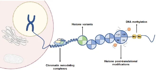
Figure 6. Epigenetic regulatory mechanisms in gene expression. Epigenetic
mechanisms comprise DNA methylation, histone post-translational modifications, histone variants and chromatin remodeling complexes. Kindly provided by Ana Lameirinhas. ... 16
Figure 7. Transcript alterations of CDH1, CDH2, CDH3 and CDH4 of a 413 MIBC patients’ cohort from cBioPortal. ... 37 Figure 8. In silico analysis of cadherins gene expression in normal and tumor bladder tissues from TCGA data. Graphical representation of CDH1 (A), CDH2 (B), CDH3 (C) and
CDH4 (D) expression in normal (n=19) and in tumor cases(n=408). ... 38
Figure 9. Distribution of membrane cadherins expression in normal and tumor bladder tissues. Graphical representation of E-cadherin (A), N-cadherin (B), P-cadherin (C) and R-cadherin (D) positive and negative cases. ... 40 Figure 10. Cytoplasmic and membrane location in the bladder cell lines.
Immunofluorescence assay for E-cadherin (A), N-cadherin (B), P-cadherin (C) and R-cadherin (D). Photograph taken in microscope Olympus IX51 with a digital camera Olympus XM10. ... 43
Figure 11 Characterization of cadherins in bladder cell lines. Graphical quantification
of protein expression and corresponding illustrative Western blot images for E-cadherin (A), N-cadherin (B), P-cadherin (C), and for R-cadherin (D). ... 44
Figure 12: Relation of cadherins genes methylation and cadherins expression in bladder cell lines. Graphical quantification for CDH1 vs ECAD (A), CDH2 vs NCAD (B),
CDH3 vs PCAD (C). ... 45
Figure 13. Relation between CDH1 methylation levels (A) and ECAD membrane expression (B) in bladder normal tissues and in bladder tumor tissues. ... 46 Figure 14. Characterization of E-cadherin, N-cadherin, P-cadherin and R-cadherin expression in bladder tumor cell lines treated with 20ng/mL TGF-β1 to induce EMT.
Illustrative images of morphology alterations of these same bladder tumor cells lines (A). Graphical representation of E-cadherin, N-cadherin, P-cadherin and R-cadherin expression
xvii
in SV-HUC1, RT- 112 and 5637 cell lines exposed to TGF-β1 (B). Corresponding illustrative western blot images of SV-HUC1, RT-112 and 5637 cell lines exposed to TGF-β1 (C). ..47
Figure 15. Cadherin switch along normal bladder and tumor tissues. Regarding IHC
results, different expression of E-cadherin, N-cadherin, P-cadherin and R-cadherin membrane occurred between normal bladder, NMIBC and MIBC tissues. Moreover, a co-cadherins expression was found which corroborates the partial EMT definition. Abbreviation: EMT- Epithelial-mesenchymal transition; IHC – Immunohistochemistry; MIBC – Muscle-invasive bladder cancer; NMIBC – Non-muscle invasive bladder cancer. ...54
xix
TABLE INDEX
Table 1 Clinical pathologic characterization of human bladder cell lines. ...26
Table 2. Primer used in qMSP. ...29
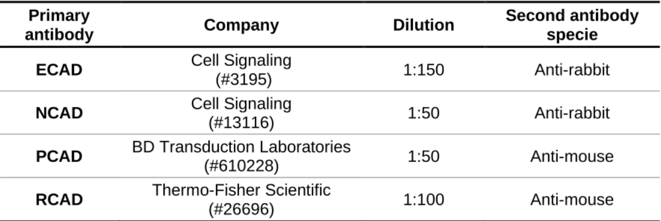
Table 3. Primary antibodies used in IHC. ...30
Table 4. Primary antibodies used in IF. ...31
Table 5. Primary antibodies used in WB. ...32
Table 6. Clinical and pathological features of patients included in this study. ...38
Table 7. Membrane E-, N-, P- and R-cadherins protein expression in normal and tumor (NMIBC and MIBC) tissues. ...40
Table 8. Spearman´s correlation between membrane cadherins in bladder tumor tissues. ...41
Table 9. Spearman´s correlation between cytoplasmic and membrane cadherins in bladder tumor tissues. ...41
Table 10. Protein expression of cytoplasmic E-, N-, P- and R-cadherins in normal and tumor (NMIBC and MIBC) tissues ...42
Table 11. Spearman´s correlation between cytoplasmic cadherins in bladder tumor tissues. ...42
xxi
LIST OF ABBREVIATIONS
5hmC - 5-hydroxymethyl-cytosine
AJCC - American Joint Committee on Cancer AJs - Adherens junctions
AKT - Protein cinase B
AMH - Anti-mullerian hormone As- Arsenic
ATCC - American Type Culture Collection AUA - America Urological Association BC- Bladder cancer
BCG - Bacillus Calmette-Guérin bHLH - Basic helix–loop–helix BMP - Bone morphogenetic protein BSA - Bovine serum albumin
BTA - Bladder tumor antigen BUC - Bladder urothelial carcinoma CAD - Cadherin
CAMs - Cell adhesion molecules Cat - Catenin
CDKN2A - Cyclin-dependent kinase inhibitor 2A Cis - Carcinoma in situ
CpG - Cytosine-phosphate-guanine CS - Cadherin switch
DAB - 3,3-Diaminobenzidine
DAPI - 4’,6-diamidino-2-phenylindole DBPs - Disinfection by-products
EAU - European Association of Oncology ECAD - E-cadherin
EDTA - Ethylenediamine tetraacetic acid
EGF - Epidermal growth factor
EMT - Epithelial-mesenchymal transition Erk -Extracellular signal-regulated kinase FBS - Fetal bovine serum
FFPE - Formalin-fixed and paraffin-embedded FGFR3 - Fibroblast growth factor receptor 3
xxii FITC - Fluorescein isothiocyanate
GDFs – Growth differentiation factors GSTM1 - Glutathione S-transferase H&E - Hematoxylin and eosin HG - High-grade
IgG - Immunoglobulin G IHC - Immunohistochemistry
IPO - Portuguese Oncology Institute JNK - c-Jun NH2-terminal kinase LG - Low-grade
LOH - Loss of heterozygosity m5C – 5-methylcytosine
MBD - Methyl-CpG-binding domain MET - Mesenchymal-epithelial transition MIBC – Muscle-invasive bladder cancer MMC - Mitomycin C
MMP - Matrix metalloproteinase NAC - Neoadjuvant chemotherapy NAT2 - N-acetyltransferase 2 NCAD - N-cadherin
NMIBC - Non-muscle invasive bladder cancer NMP22 - Nuclear matrix protein 22
NUT - Normal urothelial tissue
PAH - Polycyclic aromatic hydrocarbons PBS - Phosphate-buffered saline PCAD – P-cadherin
PFA - Paraformaldehyde PIC - Protein inhibitor cocktail
PIK3CA - Phosphatidylinositol-4,5-biphosphate 3-kinase, catalytic subunit alpha PIP3 - Phosphatidylinositol 3,4,5 trisphosphate
PLND - Pelvic lymphadenectomy PRC2 – Polycomb repressive complex 2 PVDF - Polyvinylidene difluoride (PVDF) RB1 - Retinoblastoma1
RCAD - R-cadherin
RIPA - Radio immuno precipitation assay R-SMADs - Receptor regulated SMADs
xxiii SAM - S-adenosyl-methionine
SARA - Smad anchor for receptor activation SDS - Sodium dodecyl sulfate
SDS-PAGE - Sodium dodecyl sulfate polyacrylamide gel electrophoresis Shh - Sonic hedehog
STAG2 - Stromal antigen 2
TBS-T – Tris-buffer saline-tween 20 TCGA - The Cancer Genome Atlas
TET - Ten-eleven methylcytosine dioxygenase TFs - Transcription factors
TGF-β - Transforming growth factor-β TGF-βRI - Type I TGF-β receptor TGF-βRII - Type II TGF-β receptor THMs - Trihalomethanes
TJs - Tight junctions
TMT - Bladder-sparing trimodal therapy TNM - Tumor-node-metastasis
TP53 - Tumor protein p53 TRITC - Tetramethylrhodamine
TURBT - Transurethral resection of bladder tumor VIM - Vimentin
WB - Western blot
WHO - World Health Organization WLC - White light assisted cystoscopic ZO - Zonula occludens
INTRODUCTION ‖ 3
EPIDEMIOLOGY
Bladder cancer (BC) is the tenth most common malignancy worldwide with approximately 549,000 new cases in 2018. Regarding mortality data, this cancer was responsible for 200,000 deaths, according to GLOBOCAN 2018. Despite contributing to high morbidity rates, this disease is not among the ten deadliest cancers. This type of cancer is the sixth most common malignancy in men and the seventeenth in females. In 2018, the estimated incidence in males and in females was 9.6 and 2.4 per 100.000 new cases, respectively [1]. Therefore, the incidence is approximately 4 times higher in men, compared to women [1, 2].
BC is most common in older people, being the mean age of diagnosis in men 69 and in women 71. Although this type of cancer is rare in younger people, it can occur at any age [3, 4]. Furthermore, for both genders, this disease is more frequent in developed regions such as Northern America, Western and Southern Europe, when comparing to non-developed nations [5, 6] (Figure 1). This could be explained due to the higher accessibility to better diagnosis methods in developed countries [6]. In Portugal, BC was responsible for 2,340 new cases and for 1,106 deaths, being the sixth most common in both genders. This disease is the fourth most frequent cancer in males and the eleventh most incident in women [2].
Figure 1. Representation of world map with estimated age-standardized incidence rates in
INTRODUCTION ‖ 4
RISK FACTORS
There are some factors that contribute for an increased risk of BC carcinogenesis. Tobacco is the main risk factor with an attributable risk of nearly 50% [5]. Approximately thirds of total cases are associated with smoking [7]. Furthermore, smokers have a two-to-fourfold higher risk of developing this disease compared with non-smokers [8]. Additionally, the number of smoking years, the smoke inhalation degree and the number of cigarettes smoked are associated with a higher increase of BC developing [7-9]. Some aromatic amines and polycyclic aromatic hydrocarbons (PAHs) seem to be excreted by the renal system and reach the urinary tract, playing an important role in carcinogenesis [7, 8, 10].
Occupation is the second most important risk factor with an attributable risk of less than 8% [8]. According to previous studies, painters, rubber workers and petroleum industry workers display higher BC tumorigenesis risk, however other professions also present an increased risk [11]. This occurs due to occupational exposure to some carcinogenic agents, such as, aromatic amines, PAHs, certain aldehydes and some nitrites and nitrates[7, 9, 12].
Another risk factor is exposure to ionizing radiation, which has an attributable risk of approximately 2%. Many studies have already established that patients undergoing radiotherapy to treat other malignancies, present a higher risk of developing BC [7].
Additionally, drinking arsenic (As) and chlorine contaminated water contribute to an increase of BC risk [12]. Due to the presence of As in the soil surface, contamination of drinking-water sources occurs frequently through infiltration [13]. Furthermore, water chlorination, the process of adding some disinfecting agents such as chlorine to the water, can lead to the formation of disinfection by-products (DBPs), being the trihalomethanes (THMs) the most common with a potential carcinogenic role [8, 14].
Urinary tract diseases and other conditions also contributes to increase BC risk. The most known is schistosomiasis, a chronic endemic cystitis caused by Schistosoma
haematobium, which mainly relates with squamous cell carcinoma [7, 15].
UROTHELIAL CELL CARCINOMA
Urothelial cell carcinoma is the most frequent type of BC, corresponding to, approximately, 90% of the total cases [8, 16]. Tumorigenesis results from the transitional cells that are in bladder lining, also known as urothelium [8]. Regarding these, 70-80% are non-muscle invasive bladder cancer (NMIBC), whereas the remaining 20-30% are muscle-invasive bladder cancer (MIBC) at the time of diagnosis [16, 17]. Although MIBC cases are less frequent, they are accountable for the most of BC deaths [16]. NMIBC patients have more promising outcomes than MIBC patients, with 10-year survival rates of 80% and 50%, respectively [18]. Though the mortality rates are not among the highest, 50% of NMIBC
INTRODUCTION ‖ 5
patients, who are surgically treated, exhibit recurrence once or several times. Furthermore, after some recurrences, 5-25% of NMIBC progress to MIBC [19]. These recurrences could be explained due to incomplete resections, reimplantation of tumor cells after treatments or tumor occurrence in high risk urothelium [8]. Therefore, as a result of these high recurrence rates it is of utmost importance to study BC.
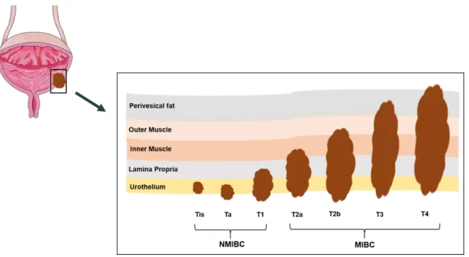
For BC, the tumor-node-metastasis (TNM) classification is followed, assessing tumor size (T), lymph node involvement (N) and distant metastasis (M) [20]. NMIBC and MIBC stage classification is related to the invasion of the tumor into the bladder wall (Figure 2). NMIBCs do not invade the bladder muscle layer, being limited to the bladder inner epithelial, although present a variable risk of local recurrence [8, 17]. NMIBC include carcinoma in situ (Cis) (stage Tis), which is a flat lesion confined to urothelium [21]. Although Cis is a NMIBC, it is considered a high-grade tumor, because it easily progresses to muscle invasion disease [22]. Other NMIBC types are non-invasive papillary carcinomas (stage Ta), which are restricted to the mucosa, as well as invasive lamina propria tumors (stage T1) [23]. On the other hand, MIBCs comprise a group of more aggressive tumors that invades the muscle layer or extend through the bladder reaching the surrounding tissues, which can promote metastization [17]. Furthermore, the tumors can either invade the superficial muscle (stage T2a) or the deep muscle (stage T2b). Additionally, the tumors can also invade the perivesical fat layer (stage T3) or extend into the adjacent organs (stage T4) [23] (Figure 2).
Recently, a similar breast tumors molecular classification emerged for BC. Briefly, bladder tumors can be divided into luminal and basal subtypes. Basal subtype cluster advanced stage tumors and metastatic disease, being enriched in inactivating mutations and deletions of tumor protein p53 (TP53) and Retinoblastoma1 (RB1) [24].On the other hand, luminal subtypes are associated with papillary histopathological features and enriched in fibroblast growth factor receptor 3 (FGFR3) mutations[24].
INTRODUCTION ‖ 6
Figure 2. Bladder cancer staging. Urothelial cell carcinoma can be divided in 2 subtypes, non-muscle invasive bladder cancer (NMIBC) and non-muscle-invasive bladder cancer (MIBC). Adapted from [23].
DIAGNOSIS
The main symptom of this disease is hematuria. Patients can have other clinical signals, such as pain or burning sensation during urination. Nevertheless, these symptoms are not specific of BC. Lately, due to diagnostic techniques development, early diagnosis has become a reality. The current detection standard method is cystoscopy supplemented by urine cytology, that can also be used in surveillance [18, 25]. Although the cystoscopy assay is invasive and expensive, it allows to have a clear image of the entire bladder [18]. Usually, BC standard cystoscopy is white light assisted cystoscopy (WLC) assay [26]. However, this method has low sensitivity in the detection of surgical margins, small tumors and satellite tumors. Besides that, residual tumors are detected late when using WLC [19].
The urine cytology exam is a less invasive process with a higher specificity (90-96%) and a reasonable sensitivity (>60) in high-grade tumor and Cis lesions [27]. However, the sensitivity, mostly in low-grade tumors, decreases to 4-31% [8, 18, 28]. In the past years, urine tumor markers assays have been developed to improve the BC detection, surveillance, and staging [8, 25]. Bladder tumor antigen (BTA) TRAK or BTA stat, UroVysion, ImmunoCyt/uCyt, nuclear matrix protein 22 (NMP22) are some examples of these assays [18, 29]. These demonstrate a higher false positive and a lower specificity and/or sensitivity, compared with the standard exam, which justifies why tumor markers tests are not present in clinical practice [18, 27, 30]. Until the development of a better assay, urine cytology will continue to be the standard procedure [30].
INTRODUCTION ‖ 7
CURRENT TREATMENTS
BC has the highest cumulative cost of treatment compared to other types of cancers [31]. BC treatments depend on the histological types. The standard treatment for NMIBCs, except for Cis, is transurethral resection of bladder tumor (TURBT) [8, 32]. The quality of the procedure influences diagnosis, treatment and prognosis [19]. Usually, adjuvant treatments are applied to decrease the recurrence and progression probability [22]. Intravesical immunotherapy Bacillus Calmette-Guérin (BCG) is the main adjuvant treatment. According to previous studies, it presents better results in tumor elimination [19, 22]. Moreover, in cases that TURBT and BCG do not result and there are no evidences of progression, alternative treatments could be apply, such as, intravesical chemotherapy [19]. Here, the most chemotherapeutic agent used is mitomycin C (MMC) [33]. However, when all of these treatments do not result, cystectomy is recommended [34].
Due to the fact that Cis lesions can only be detected by microscopy, TURBT is not an option. Therefore, in these cases BCG is the first option treatment, according to the American Urological Association (AUA) and the European Association of Oncology (EAU) guidelines [35].
When tumors progress to MIBC or the primary tumor is MIBC, the standard treatment is radical cystectomy followed by pelvic lymphadenectomy (PLND) [17, 34]. Also, this can be complemented with cisplatin-based neoadjuvant chemotherapy (NAC).The alternative treatment option is bladder-sparing trimodal therapy (TMT), which is a combination of TURBT, chemotherapy and external beam radiotherapy [36]. Moreover, in patients that present or develop metastases, cisplatin-based chemotherapy is the standard treatment applied, although immune checkpoint inhibitors have also been used [17].
UROTHELIAL CARCINOGENESIS
The loss of heterozygosity (LOH) of chromosome 9 in the normal urothelium is associated not only with BC, but also with hyperplasia and dysplasia lesions formation, which may suggest that this alteration participates in the earliest phases of bladder tumorigenesis [37]. In line with behavior and molecule profile, BC formation can be divided into 2 pathways, according to the classical views, the papillary pathway and the Cis pathway [17].
Papillary pathway begins with hyperplasia formation resulting of chromosome 9 deletion as well as FGFR3/RAS mutations. The FGFR3 and RAS mutations are mutually exclusive of the urothelial hyperplasia, which may propose that these genes have analogous functions and their mutations grant the same phenotype [18]. Approximately 80% of NMIBC cases
INTRODUCTION ‖ 8
present mutations in FGFR3, which are associated to a higher risk of recurrence [38]. Likewise, this lesion progresses to a low-grade (LG) Ta when phosphatidylinositol-4,5-biphosphate 3-kinase, catalytic subunit alpha (PIK3CA)/ stromal antigen 2
(STAG2) mutations occur. In its turn, LG Ta can progress to high-grade (HG) Ta, although
the genomic mechanisms are still unknown [37, 38] (Figure 3).
Cis pathway initiates due to dysplasia formation. The normal urothelium go through LOH on chromosome 9 and TP53 mutations. Additionally, tumor protein p53 (TP53) and
RB1 tumor suppressor genes encode RB and TP53 proteins, respectively, which participate
in proliferation, differentiation and apoptosis [38]. Moreover, RB1 deletion is related to Cis formation from dysplasia [37]. According to some datasets, such as Cancer Genome Atlas (TCGA) analyses, it is known that TP53 mutations are common in Cis and MIBCs, whereas only 8% of NMIBCs presents these [37, 38]. Furthermore, RB1 inactivation has also been observed predominantly in MIBC, usually with simultaneous TP53 mutations (Figure 3) [37].
In some cases, HG Ta mainly presents cyclin-dependent kinase inhibitor 2A (CDKN2A) inactivation, which leads to HG T1. However, TP53 and RB1 inactivation can also occur in the progression of Ta to T1. Furthermore, Cis can equally progress to T1. After several genomic modifications, usually alterations in the p53 pathway, T1 progresses to MIBC (T2-T4), which, in some cases, can lead to metastases [37, 38]. One biological process that is involved in invasive carcinoma formation is epithelial mesenchymal transition (EMT).
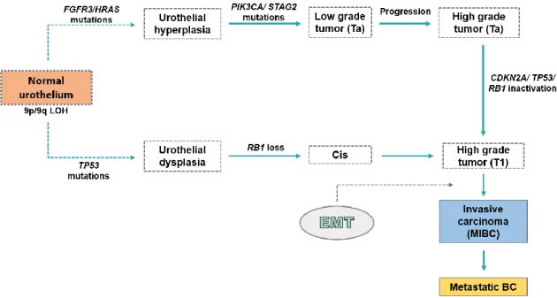
Figure 3. Urothelial carcinogenesis classical pathways. Regarding behavior and molecule
profile, BC formation can be divided in papillary pathway and the Cis pathway. The first one start with chromosome 9 deletion and FGFR3/RAS mutations, promoting hyperplasia lesion formation. Then, after the PIK3CA/STAG2 mutations hyperplasia progresses to a low-grade Ta. Lastly, low-grade Ta might progress to high-grade Ta. Cis pathway initiates with chromosome 9 deletion and TP53
INTRODUCTION ‖ 9
mutations, promoting urothelial dysplasia formation, which can progress to Cis when RB1 deletion occurs. High grade Ta, providing from papillary pathway, can suffer a CDKN2A/TP53/RB1 inactivation, which results in a high-grade T1. Moreover, Cis can also progress to T1. Finally, with EMT involvement, the non-muscle invasive tumors can progress to MIBC and, subsequently, can metastasize. Abbreviation: BC - Bladder cancer; CDKN2A - Cyclin-dependent kinase inhibitor 2A; Cis - Carcinoma in situ; EMT - Epithelial-mesenchymal transition; FGFR3 - Fibroblast growth factor receptor 3; MIBC Muscle-invasive bladder cancer; PIK3CA - Phosphatidylinositol-4,5-biphosphate 3-kinase, catalytic subunit alpha; RB1 - Retinoblastoma1; STAG2 - Stromal antigen 2; TP53 - Tumor protein p53; Adapted from [37].
EPITHELIAL-MESENCHYMAL TRANSITION (EMT)
EMT is defined as a biological process that undergo genetic, molecular and phenotypic alterations, which results in the transdifferentiation of the epithelial cells into mesenchymal cells [39, 40]. Therefore, epithelial cells lose their epithelial features and acquire mesenchymal and fibroblast-like properties features [41]. Usually, this is not an irreversible process, because the cells that underwent EMT can recover their original epithelial characteristics. This is known as mesenchymal-epithelial transition (MET) [39].
EMT is divided in 3 different types, according to the biological context [40]. Type 1 EMT, occurs in implantation, embryo formation and organ development, whereas this transition promotes the development of new tissues with different functions. Type 2 EMT relates with the beginning and the end of the inflammation process and can be associated with wound healing and tissue regeneration [16, 40]. Finally, type 3 EMT, also known as oncogenic EMT, happens in neoplastic cells that earlier suffered genomic and epigenetic alterations. Hence, cells develop the abilities of invasion and metastization [40].
Epithelial cells compose the transitional epithelium, also known as the urothelium [42]. These cells exhibit apical-basal polarity, whereupon the apical side is aimed at the lumen and the basolateral surface is anchored to the basement membrane. Moreover, they have the ability to adhere at this membrane and adjacent cells (cell-to-cell interactions) by intercellular junctions, such as tight junctions (TJs), adherens junctions (AJs) and desmosomes. Consequently, the cells can communicate with each other [39, 43-45].Throughout EMT, epithelial cells lose apical-basal polarity and cell-cell adhesion structures. Besides, also occurs signaling programmed changes, such as cadherin switch (CS). On the other hand, the modulation of the organization of cytoskeleton and the gain of front–rear polarity takes place, which makes cells take on a spindle-shape. As a result, the cells become isolated, motile and resistant to apoptosis, which allows them to invade the tissue parenchyma and go to blood stream, resulting in cancer cell invasion and metastasis [39, 43, 44]. Particularly, early work established that epithelial-cadherin, also known as E-cadherin (ECAD) loss and matrix metalloproteinase (MMP) 9 increased expression was associated with a poor clinical outcome in patients with urothelial tumors, suggesting that EMT might also be associated with bladder cancer progression and metastasis [46].
INTRODUCTION ‖ 10
Briefly, the main epithelial markers associated with adhesion are adherens junction proteins, such as ECAD and β-catenin and tight junction proteins, like occludins or cludins. Moreover, also cytokeratin, specially 5 and 6, and finally Zonula occludens (ZO)-1. Nevertheless, the main mesenchymal markers which promote invasion and mobility are neuronal-cadherin, also known as N-cadherin (NCAD), vimentin (Vim) and fibronectin, MMPs (Figure 4) [38, 43].
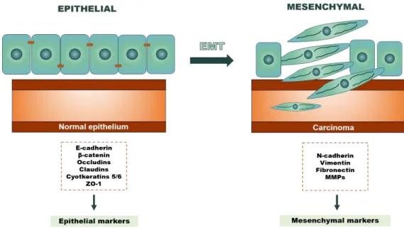
Figure 4. Cellular features alterations associated with EMT biological process. During EMT the
cells lose epithelial features and acquire mesenchymal features. Epithelial cells have an apical-basal polarity and exhibit cell-to-cell interactions through tight junctions, adjacent cells and desmosomes. Currently, these cells express, mainly, adherens junction proteins (E-cadherin and β-catenin), tight junction proteins (occludins and claudins), cytokeratin, such as 5 and 6 and ZO-1. Throughout EMT, these epithelial markers are replaced by mesenchymal markers, such as N-cadherin, vimentin fibronectin and MMPs. Due to this, cells acquire front-rear polarity and fibroblast-like phenotype, losing their adhesion ability. Consequently, they gain motility and invasion capacity. Abbreviation: EMT - Epithelial-mesenchymal transition; MMPs - Matrix metalloproteinases; ZO-1 - Zonula occludens-1; Adapted from [47].
CADHERINS
Cell adhesion molecules (CAMs) play an important role in cell shape and integrity of cell-cell interactions. CAMs can be classified as calcium binding-dependent, which includes selectins and cadherins, or as calcium binding-independent, whereas molecules such as integrins and immunoglobulins belong to this group [48]. Cadherins are a superfamily of transmembrane glycoprotein molecules that usually tend to be in AJs. Furthermore, they are the main mediators of cell-cell adhesion in epithelial tissues [7, 48]. This superfamily can be divided into 4 subfamilies, namely classical cadherins, protocadherins, desmosomal cadherins and atypical cadherins [48].
The type 1 classical cadherins family, include E-, N-, placental-cadherin, also known as P-cadherin (P-CAD) and retinal-cadherin, also known as R-cadherin (RCAD) [48].
INTRODUCTION ‖ 11
Moreover, all of them include a cytoplasmic carboxyl-terminal, a transmembrane and an extracellular N-terminal domain. Due to this last domain, it is possible that the homophilic interactions that occur between cadherins either in the same cell or in adjacent cells, allows the cell-cell adhesion [48-50]. Apparently, these four cadherins play an important role in the EMT development.
ECAD is a tumor suppressor protein encoded by CDH1 gene. This protein is expressed specially in normal epithelial tissues [51]. Besides, it is considered the most important regulator of EMT, because it is responsible for mediate the interactions between adjacent epithelial cells and the maintained of apical-basal polarity [16]. In several carcinomas, such as, colorectal, breast cancer and BC, it was reported loss or low levels of ECAD, compared to normal tissue [52]. In BC the role of this glycoprotein is well established. The lower ECAD levels promote an alteration in cell morphology and increases migration and invasion capacity, leading to increased biological aggressiveness [7, 53-55]. Previous studies, based in immunohistochemistry, showed that normal urothelium presented a high ECAD expression. On the other hand, both Ta (20% of cases) and T1 (60% of cases) presented a reduction of ECAD expression and in MIBCs (80% of cases) the amount of this protein was either low or absent [16]. Therefore, lower ECAD levels contribute to poor survival rates and facilitates tumor progression [54, 56]. Moreover, other studies demonstrated an association between lower ECAD immunoexpression and higher recurrences in patients with low grade BC [54].
Loss of ECAD expression is normally replaced by an increase of NCAD. This biological process is named CS, which is an important feature in EMT [51, 57, 58]. In BC, CS occurs, so ECAD is replaced by a novel NCAD expression. However, CS also can happen when ECAD levels stay equal but the cells acquire de novo or increased N-CAD expression [59]. Nevertheless, the opposite can also happen in some situations such as ovarian tumorigeneses, where tumor cells experience a decrease on NCAD, being replaced by an increase of E- and P-CAD expression [51].
NCAD is considered a mesenchymal marker encoded by CDH2 gene [64]. This protein is expressed in neural, endothelial and muscle cells. Nonetheless, apparently no expression or low NCAD expression was found in epithelial cells [60, 61]. The NCAD upregulation has been associated with prostate, melanoma, colon, breast and urothelial malignancies as well as progression [61]. In BC, high levels of NCAD are related with tumor aggressiveness and, consequently, with more advanced tumor stage [62]. Therefore, NCAD´s higher expression promotes an invasive phenotype, such as migration and motility events of tumor cells from the bladder to other structures and organs [58]. Indeed, previously studies demonstrate that glycoprotein over-expression is associated with lymph node metastasis and lymphovascular invasion [62]. Consequently, this leads to a worse
INTRODUCTION ‖ 12
clinical outcome and poor overall survival, as well as recurrence-free survival [59, 61]. Former immunohistochemistry studies demonstrate that normal urothelium do not have NCAD expression. By the same token, a high number of Ta and T1 (80% cases) may have a lack of this protein. Although, 60% of advanced MIBCs express NCAD [16, 52, 58]. Likewise, NCAD can function as a predictive biomarker of disease-recurrence and cancer-specific mortality and as a prognostic marker of progression in pT1 tumors [59, 62].
In CS, ECAD expression could also be replaced by PCAD, according to some authors [16, 63]. This biological process can happen in colorectal, endometrial, breast, pancreatic, gastric and specially in BC. Nevertheless, this alternative CS is not well explored [58, 64].
PCAD is encoded by CDH3 gene. This glycoprotein is usually weakly expressed in some basal layers of stratified epithelium tissues, such as esophagus, skin and especially in bladder urothelium [16, 58]. According to former studies PCAD expression seems to play a dual role, depending of tumor model. On the one hand, PCAD expression in melanoma, appears to be associated with suppressed invasion [64]. On the other hand, in breast or endometrial carcinoma, this same protein seems to be related with an aggressive phenotype. In BC, PCAD's role is less established compared with ECAD or NCAD, although it seems to be associated with tumorigenesis [58]. Previous IHC studies prove that PCAD is expressed in normal urothelium, even though it shows a weak expression [63]. Similarly to NCAD, the majority of Ta and T1 (80% cases) demonstrate weak PCAD expression, while 50% to 88% of advanced MIBCs present a PCAD overexpression [16]. Regarding PCAD high expression it seems to participate in recurrence disease [57]. Furthermore, it seems to be associated with a more aggressive phenotype, since might contributes for invasion and migration [63]. It is possible that the PCAD overexpression may contributes to the basal cells migration to more superficial bladder layers, such as intermediate cell layer [53, 63]. Consequently, BC patients with normal PCAD expression have a better survival rate compared with patients with higher PCAD levels [31, 57, 63].
Although, PCAD and ECAD can be co-expressed in urothelium, only ECAD seems to participate in cell-cell adhesion [58, 63, 65]. However, even in the presence of ECAD, PCAD seems to be capable to promote a malignant and invasive phonotype [31]. In the same way as NCAD, PCAD seems not to display an invasive-suppressor role [52]. Additionally, in bladder tumorigeneses, during cadherin switch, PCAD´s overexpression may happen in an independent or synchronized way to NCAD´s expression [16].
RCAD is encoded by CDH4 gene. This protein is highly expressed in the brain and plays an important role in the induction of type 1 EMT in kidneys and striated muscle embryogenesis [66, 67]. The knowledge of RCAD´s role in tumors is still reduced, compared with ECAD, NCAD and PCAD information. Regarding RCAD role, an opposite behavior in
INTRODUCTION ‖ 13
sarcomas and in carcinomas was observed, since in breast, colorectal or nasopharyngeal carcinoma occurred a downexpression with tumor development [68]. However, in osteossarcoma, an overexpression of RCAD is essential for tumor development, as well as cell migration and invasion. Nevertheless, when CDH4 knockdown happens, it is observed an inhibition of the tumor, as well as of the metastasis [68]. Currently, there are no studies of RCAD´s role in BC. Due to the lack of information of RCAD in BC it is important to study the role that might plays in normal urothelium. Besides, it is essential understand if this protein might be a tumor suppressor or a tumor induction. Furthermore, it is necessary understand the role that may has in the establishment of EMT.
CADHERINS REGULATORY MECHANISMS
TGF-β signaling pathways
The induction of EMT is related with alterations in some signaling pathways, such as transforming growth factor-β (TGF-β), bone morphogenetic protein (BMP), epidermal growth factor (EGF), Wnt, sonic hedehog (Shh), notch or integrin signaling [44].
Cytokines superfamily comprises TGF-β family. In its turn, TGF-β family includes a higher number of proteins, such as activins family, BMP, growth differentiation factors (GDFs), nodal, lefty, myostatin, anti-mullerian hormone (AMH) and TGF-β subfamily [69]. TGF-β subfamily is constituted by three isoforms, such as TGF-β1, β2 and β3. These proteins play an important role as ligands in TGF-β signaling, which is the most well described EMT-induced pathway [69-71]. The most study isoform is TGF-β1, which promotes embryonic development, wound healing and fibrosis [39, 72]. On the other hand, this isoform plays a double role in tumorigeneses. In the normal cells and the beginning or early stage of tumor formation it can function as tumor suppressor, causing apoptosis and growth tumor inhibition. However, in advanced stages, it causes EMT induction and, consequently, promotes cells motility and invasion. Moreover, tumor progression leads to an overexpression and higher segregation of TGF-β1 by tumor cells, which is then released for adjacent cells [73]. Therefore, TGF-β1 acts both in tumor cells and in nearby stroma, promoting immune surveillance suppression, angiogenesis induction, degradation of the extracellular matrix and recruitment inflammatory cells for the tumor microenvironment [69, 72, 73].
The TGF-β signaling pathways can be divided in independent signaling or Smad-dependent signaling[74]. In Smad-dependent signaling occurs a complex formation between TGF-β ligands and heteromeric complex of serine/threonine kinase receptors, named as type I TGF-β receptor (TGF-βRI) and type II TGFβ receptor (TGFRβII).
INTRODUCTION ‖ 14
Regarding this interaction, TGF-βRI it will be phosphorylated, which results in the recruitment and phosphorylation of receptor regulated SMADs (R-SMADs) transcription factors, SMAD2 and SMAD3 [44, 47, 69, 75]. Then, SMAD4 is recruited, forming a trimeric complex capable to translocated to the nucleus, with help of importins [44, 72, 75]. Here, SMAD complex interact with DNA-binding transcription factors. Consequently, occurs activation or repression of the gene’s transcription important for EMT establishment. The transcription factors (TFs) involved are the zinc-finger transcription factors, which includes Snail and Slug. Moreover, basic helix–loop–helix (bHLH) factors, such as Twist and E12/E47 and zinc-finger factors, like ZEB1 and ZEB2 are also involved [69, 76]. Herewith, usually these transcription factors promote repression of genes that encodes epithelial proteins, such as CDH1 [70]. Concurrently, contribute to activation of genes that encodes mesenchymal proteins, for example, CDH2, although the relation TGF-β and CDH2 remains unclear (Figure 5) [70].
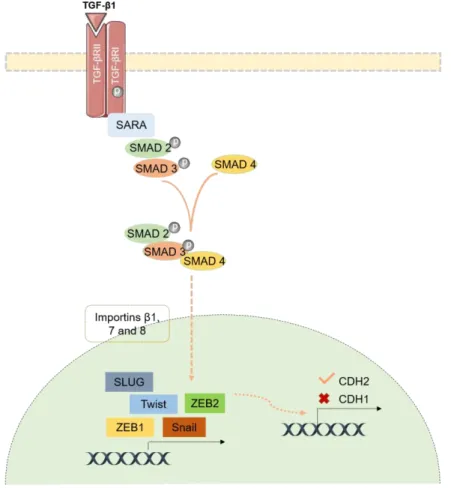
Figure 5. Smad-dependent signaling in EMT. TGF-β1 ligand binds to the heteromeric complex of
TGF-βI and TGFRβII. Then, the cofactor SARA interacts with R-SMADs transcription factors, SMAD2 and SMAD3, in order to be recruited to the TGF-βR complex. Hereafter, SMAD4 is also recruited, resulting in a trimeric complex formation, being capable to translocated into the nucleus, with the help of importins-β1, importins 7 and 8. Then, the SMAD2/3/4 complex can activate the DNA-binding transcription factors expression, such as zinc-finger transcription factors (Snail and Slug), bHLH factors (Twist and E12/E47) and zinc-finger factors (ZEB1 and ZEB2). The Snail, Slug, Twist, ZEB1 and ZEB2 are EMT transcription factors that are capable of CDH1 downregulation and CDH2
INTRODUCTION ‖ 15
upregulation. Abbreviation: bHLH - basic helix–loop–helix; R-SMADs - Receptor-regulated SMADs; SARA - Smad anchor for receptor activation; TGFRβII - Type II TGF-β receptor; TGF-β1 - Transforming growth factor β1; TGF-βI- Type I TGF-β receptor; Adapted from: [44].
Smad-independent signaling also participates in the gene reprogramming and, consequently, in EMT. Here, the ligand still being β1, as well as the receptors are TGF-βRI and TGF-TGF-βRII. However, the downstream targets activated are different. The signaling pathways are c-Jun NH2-terminal kinase (JNK) and p38, phosphatidylinositol 3,4,5 trisphosphate (PIP3) - Protein Kinase B (AKT), RhoA and Ras- extracellular signal-regulated kinase (Erk) [39, 47].
Currently, the role that TGF-β might play in BC is less detailed. Therefore, it is important study the impact of TGF-β family in EMT induction in bladder carcinogenesis [75].
Other regulatory mechanisms are associated with cadherins expression. Indeed, Snail1 can repress CDH1 by polycomb repressive complex 2 (PRC2) recruitment, which promote histone modifications [77, 78]. Moreover, micro-RNAs might also influence cadherins genes regulation, since miR-200 regulates CDH1 through ZEB1 and ZEB2 [79]. Additionally, methylation is other cadherins regulatory mechanism [77].
Epigenetics
In 1942, Conrad Waddington defined the term “Epigenetics” as alterations in the expression of proteins without altering the DNA sequence[80-82]. Currently, it is known that epigenetic can have a role in normal biological events, such as embryonic processes, gene imprinting, stability of genome as well as cell differentiation [83, 84]. However, it can also play an important role in pathogenic events, like tumorigenesis, cancer progression and resistance to therapies [81]. Epigenetic alterations are reversible and can precede genomic alterations. Furthermore, they frequently happen in the early stages of neoplastic process. Epigenetic mechanisms can be mainly divided in DNA methylation, histone post-translational modifications, histone variants and chromatin remodeling complexes (Figure 6) [85].
INTRODUCTION ‖ 16
Figure 6. Epigenetic regulatory mechanisms in gene expression. Epigenetic mechanisms
comprise DNA methylation, histone post-translational modifications, histone variants and chromatin remodeling complexes. Kindly provided by Ana Lameirinhas.
Cadherins promoter methylation
The DNA methylation is the most well researched epigenetic modification in BC [81]. This chemical modification takes place when a methyl (CH3) group, donated by
S-adenosyl-L-methionine (SAM), is transferred to the 5´position of a cytosine ring. The DNA methyltransferases enzymes, which in mammals are DNMT1, DNMT3a and DNMT3b, catalyze the donation of CH3. The final product of this reaction is the 5-methylcytosine (m5C)
formation [83, 86]
The DNMT3a and DNMT3b are de novo DNA methyltransferases. They are important to establish the DNA methylation patterns during embryonic development [87]. Contrarily, DNMT1 is the enzyme responsible for preserving the methylation patterns throughout the DNA replication, as it completes the hemi-methylated regions of DNA. This allows the transfer of the methylome from the maternal to the daughter cells [87, 88]. Oppositely, there are demethylases such as ten-eleven methyl cytosine dioxygenase (TETS) that allows the oxidation of m5C, promoting the formation of the
5-hydroxymethyl-cytosine (5hmC) [18].
DNA methylation usually occurs in cytosine-phosphate-guanine (CpG) islands. These are defined as regions with 200 bases, enriched at least 50% in cytosines followed by guanines, and with a ratio of “observed to statistically excepted CpG frequencies of at least 0.6”. They are commonly located upstream of genes promoters [89]. Approximately 60% of human gene promoters are in relation to CpG islands. In the normal tissues, these islands are normally unmethylated. However, a minor amount of them (approximately 6%) present methylation, in order to avert chromosome instability as well as to promote tissue
INTRODUCTION ‖ 17
differentiation processes or inactivation of the X chromosome in females [81, 90]. Still, abnormal methylation of CpG islands is commonly associated to transcription inhibition of some genes, such as tumor suppressor genes. Therefore hypermethylation is usually related to carcinogenesis [91]. This silencing can occur in a direct or an indirect way. On one hand, the first event takes place when the methylation prevents the recruitment of transcription factors. On the other hand, the indirect inhibition occurs due to the recruitment of methyl-CpG-binding domain (MBD) proteins, that consequently provide the recruit of histone deacetylating complexes and chromatin-remodeling complexes [89].
DNA methylation alterations have been involved in the initiation of bladder carcinogenesis as well as in the progression [92]. Apparently, urothelial carcinogenesis differs not only genetically but also epigenetically, since less loci were hypermethylated in non-invasive (10%) compared with invasive tumors (38%) [92]. Indeed, DNA hypermethylation happened in, approximately, 50-90% of BC cases, causing the silencing of several genes such as tumor suppressors, DNA repair genes and cell cycle control, which contributes for BC formation or progression [81].
CDH1 methylation role´s has been well studied in epithelial carcinomas, such as
breast and prostate carcinoma [77, 93]. In BC, CDH1 usually exhibits higher methylated frequencies, approximately, among 40 and 60% [94, 95]. Previous studies demonstrated that CDH1 promoter hypermethylation in BC as an important mechanism to regulate ECAD expression [42, 96, 97]. Indeed, normal bladder tissues present absence of methylation (24% of methylated cases) and consequently present higher ECAD immunostaining. Contrarily, bladder tumor tissues are hypermethylated (84% of methylated cases), being ECAD downexpressed or absence [96].
For CDH2 methylation no data have been reported in BC. However, previous reports only refer methylation as a possible regulatory mechanism of CDH2 promoter [98] .
Regarding CDH3 methylation, there are no studies in BC. However, according to former results performed in other model tumors, such as in breast cancer. In this malignancy, PCAD aberrant expression seems to be regulated by promoter CDH3 hypomethylation, since 71% of PCAD negative staining cases presented CDH3 methylation, while 65% of PCAD positive cases showed CDH3 unmethylated [99].
Finally, there is no data concerning the CDH4 methylation role in BC. Nevertheless, previous studies in epithelial cancers have demonstrated CDH4 promoter gene regulation by hypermethylation [68]. In gastrointestinal carcinomas, CDH4 might have a tumor suppressor gene role. Specifically, 78% of colorectal and 95% of gastric carcinomas were showed to present CDH4 promoter hypermethylation, which associated with RCAD downregulation. Moreover, this gene’s promoter methylation might be implicated in early event in gastrointestinal tumor progression [100].
AIMS ‖ 21
It is currently acknowledged that approximately half of NMIBC patients have a major risk of recurrence, whereas 5-25% can progress to MIBC. Therefore, MIBC patients have a higher rate of metastasis and, consequently, a poor prognosis. Hence, it is of major importance to unveil the mechanisms that lead to bladder cancer progression. Moreover, in the future, it may be possible to develop new target therapies that might slow or even inhibit the NMIBC to MIBC progression.
Thus, the aim of this project consists in investigate whether E-cadherin, N-cadherin, P-cadherin and R-P-cadherin differential expression might be implicated in epithelial-mesenchymal transition and, consequently, in bladder tumors progression.
Specifically, the tasks for the project are:
1. In silico analysis of cadherins expression in normal and bladder cancer tissues in TCGA dataset.
2. Evaluate E-Cadherin, N-Cadherin, P-Cadherin and R-Cadherin expression in primary bladder tumors samples surgically removed as well as in normal bladder tumor tissues.
3. Associate cadherins immune-expression patterns with clinicopathological data. 4. Characterize cadherins’ expression in one normal bladder cell lines and in seven
bladder cancer cell lines.
5. Uncover TGF-β1 and DNA promoter methylation role in cadherins expression regulation.
![Figure 1. Representation of world map with estimated age-standardized incidence rates in 2018 of bladder cancer in both sexes in all ages [2] .](https://thumb-eu.123doks.com/thumbv2/123dok_br/15758191.1074510/28.892.153.757.677.985/figure-representation-world-estimated-standardized-incidence-bladder-cancer.webp)