Increased expression levels of Syntaxin 1A and Synaptobrevin
2/Vesicle-Associated Membrane Protein-2 are associated with the
progression of bladder cancer
Sadaf Azad Raja
1*, Seher Abbas
1*, Syed Tahir Abbas Shah
1, Aamira Tariq
1, Nazia Bibi
1, Arzu Yousuf
2,
Athar Khawaja
2, Muhammad Nawaz
3, Arshad Mehmood
3, Muhammad Jadoon Khan
1Alamdar Hussain
1 1Department of Biosciences, COMSATS Institute of Information Technology Islamabad, Pakistan.
2Department of Urology and Kidney Transplant, Shifa International Hospital, Islamabad, Pakistan.
3
Armed Forces Institute of Urology (AFIU), Rawalpindi, Pakistan.
Abstract
Gene expression is tightly regulated in time and space through a multitude of factors consisting of signaling mole-cules. Soluble N-ethylmaleimide-sensitive-factor attachment protein receptors (SNARE) are membrane proteins responsible for the intercellular trafficking of signals through endocytosis and exocytosis of vesicles. Altered expres-sion of SNARE proteins in cellular communication is the major hallmark of cancer phenotypes as indicated in recent studies. SNAREs play an important role in maintaining cell growth and epithelial membrane permeability of the blad-der and are not only involved in cancer progression but also metastatic cell invasion through SNARE-mediated traf-ficking. Synaptobrevin2/Vesicle associated membrane protein-2 (v-SNARE) and Syntaxin (t-SNARE) form a vesicular docking complex during endocytosis. Some earlier studies have shown a critical role of SNARE in colon, lungs, and breast cancer progression and metastasis. In this study, we analyzed the relative expression of the STX1A and VAMP2 (SYB2) for their possible association in the progression and metastasis of bladder cancer. The profiling of the genes showed a significant increase inSTX1A and VAMP2 expression (p < 0.001) in high-grade tu-mor cells compared to normal and low-grade tutu-mors. These findings suggest that elevated expression ofSTX1A and VAMP2 might have caused the abnormal progression and invasion of cancer cells leading to the transformation of cells into high-grade tumor in bladder cancer.
Keywords: SNARE, bladder cancer, vesicle fusion, gene expression.
Received: November 10, 2017; Accepted: July 9, 2018.
Introduction
Transitional cell carcinoma is the common form of histologic bladder cancer (90% cases) and has significant mortality rate (77.89% 5-year relative survival) (American Cancer Society, 2017). High grade tumors have high proba-bility of recurrence with high percentage of progression while low grade tumors have low frequency of recurrence and are less progressive (Miyamotoet al., 2010). Accord-ing to WHO in 2004 (WHO/International Society of Uro-logical Pathology (ISUP) classification), the classification of bladder cancer is useful in differentiating carcinomas for prognostic evaluation (Panet al., 2010). Low grade lary urothelial carcinomas (LPUCs) and high grade papil-lary urothelial carcinomas (HPUCs) have distinct cancer progression categories and recurrence, and therefore, WHO
recently recommended the staging of bladder cancer into only two categories: low grade and high grade (Miyamoto et al., 2010; Panet al., 2010).
In almost all of the cancers, signal transduction dysre-gulation has a key role in triggering the cell for survival in malignant conditions (Bartsch et al., 2010). The tumor microenvironment plays a crucial role in maintaining the tumor growth, progression, and metastasis via exploiting growth factors, enzymes, and other signaling molecules that are preferably transported via exosomes (Kanget al., 2017). Proteome analysis of extracellular vesicles (EVs) secreted by the epithelial membrane in muscle invasive bladder cancer (MIBC) showed that these vesicles contain a number of proteins and signaling molecules that are trans-ported to extracellular matrix (ECM) (Silverset al., 2017). The vesicle trafficking is basically controlled by regulatory receptor proteins present on the membrane of the targeting cell, functioning with the aid of gated channels (Palfreyman and Jorgensen, 2008). Membrane trafficking in the euka-ryotic cell is mediated by a SNARE complex [soluble
Send correspondence to Alamdar Hussain. COMSATS Institute of Information Technology– Biosciences, Chak Shahzad, Park Road, 45550 Islamabad, Pakistan. E-mail: [email protected]. * These authors contributed equally to this work.
synaptobrevin2 (VAMP2) that mediate final vesicle fusion (Fang and Lindau, 2014).
SNARE proteins are known to actively derive vesicu-lar trafficking between the cells to maintain the cell integ-rity via cell growth, migration, and wound healing in a regulative manner (Tianet al., 2014). Delivery of extra-cellular matrix (ECM) and integrins through vesicular transport is the fundamental function of SNARE proteins during cell proliferation and motility. Though SNARE-mediated exosome transport of integrins is critical for can-cer development, epidermal growth factors at the cell sur-face have a major role in ECM regulation, cell survival, and progression (Enrichet al., 2015). SNARE proteins regulate matrix degradation and allow cell migration/invasion (Wil-liamset al., 2014). Functional silencing of SNARE proteins decreases the ability of breast cancer cells to invade and mi-grate (Steffenet al., 2008). Inhibition of SNARE proteins impairs the development of invadopodium, disrupts cell in-vasion, and inhibits migration in tumors (Williams and Coppolino, 2014). Altered expression of the SNARE com-plex has been found critical for various cancers as they are the core signaling proteins involved in vesicular fusion and known to be good targets for cancer therapy (Meng and Wang, 2015).
STX1A and VAMP2 are known neuronal SNAREs that mediate synaptic vesicular fusion (Ramakrishnan et al., 2012).STX1Aoverexpression has also been observed in primary brain tumor and colorectal, lung, and breast can-cers (Grabowskiet al., 2002; Arsenaultet al., 2013; Fer-nández-Nogueiraet al., 2015). Blocking ofSTX1Ainhibits tumor growth in glioblastoma (Ulloaet al., 2015). Little is known about the expression pattern ofVAMP2 in breast and lung cancers and also in bladder cancer. However, loss ofVAMP2in neuronal tissue leads to endolysosomal degra-dation. VAMP2 relies on its sorting behavior for vesicle exocytosis and fusion with target sites. Decreased expres-sion of VAMP2 causes abnormalities in the degradation pattern of useless proteins (Habermanet al., 2012). Hetero-genic expression of VAMP2 and other SNARE proteins was found in undifferentiated colorectal carcinomas (Gra-bowskiet al., 2002). Importantly,VAMP2is known to be involved in the integrin trafficking and critical for cancer
Material and Methods
Tumor sampling
Tumor and normal tissue samples were collected from post-surgical bladder cancer specimens. The study was approved by the ethical review board (ERB) of COMSATS Institute of Information Technology (No. CIIT/Bio/ERB/18/76). The data were obtained with the written consent of the patients involved in the study. Dis-ease histories were confirmed by the Department of Pathol-ogy, Pakistan Institute of Medical Science (PIMS). The total number of samples was 55, out of which 26 were paired. Surrounding normal tissue samples were used as controls. The histopathology reports of the patients were obtained from the Department of Urology PIMS and Shifa International Hospital, Islamabad, Pakistan, for categoriz-ing the tumor samples accordcategoriz-ing to their grades and cancer stage.
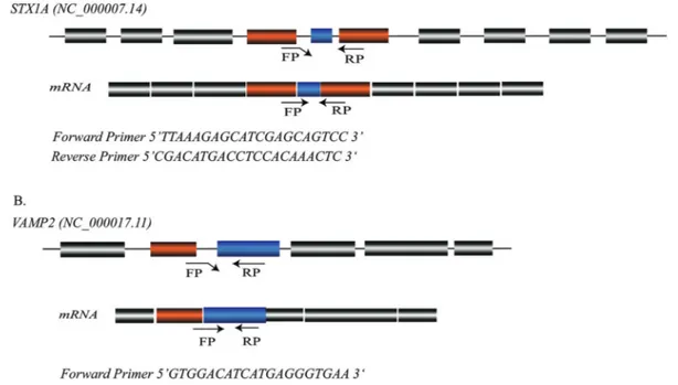
Quantitative PCR
each sample. The relative fold-increase in the expression of theSTX1AandVAMP2genes was analyzed using the 2-DDCT method (Livak and Schmittgen, 2001). The data were nor-malized with the internal controlTUB3 and the average fold-increase was determined by calculating the relative ex-pressions of each tumor sample.
Statistical analysis
Statistical analyses were performed with OriginPro 2017 (OriginLab, Northampton, MA). For expression data, the Ctvalues of the target genesVAMP2 and STX1Awere
normalized with the control gene (TUB3) Ct. Normality of
the data was assessed by the Shapiro-Wilk test. The correla-tion among different factors was assessed by the Spearman Correlation Coefficient test. Depending on the experiment, the statistical significance was determined using the Wil-coxon, Mann-Whitney, or Kruskal-Wallis ANOVA test, and specific comparisons were made by the Tukey’s Hon-estly Significant Difference (HSD) test. Fisher’s exact two-tailed test was performed to calculate patient data con-tingency, withp< 0.05 considered as significant.
Results
Tumor grade and stage
Out of 55 bladder tumor samples, 31 were high grade and 24 were low grade according to the WHO/ISUP classi-fication system (Miyamotoet al., 2010). The
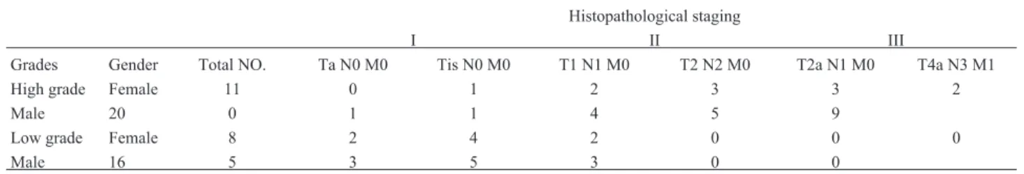
histopatho-logy reports of the patients were obtained from the hospi-tals (Pakistan Institute of Medical Sciences and Shifa Inter-national Hospital, Islamabad Pakistan) and, according to the histopathology examination; the tumors that had recur-rent behavior were categorized as high grade tumors. The samples were confirmed as transitional cell carcinomas by the Department of Histopathology (PIMS). There were 10 high grade tumors that had metastasized to the pelvic wall and prostate gland. Among the low grade tumors there were seven tumors that had spread only to the sub-epithelial con-nective tissue of the bladder. The staging of tumors was based on the TNM staging system (Table 1). The signifi-cance and distribution of tumor grade among age intervals was calculated using the Chi-square test (Table 2).
Expression ofSTX1AandVAMP2
The relative fold-change in the expression of the STX1Awas analyzed using the 2-DDCT method (Livak and Schmittgen, 2001). The data were normalized with the in-ternal control gene tubulin (TUB3),and the average fold-change was determined by calculating the relative expres-sions of each tumor sample (Table S1). The relative RNA level ofSTX1Ashowed a five-fold increase in tumors com-pared to their controls (p< 0.005). Similarly, the expression ofVAMP2was 2.9-fold higher in tumor samples compared to their controls (p< 0.001) (Figure 2).
Expression ofSTX1AandVAMP2relative to tumor grades
Expression levels of the genes were correlated to tu-mor grades (Table S2). According to the pathological grad-ing of bladder cancer, the expression ofSTX1Awas highly
in high grade tumors and 2.5-fold higher in low grade tu-mors. Therefore, a significant difference ofSTX1A expres-sion was observed between the high grade and low grade tumors (p < 0.001).VAMP2 expression was also signifi-cantly increased in high grade tumors compared to the low grade tumors (p< 0.001). Low grade tumors had a lesser fold-increase in the expression of STX1A and VAMP2, whereas high grade tumors showed relatively higher fold-increases in the expression of both genes (Figure 3). These results suggest that the genes had higher expression in higher grades tumors. The expression of both genes in the controls was normal, indicating that there wass no genetic aberration that might have caused the tumors to progress to high grade.
Expression ofSTX1AandVAMP2relative to tumor stages
The increased expression of both genes was posi-tively correlated to tumor stage (Table S3). Our results showed that the expression of STX1A and VAMP2 in-creased progressively according to the stage of the tumor (Figure 4A, C). Stage II tumors are invasive and show inva-sion in the bladder muscles, while stage III tumors are highly invasive and tend to spread in adjacent organs. In an-other study, no change in expression levels was found for both genes between stage II and III (Lopez-Beltran 2008). In our study we observed a significant difference in the ex-pression levels of the two genes between stage I and III, suggesting that the expression ofSTX1AandVAMP2 in-creases in a tumor in a stage-dependent manner (Figure 4B and 4D).
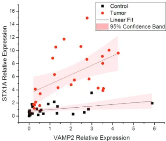
Expression correlation betweenSTX1AandVAMP2
Both genes are a crucial part of SNAREs, as vesicle fusion only takes place followed by their interaction form-ing a vesicle fusion complex. It was previously reported that cancer progression might have a role in the increased expression of the two genes that mediate cell communica-tion through vesicle fusion (Meng and Wang, 2015). These results suggest that the enhanced expression ofSTX1Aand VAMP2might have role in triggering tumor progression in high grade stage III tumors. In our correlation analysis
re-Patient data Age interval p-value
35-40 years > 40 years Gender
Male 24 12 0.5655
Female 11 8 0.5655
High grade tumor
Muscle invasive 17 9 0.1313
Non-muscle invasive 4 1 0.1313
Low grade tumor
Muscle invasive 1 2 0.1937
Non-muscle invasive 16 5 0.1937
sults, the increase in the expression of both genes was linear (Table S4) according to tumor grade and stage, which de-termines the strongly positive linear correlation between the two genes (Figure 5)
Discussion
Epithelial cells along the inner surface of organs form a primary barrier where absorption, secretion, fusion of
extracellular vesicles, and exocytosis of exosomes takes place. These cells have a regulatory vesicular communica-tion that is accomplished by a complex of SNARE proteins. SNARE proteins are responsible for maintaining the per-meability of the bladder epithelium (Bornet al., 2003). The inner luminal membrane exposed to urine releases small
Figure 3- Expression of Synaptobrevin2 and Syntaxin1A in low and high grade tumor tissues. Boxplots of normalized (relative) gene expression of Synaptobrevin2 and Syntaxin1A showing significantly higher expression in high grade tumors compared to low grade tumors.
Figure 4- Tumor stage-dependent gene expression. (A and C) Shown is the increase in expression of Synaptobrein2 and Syntaxin1A in a tumor stage-dependent manner (lowest in I and highest in III). (B) A significant difference in the expression of Synaptobrevin2 is seen between stages I and II, and stages I and III, whereas no difference is observed between stage II and III. (D) A significant difference is seen in the expression of Syntaxin1A be-tween stages I and II, and stages I and III, whereas no difference is observed bebe-tween stages II and III.
mation. Increased expression ofSTX1Ain neuronal cells was reported to be responsible for tumor formation in pri-mary brain tumors (Ulloaet al., 2015). However, in some other cancers, like breast cancer, the expression ofSTX1A has been shown to be variable (Fernández-Nogueiraet al., 2015). Our results showed the increased expression of both genes in bladder tumors compared to their normal adjacent tissue.
SNARE proteins are not only involved in the trans-port of neurotransmitters and neuropeptides, but also in the transfer of growth factors, recycled receptors, or integrins, and are involved in the secretion of matrix proteases that give the cell the capacity for invasion and migration. Thus, they are involved in cell progression in a regulative manner (Enrichet al., 2015). Apart from cell progression, STX1A and VAMP2 have been reported to be regulatory proteins of the SNARE complex, involved in cell navigation and mi-gration and, hence, metastasis of cancer cells (Zylbersztejn and Galli, 2011; Friedl and Alexander, 2012). The Cancer Genome Atlas (TCGA) dataset of 406 bladder tumor sam-ples revealed an average FPKM value of 3.3 for STX1A expression and 14.9 for VAMP2 expression (https://cancergenome.nih.gov). The Human Protein Atlas (HPA) and Genotype-Tissue Expression Dataset (GTEx) demonstrate a similar trend of expression for both genes in bladder cancer. Our data suggests that the expression of STX1AandVAMP2was higher in high grade tumors exhib-iting aggressive behavior. Overexpression of both genes in tumor cells suggests enhanced vesicular exocytosis that might have caused increased recycling of integrins and ex-cretion of matrix proteases, resulting in a favorable tumor microenvironment for cancer cell development and metas-tasis.
Conclusions
As an important part of the core SNARE complex, STX1A and VAMP2 are associated with vesicular traffick-ing of growth factors, integrins, and proteases. Dysregu-lation of vesicular trafficking might cause multiple malig-nancies, more importantly cancer cell formation, altered cell adhesion, and alteration of the extracellular matrix, fa-voring tumor growth (Raineroet al., 2013). Vesicular
traf-Sciences (PIMS), and SHIFA International Hospital (Paki-stan). This research was supported by a grant (CRGP) funded by COMSATS Institute of information Technology (Pakistan).
Conflict of Interest
The authors declare that there is no conflict of interest that could be perceived as prejudicial to the impartiality of the reported research.
Authors Contributions
SAR, SA, MJK and AH conceived the idea and de-signed the project. SAR, SA and MJK helped in experimen-tation and data acquisition. AY, AK, MN, AM contributed to clinical evaluation and sample provision. SAR, SA, STAS, AT, NB contributed to data analysis and the inter-pretation of the results. AH took the lead in writing the manuscript along SAR, SA, STAS, AT and NB. AH and MJK supervised the research. All authors reviewed and ap-proved the manuscript.
References
American Cancer Society (2017) Cancer Facts & Figures, Ameri-can Cancer Society, Atlanta, 71 p.
Arsenault J, Ferrari E, Niranjan D, Cuijpers SAG, Gu C, Vallis Y, O’Brien J and Davletov B (2013) Stapling of the Botulinum Type A Protease to growth factors and neuropeptides allows selective targeting of neuroendocrine cells. J Neurochem 126:223–233.
Bartsch G, Mitra AP and Cote RJ (2010) Expression profiling for bladder cancer: Strategies to uncover prognostic factors. Ex-pert Rev Anticancer Ther 10:1945–1954.
Born M, Pahner I, Ahnert-Hilger G and Jöns T (2003) The mainte-nance of the permeability barrier of bladder facet cells re-quires a continuous fusion of discoid vesicles with the apical plasma membrane. Eur J Cell Biol 82:343–350.
Chang CW and Jackson MB (2015) Synaptobrevin transmem-brane domain influences exocytosis by perturbing vesicle membrane curvature. Biophys J 109:76–84.
Enrich C, Rentero C, Hierro A and Grewal T (2015). Role of cho-lesterol in SNARE-mediated trafficking on intracellular membranes. J Cell Sci 128:1071–1081.
Fang Q and Lindau M (2014) How could SNARE proteins open a fusion pore? Physiology 29:278–285.
Fernández-Nogueira P, Bragado P, Almendro V, Ametller E, Rios J, Choudhury S, Mancino M and Gascón P (2015) Differen-tial expression of neurogenes among breast cancer subtypes identifies high risk patients. Oncotarget 7:5313-5326. Friedl P and Alexander S (2012) Cancer invasion and the
micro-environment: Plasticity and reciprocity. Cell 147: 992–1009.
Grabowski P, Schönfelder S, Ahnert-Hilger G, Foss HD, Heine B, Schindler I, Stein H, Berger G, Zeitz M and Scherübl H (2002) Expression of neuroendocrine markers: A signature of human undifferentiated carcinoma of the colon and rec-tum. Virchows Archiv 441:256–263.
Haberman A, Williamson WR, Epstein D, Wang D, Rina S, Meinertzhagen IA and Hiesinger PR (2012) The synaptic vesicle SNARE neuronal synaptobrevin promotes endolyso-somal degradation and prevents neurodegeneration. J Cell Biol 196:261–276.
Hasan N and Hu C (2010) Vesicle-associated membrane protein 2 mediates trafficking ofa5b1 integrin to the plasma mem-brane. Exp Cell Res 316:12-23.
Hurst RE, Van Meerveld B, Wisniewski AB, VanGordon S, Lin H, Kropp BP, and Towner RA (2015) Increased bladder per-meability in interstitial cystitis/painful bladder syndrome. Translat Androl Urol 45:563–571.
Kang HW, Kim WJ and Yun SJ (2017) The role of the tumor microenvironment in bladder cancer development and pro-gression. Translat Cancer Res 6 Suppl 4: S744-S758. Livak KJ and Schmittgen TD (2001) Analysis of relative gene
ex-pression data using real-time quantitative PCR and the 2-DDCTmethod. Methods 25:402-408.
Lopez-Beltran A (2008) Bladder cancer: Clinical and pathologi-cal profile. Scand J Urol Nephrol 42:95–109.
Meng J and Wang J (2015) Role of SNARE proteins in tumou-rigenesis and their potential as targets for novel anti-cancer therapeutics. Biochim Biophys Acta 1856:1–12.
Miyamoto H, Miller JS, Fajardo DA, Lee TK, Netto GJ and Ep-stein JI (2010) Non-invasive papillary urothelial neoplasms: The 2004 WHO/ISUP classification system. Pathol Int 60:1–8.
Nevins AK and Thurmond DC (2005) A direct interaction be-tween Cdc42 and vesicle-associated membrane protein 2 regulates SNARE-dependent insulin exocytosis. J Biol Chem 280:1944-1952.
Palfreyman MT and Jorgensen EM (2008) Roles of SNARE pro-teins in synaptic vesicle fusion. In: Wang ZW (ed) Molecu-lar Mechanisms of Neurotransmitter Release. Humana Press, Totowa, NJ, pp 35–59.
Pan CC, Chang YH, Chen KK, Yu HJ, Sun CH and Ho DNT (2010) Prognostic significance of the 2004 WHO/ISUP clas-sification for prediction of recurrence, progression, and can-cer-specific mortality of non-muscle-invasive urothelial tu-mors of the urinary bladder: A clinicopathologic study of 1,515 cases. Am J Clin Pathol 133:788–795.
Polchi A, Tancini B and Emiliani C (2013) Signaling pathways in exosome biogenesis, secretion and fate. Genes 4:152–170.
Rainero E, Van den Berghe P and Norman JC (2013) Inter-nalisation, endosomal trafficking and recycling of integrins during cell migration and cancer Invasion. In: Yarden Y and Tarcic G (eds) Vesicle Trafficking in Cancer. Springer, New York, pp 327-359.
Ramakrishnan NA, Drescher MJ and Drescher DG (2012) The SNARE complex in neuronal and sensory cells. Mol Cell Neurosci 50:58-69.
Rio DC, Ares M, Hannon GJ and Nilsen TW (2010) Purification of RNA using TRIzol (TRI Reagent). Cold Spring Harb Protoc 5: 5440–5441.
Silvers CR, Miyamoto H, Messing EM, Netto GJ and Lee YF (2017) Characterization of urinary extracellular vesicle pro-teins in muscle-invasive bladder cancer. Oncotarget 8:91199-91208.
Shukla AL, Berglund L, Nielsen LP, Nielsen S, Hoffmann HJ and Dahl R (2000). Regulated exocytosis in immune function: Are SNARE-proteins involved? Respir Med 94:10–17. Steffen A, Le Dez G, Poincloux R, Recchi C, Nassoy P, Rottner
K, Galli T and Chavrier P (2008) MT1-MMP-dependent in-vasion is regulated by TI-VAMP/VAMP7. Curr Biol 18:926–931.
Tian AG, Tamori Y, Huang YC, Melendez NT and Deng WM (2014) Efficient EGFR signaling and dorsal-ventral axis pat-terning requires syntaxin dependent Gurken trafficking. Dev Biol 373:349–358.
Ulloa F, Gonzàlez-Juncà A, Meffre D, Barrecheguren PJ, Martí-nez-Mármol R, Pazos I, Olivé N, Cotrufo T, Seoane J and Soriano E (2015) Blockade of the SNARE protein Syntaxin 1 inhibits glioblastoma tumor growth. PLoS One 10:e0119707.
Williams KC and Coppolino MG (2014) SNARE-dependent in-teraction of Src, EGFR and 1 Integrin regulates invadopodia formation and tumor cell invasion. J Cell Sci 127:1712–1725.
Williams KC, McNeilly RE and Coppolino MG (2014) SNAP23, syntaxin 4, and vesicle-associated membrane protein 7 (VAMP7) mediate trafficking of membrane type 1–matrix metalloproteinase (MT1-MMP) during invadopodium for-mation and tumor cell invasion. Mol Biol Cell 25:2061-2070.
Zylbersztejn K Galli T (2011) Vesicular traffic in cell navigation. FEBS J 278:4497–4505.
Internet resources
The Human Protien Atlas (HPA),
https://www.proteinatlas.org/ENSG00000220205-VAMP2 /pathology/tissue/urothelial+cancer (accessed 12 March 2018).
The Human Protien Atlas (HPA),
https://www.proteinatlas.org/ENSG00000106089-STX1A/ pathology/tissue/urothelial+cancer (accessed 12 March 2018).
The Cancer Genome Atlas (TCGA),
https://cancergenome.nih.gov/cancersselected/UrothelialBl adderCarcinoma (accessed 12 March 2018).