1. Department of Pulmonary and Critical Care Medicinet, The First Affiliated Hospital of Wenzhou Medical University;
2. Endocrinology department, The First Affiliated Hospital of Wenzhou Medical University.
this study might provide further insights into potential molecular mechanism of the disease.
Keywords: Interstitial lung disease; Systemic sclerosis; Biomarker.
INtrODUctION
Systemic sclerosis (SSc) is a rare chronic progressive au-toimmune disease of the connective tissue, character-ized by immune dysfunction, vascular disease, cellu lar inflammation, and skin fibrosis1. SSc could affect mul-tiple internal organs leading to disorders, such as inter-stitial lung disease (ILD)1, 2, but in SSc, the lung in-volvement might occur without skin inin-volvement3. Likewise, ILD is a group of diseases associated with con-nective tissue disease, which is regarded as a potential inflammation caused by the thickening of the intersti-tial tissue around the alveolar wall4. According to a re-view by Denton and Khanna (2017), four-fifths of SSc patients have pulmonary fibrosis or interstitial lung dis-ease, but only 25-30% develop progressive ILD5, often accompanied by a histopathological pattern of non-spe-cific interstitial pneumonia (NSIP)6. The results of the EULAR Scleroderma Trials and Research (EUSTAR) co-hort showed that lung diseases were the main cause of death in SSc patients, with pulmonary fibrosis, ac-counting for more than one-third of mortality7. In a Multinational Systemic Sclerosis Inception Cohort study, including patients from Australia, Canada, and Spain, ILD accounted for 20.7% of SSc-related deaths8. In addition to increasing the utilization of me dical re-sources and related direct costs, systemic sclerosis-as-sociated interstitial lung disease (SSc-ILD) also placed a heavy burden on the medical system with patients hav-ing poor health-related quality of life (HRQoL) in Aus-tralia9. The course of SSc-ILD from localized pulmonary infiltration to severe disease might usually be
non-pro-Matrix metalloproteinase 7 is a candidate
biomarker in systemic sclerosis-associated
interstitial lung disease
Xu Z1, Chen W2, Chen C1
ACTA REUMATOL PORT. 2020;45:191-200
AbstrAct
Background: Pulmonary complications, including pul-monary fibrosis, are the leading causes of death in sys-temic sclerosis (SSc). However, the aetiology and patho-physiologic mechanisms of the disease have not been comprehensively investigated, and drugs for treating systemic sclerosis-associated interstitial lung disease (SSc-ILD) are limited. The objective of this study was to identify key novel genes and pathways linked to SSc-ILD and decipher the molecular mechanisms in-volved in the disease.
Methods: We compared three microarray datasets in the GEO database including 42 SSc-ILD samples and 18 normal samples to obtain differentially expressed genes (DEGs). Gene Ontology (GO) analysis and the Kyoto Encyclopaedia of Genes and Genomes (KEGG) pathway analysis were performed, and a protein-pro-tein interaction network was constructed. After valida-tion, gene set enrichment analysis (GSEA) was applied to obtain further insights into the function of the se-lected hub genes.
Results: A total of 25 DEGs were filtered. The GO anal-ysis revealed genes that were mainly enriched in im-mune response, chemokine activity, and extracellular regions. KEGG pathway analysis of the DEGs revealed that SSc-ILD was associated with the tumour necrosis factor (TNF) signalling pathway and cytokine-cytokine receptor interaction. Matrix metallopeptidase 7 (MMP7) expression was consistently increased in all the three datasets, and results of the GSEA indicated that MMP7 might play a role in the regulation of the G-protein coupled amine receptor activity.
Conclusions: In summary, the novel DEGs, especially MMP7 and the SSc-ILD pathway genes identified in
gressive, with the event occurring primarily in the first years of the disease onset, which might eventually progress to respiratory failure or death10.
Due to the widespread use of angiotensin-convert-ing enzyme (ACE) inhibitors, the mortality of sclero-derma nephropathy and its sequelae might be marked-ly reduced11, but effective SSc-ILD treatment is still being explored. Although many SSc-ILD treatments have been evaluated, the current management is large-ly limited to immune regulation, and non-selective im-munosuppressants are still the most commonly used10. However, nonselective immunosuppressants, parti -cularly cyclophosphamide, might have been linked to acute toxic accidents such as hematopoietic suppres-sion12, and could increase the risk of diseases like oth-er rheumatism, cancoth-er, and gonadal failure with cu-mulative doses13. Although mycophenolate mofetil could be identified as a safe drug, unfortunately it has a long-term effect, with unaffordable management costs for many patients in limited resourced economies 14. Likewise, biologicals such as belimumab, tocilizu mab, and abatacept appeared promising as they exhi -bited some skin benefit15, while Nintedanib showed a positive effect in ILD16.
High-throughput platform-based microarrays have become a reliable and efficient tool for exploring vital genetic or epigenetic changes in disease and identify-ing promisidentify-ing biomarkers for disease diagnosis and prognosis. Efficient microarray and bioinformatics analysis could help unravel the molecular mechanisms of pathogenesis and disease progression. This is a rec-ommended pathway to explore genetic variation and identify potential diagnostic biomarkers. Therefore, we aimed to identify differentially expressed genes (DEGs) and pathways, as well as decipher the poten-tial molecular mechanisms involved in SSc-ILD.
mAterIAls AND methODs
GeNe exPressION DAtA AND DAtA PrePrOcessING
The transcription profiles for the lung tissue of SSc-ILD and normal samples dataset were retrieved from the NCBI GEO database (http://www.NCBI.nlm.nih. gov/GEO/) using the keywords “systemic sclerosis in-terstitial lung disease”, “systemic sclerosis”, and “in-terstitial lung disease”. “Homo sapiens”, “lung tissue” and “Expression profiling by array” were included in the next round of screening. The dataset included
GSE48149, GSE76808, and GSE81292. The microar-ray data of GSE48149 was based on GPL16221 plat-form [Illumina HumanRef-8 v3.0 expression bead chip (Search Key version)], GSE81292 was based on GPL18991 platform [HG-U133A_2; Affymetrix Hu-man Genome U133A 2.0 Array; HGU133A2_Hs_EN-TREZG_16.0.0], and GSE76808 microarray data was based on GPL571 platform [HG-U133A_2; Affymetrix Human Genome U133A 2.0 Array]. The extracted datasets were from the total lung RNA and were for the RNA gene expression profiling. The samples included 42 SSc-ILD lung tissues and 18 normal controls (all from cancer-free patients).
The R software (version 3.4.2; https://www.Rproject.org/) was used for data mining and statistical analy -sis. If multiple probes corresponded to the same gene symbol, the “aggregation” package (https://CRAN.R-project.org/package=aggregation) in R was used as the expression value of that specific gene to calculate the average. If there was no expression value for the probe, it was supplemented with the nearest neighbour mean (KNN) of the impute package17in R. Finally, the “Lim-ma” package18was used for background correction and normalization.
IDeNtIfIcAtION Of DeGs
The DEGs for the SSc-ILD samples compared with the normal controls were screened using the t-test method in the R “Limma” package. The | log2 FC |> 1 and the adjusted P-values <0.05 were selected as the cut-off cri-teria. Also, we compared 10 samples with SSc without ILD in the GSE48149 with the normal controls. DeGs eNrIchmeNt ANAlysIs
The Database for Annotation, Visualization, and Inte-grated Discovery (DAVID) 6.8 (https://david.ncifcrf. gov/)19was used for Gene Ontology (GO) analysis and the Kyoto Encyclopaedia of Genes and Genomes (KEGG) pathway analysis. The GO analysis included molecular function (MF), biological processes (BP), and cellular component (CC). The KOBAS 3.020 (http://KOBAS.cbi.pku.edu.cn) was used to evaluate the KEGG pathway analysis of the DEGs.
PrOteIN-PrOteIN INterActION (PPI) NetwOrk ANAlysIs
The PPI network analysis is a crucial tool for unders -tanding biological responses in health and disease. DEGs were imported into the STRING (version: 10.5) online database (http://string-db.org), a biological
database and network resource of proteprotein in-teraction, to analyse the functional interactions be-tween proteins (parameters were the default in the STRING database). This was combined with the Gen-eMANIA online platform (http://GenGen-eMANIA.org), and the file was subsequently imported into the Cytoscape software (version 3.7.1; http://www.Cytoscape.org/). The hub gene was identified through the CytoHubba plug with a maximum degree.
hUb GeNes vAlIDAtION
The hub genes of interest were further validated in the GSE48149, GSE81292, and GSE76808. Additionally, the curve was plotted with the “ggstatsplot” package (https://CRAN.R-project.org/package=ggstatsplot) to evaluate the capability of the selected genes to distin-guish the SSc-ILD patients and controls.
GeNe set eNrIchmeNt ANAlysIs
To investigate the potential functions of the selected hub genes in SSc-ILD, the gene set enrichment analy-sis (GSEA) was performed. The GSEA is a free chip data analysis tool, which is based on the existing genesets. The median expression level of the hub gene was di-vided into two groups in the three datasets. The c5.all.v7.0.symbols.gmt was selected as the reference
gene setting in the Molecular Signatures Database (MSigDB), and P adjusted value <0.05, while the FDR < 0.25 was selected as the cut-off standard.
resUlts
IDeNtIfIcAtION Of DeGs
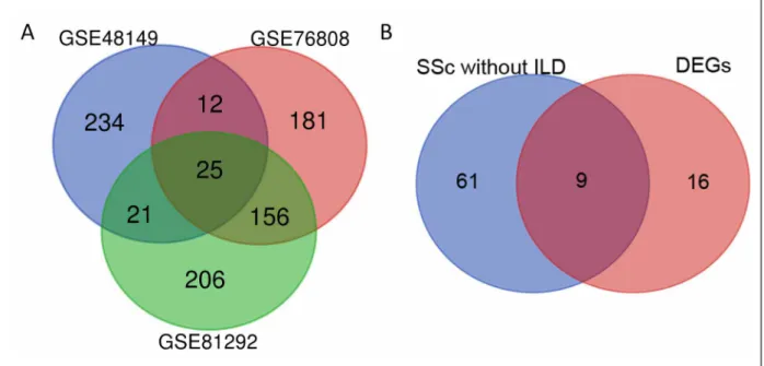
In the SSc-ILD compared with normal samples, the DEGs were identified in the three transcript profiles obtained from the GEO database, including GSE48149, GSE81292 and GSE76808. There were 25 abnormally expressed DEGs, including "TNFAIP3", "CDH3", "LIF", "CX3CL1", "APOLD1", "IL13RA2", "MMP19", "CCL2", "SLCO4A1", "IL1RL1", "CHI3L2", "CA4", "COL15A1", "MEOX1", "PTGS2", "DUSP5", "COL3A1", "HYAL1", "RGS1", "MMP7", "IGFBP2", "AGER", "EDNRB", "CXCL14", "VIPR1" (Figure 1A). Likewise, 9 DEGs (“TNFAIP3”, “IL13RA2”, “DUSP5”, “IL1RL1”, “APOLD1”, “RGS1”, “IGFBP2”, “COL15A1”, “LIF”) which were also abnormally expressed were found in the SSc without ILD samples (Figure 1B). The 25 DEGs were used for subsequent analysis because the SSc without ILD samples came from a single dataset. The calculation criteria were P <0.05 and the absolute value of log2FC> 1.
fIGUre 1. (A) The DEGs between the SSc-ILD and normal samples using Bioconductor package VennDiagram. (B) The genes
between the SSc without ILD and the DEGs (between SSc-ILD and normal samples). The DEGs were identified with the classical t-test. Statistically significant DEGs were defined at P < 0.05 and absolute log2 fold change > 1 as the cut-off criterion. GSE,gene expression series.
tokine-cytokine receptor interaction were the top canonical pathways associated with the DEGs (Figure 2D). We imported the results into R software to draw the graphics, and P < 0.05 was considered as the threshold value of significance.
PPI NetwOrk ANAlysIs
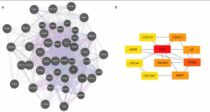
The online database STRING, GeneMANIA, and Cy-toscape software were used to analyse the DEGs. The 25 DEGs showed a complex DEGs-PPI network with a co-expression of 86.48%, a shared protein domain of 3.19%, a co-localization rate of 8.14%, and genetic in-teractions of 1.19% (Figure 3A). We identified the first six genes with the highest degree of interaction, in-cluding C-C motif chemokine ligand 2 (CCL2), keGG AND GO eNrIchmeNt ANAlyses Of DeGs
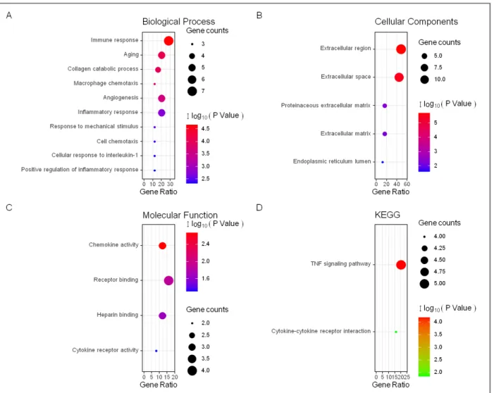
The enrichment analyses of 25 DEGs were carried out to unravel the biological classification of DEGs using DAVID (Figure 2 and Table I). The results of the GO analysis showed that variations in the BP of common DEGs were significantly enriched in immune response, aging, collagen catabolic process, macrophage chemo-taxis, angiogenesis, and inflammatory response (Fi gure 2A). For the CC, the DEGs were observably enriched in the extracellular region, extracellular space, and pro-teinaceous extracellular matrix (Figure 2B). Changes in the MF were mainly enriched in chemokine activi-ty, receptor binding, and heparin-binding (Figure 2C). The analysis of the KEGG pathway revealed that tu-mour necrosis factor (TNF) signalling pathway and
cy-fIGUre 2. The GO analyses of the common DEGs. (A) The GO analyses according to the biological process. (B) The GO analyses
according to cellular component. (C) The GO analyses according to molecular function. (D) The Kyoto Encyclopaedia of Genes and Genomes analyses of the common DEGs to identify the canonical pathways.TNF, tumour necrosis factor.
prostaglandin-endoperoxide synthase 2 (PTGS2), ma-trix metallopeptidase 7 (MMP7), TNF alpha-induced protein 3 (TNFAIP3), leukaemia inhibitory factor (LIF), and CX3C motif chemokine ligand 1 (CX3CL1) (Fi -gure 3B).
vAlIDAtION Of the hUb GeNes
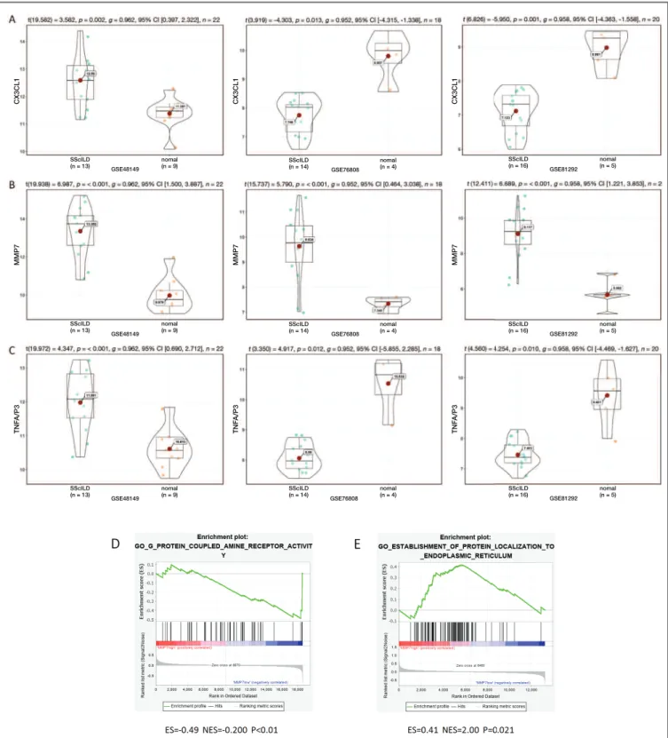
The expression levels of the six hub genes including “CCL2”, “PTGS2”, “MMP7”, “TNFAIP3”, “LIF”, and “CX3CL1” in the GSE48149, GSE76808, and GSE81292 datasets were evaluated. The P-value < 0.05 was taken as the cut off standard, only three genes were selected which were the CX3CL1, MMP7, and TNFAIP3. The expression of MMP7 in all three datasets increased significantly, whereas the CX3Cl1 and TNFAIP3 expression decreased in the GSE76808 and GSE81292 datasets but increased in the GSE48149 dataset. We observed that MMP7 was the most signi -ficant (Figure 4).
hUb GeNe GseA ANAlysIs
We executed a GSEA analysis of the MMP7 hub gene to further elucidate the possible mechanism of the
SSc-ILD-related genes. The samples were divided into high expression and low expression groups according to the median value of gene expression. The GSEA ana -lysis showed that low expression of MMP7 was en-riched in the G-protein-coupled amine receptor acti vity (GSE48149) (Figure 4E), and the high expression was enriched in the establishment of protein localization to the endoplasmic reticulum (GSE76808) (Figure 4D).
DIscUssION
At present, the etiopathogenesis of the SSc-ILD is not entirely clear; nevertheless, the principal pathogenesis of SSc-ILD was thought to sustained damage to lung cells21. The sustained damage induced fibrotic stimulation, leading to fibroblast activation and myofibro -blast transformation21. Lung injury might result in the release of fibrotic mediators, such as transforming growth factor β1 (TGF-β1) and connective tissue growth factor (CTGF)21. Furthermore, TGF-β promot-ed the increase of the myofibroblasts in the damagpromot-ed tissues and the transformation of pericytes22. Thus,
un-tAble I. the bIOlOGIcAl PrOcess IN the eNrIcheD ANAlysIs Of the DIffereNtIAlly exPresseD GeNes betweeN the ssc-IlD AND NOrmAl sAmPles (tOP 20 AccOrDING tO P-vAlUe)
Term Description Count P Value
GO:0006955 immune response 7 2.20E-05
GO:0007568 aging 5 8.19E-05
GO:0030574 collagen catabolic process 4 1.01E-04
GO:0048246 macrophage chemotaxis 3 1.51E-04
GO:0001525 angiogenesis 5 2.61E-04
GO:0006954 inflammatory response 5 0.001898
GO:0009612 response to mechanical stimulus 3 0.003187
GO:0060326 cell chemotaxis 3 0.003854
GO:0071347 cellular response to interleukin-1 3 0.004581
GO:0050729 positive regulation of inflammatory response 3 0.004836 GO:0007166 cell surface receptor signalling pathway 4 0.006749 GO:0071356 cellular response to tumour necrosis factor 3 0.010683
GO:0071222 cellular response to lipopolysaccharide 3 0.011247
GO:0010226 response to lithium ion 2 0.012793
GO:0006935 chemotaxis 3 0.013017
GO:0019221 cytokine-mediated signalling pathway 3 0.0149
GO:0002523 leukocyte migration involved in inflammatory response 2 0.015615
GO:0071318 cellular response to ATP 2 0.018428
GO:0007165 signal transduction 6 0.022087
derstanding the pathology and molecular mechanism of the SSc-ILD could facilitate and enhance clinical di-agnosis and treatment. However, analysis of the data as a single microarray dataset produced higher false-positive rates, and one-sided results were observed. In this study, we performed a comprehensive analysis of three RNA microarray datasets (GSE48149, GSE81292, and GSE76808) from which 25 DEGs were identified. Then the online website was used further to cluster the DEGs through function and pathway en-richment analyses. Furthermore, the DEGs-PPI net-work was constructed through the STRING database and Cytoscape, which revealed the molecular mecha-nism of the SSc-ILD.
Therefore, the DEGs reflected the key biomarkers related to the pathogenesis and progression of SSc-ILD, some of which had been explored and used for thera-peutic purposes. The CX3CL1 was associated with ILD progression, and its concentration in the lung tissue and serum of patients with SSc increase23. The de-creased expression of serum chemokine (C-X-C motif) ligand 14 (CXCL14) helped to maintain abnormal im-mune function and neovascularization disorders, both of which might be fundamental to the development of SSc. Furthermore, using intravenous cyclophos-phamide pulse therapy, the serum CXCL14 levels in
SSc-ILD patients who successfully treated significant-ly increased compared with the baseline24. It was ne -cessary to identify more DEGs and explore whether possible targeted genes affected the occurrence and de-velopment of SSc-ILD. Through molecular experi-ments, it was possible to identify biomarkers in SSc-ILD.
In the DEGs enrichment analysis, biological pro-cesses included immune response, aging, collagen catabolic process, macrophage chemotaxis, angioge -nesis, and inflammatory response. As in previously published studies, SSc-ILD was shown to result from an interaction among fibrotic, self-immunity, inflam-mation, and vascular injury25. The cellular components included the extracellular region, extracellular space, and the proteinaceous extracellular matrix. The onset of SSc-ILD damaged the alveolar epithelium or vascu-lar system, or both, followed by abnormal activation of the immune system25, 26. This process promoted the re-cruitment and activation of fibroblasts, the excessive production of the extracellular matrix, and the normal lung structure eventually replaced by scars25, 26. The in-creased release of the fibrotic cytokines, growth factors, peptides, and bioactive proteins, resulted in stronger signalling26.
The SSc-ILD signalling pathways were different from tAble II. the kyOtO eNcyclOPAeDIA Of GeNes AND GeNOmes ANAlyses Of the DIffereNtIAlly
exPresseD GeNes AccOrDING tO kObAs 3.0 ANAlysIs (cOrrecteD P-vAlUe < 0.95 wAs cONsIDereD As threshOlD vAlUes Of sIGNIfIcANt DIffereNce)
Corrected
ID Term Number P-Value
hsa04668 TNF signalling pathway 112 1.20E-06
hsa04060 Cytokine-cytokine receptor interaction 294 1.20E-06
hsa04657 IL-17 signalling pathway 93 0.001286
hsa04061 Viral protein interaction with cytokine and cytokine receptor 100 0.001286 hsa04933 AGE-RAGE signalling pathway in diabetic complications 100 0.001286
hsa04062 Chemokine signalling pathway 190 0.005848
hsa05163 Human cytomegalovirus infection 225 0.007972
hsa04974 Protein digestion and absorption 90 0.02281
hsa04064 NF-kappa B signalling pathway 100 0.023513
hsa04926 Relaxin signalling pathway 130 0.033497
hsa04630 Jak-STAT signalling pathway 162 0.036993
hsa04621 NOD-like receptor signalling pathway 178 0.036993
hsa00910 Nitrogen metabolism 17 0.047452
TNF, tumour necrosis factor. IL, Interleukin. AGE-RAGE, Advanced glycated end-products- receptor. NF, nuclear factor. Jak-STAT, Janus ki-nase-signal transducers and activators of transcription. NOD, Nucleotide-binding and oligomerization domain.
the signal transduction pathways of primary tumours, such as the TNF signalling pathway, and cytokine-cy-tokine receptor interaction. Scleroderma was an in-flammatory disease, and TNF played a critical role in the pathogenesis of inflammatory diseases. Previous studies showed that high TNF-alpha was the best pre-dictor of progressive disease through univariate analy-sis27. Cytokine (Interleukin-17) was necessary for the pathogenesis of lung impairment in patients with sys-temic sclerosis28. Although some pathways were iden-tified, a series of molecular experiments might help to provide more detailed and robust evidence for the like-ly phenotypes and pathway regulation of these pre-dicted SSC-ILD genes.
Besides, the six hub genes of the SSc-ILD scored highest in the protein network. Previous research had pointed out that when the experimental group inclu -ded ILD patients, some genes including CCL2 and male factors, could predict PAH with very high accuracy29. Alveolar lavage fluid from SSc-ILD patients showed higher CCL2 (30). In our study, the logFC of MMP7 was greater than 2 in the three datasets. Elevated serum MMP7 levels in patients with SSc might correlate with pulmonary involvement31, and serum MMP7 was
sig-nificantly higher in both the IPF and SSc-ILD patients32. The plasma MMP7 levels were negatively correlated with forced vital capacity (FVC) and carbon monoxide (DLCO) decline33, together with FVC and DLCO (PCMI) contribution to mortality prediction34. Zuo et
al. (2002), confirmed that MMP7 protein expression in epithelial cells increased in the bleomycin-induced fibrosis model, and also that MMP7-/- knockout could prevent bleomycin-induced fibrosis35. Unfortunately, most previous studies focused on IPF, not SSc-ILD. The co-expression and interaction between the hub genes are a new and exciting border area, requiring further re-search.
Studies of genetically targeted mice of pulmonary fibrosis revealed that most MMPs promoted the deve -lopment of pulmonary fibrosis and found multiple me chanisms. The MMP7 could promote the epithe lial-to-mesenchymal transition, increase the activity of fi-brotic mediators, or reduce the levels of anti-fifi-brotic mediators in the lung tissue36. Besides, elevated MMP7 might indicate asymptomatic ILD and reflect disease progression in patients with idiopathic pulmonary fi-brosis33. The MMP7 found on the surface of the lung epithelial cells was one of several MMPs that activated
fIGUre 3. Based on the STRING database, GeneMANIA, and Cytoscape software, the protein-protein interaction networks of the
differentially expressed genes were constructed along with the modular analyses. (A) The result of the protein-protein interaction networks according to GeneMANIA. (B) Different degree scores have different colours displayed on the graph. As the colour turned red, the score become higher.
SScILD (n = 13) nomal (n = 9) SScILD (n = 13) nomal (n = 9) SScILD (n = 14) nomal (n = 4) SScILD (n = 16) nomal (n = 5) SScILD (n = 16) GSE81292 GSE81292 GSE81292 GSE76808 GSE76808 GSE76808 GSE48149 GSE48149 GSE48149 nomal (n = 5) SScILD (n = 16) nomal (n = 5) SScILD (n = 14) nomal (n = 4) SScILD (n = 14) nomal (n = 4) SScILD (n = 13) nomal (n = 9) C X 3 C L 1 C X 3 C L 1 C X 3 C L 1 M M P 7 M M P 7 M M P 7 T N F A /P 3 T N F A /P 3 T N F A /P 3
fIGUre 4. Validation of the hub genes, including MMP7(A), CX3CL1(B), and TNFAIP3(C) in the datasets (GSE48149, GSE76808,
and GSE81292). (D) The GSEA analysis of high expression of MMP7 in GSE76808. (E) The GSEA analysis of low expression of MMP7 in GSE48149. Profile of the Running ES Score & Positions of GeneSet Members on the Rank Ordered List in (D) and (E). ES: enrichment score; NES: normalized enrichment score.
TGF-β37,38, and the TGF-β signalling pathway was a key mediator of fibroblast activation39. Nintedanib had great potential in the treatment of progressive fibrotic interstitial lung disease. It did not only increase the expression and activity of MMPs in TGFβ1 activated pe -ricytes but also inhibited TGF-β-induced fibroblast to myofibroblast transformation16, although the specific relationship between MMP7 and Nintedanib still needs further research. In the colonic mucosa, the MMP7 controlled the transepithelial influx of neutrophils by generating a chemokine gradient, which could cause tissue damage if in excess40. However, there are few studies on the related mechanisms of SSc-ILD. Since MMP7 consistently increased in all three datasets, we chose MMP7 for the GSEA analysis. It was found that the low expression of MMP7 was rich in the G-protein coupled amine receptor activity, and the high expression was enriched in the establishment of protein localization in the endoplasmic reticulum, which might pave the way for subsequent research. The G-protein-coupled receptor kinase-2 could regulate the MMP7 levels and play a key role in TNF -induced wound epi thelial cell healing in the colon epithelial cells41. Many classical het-erotrimeric G protein-coupled receptors (GPCRs) have been shown to activate the classical Wnt pathway and ultimately stabilize β-catenin42, while MMP7 might re -gulate β-catenin localization and signalling activation in injured lung epithelium43. Some agonists activate of GPCR activated MMPs and then activate the EGFR, thereby promoting vasoconstriction and growth44.
Our approach improved our understanding of the potential targets of SSc-ILD treatment. However, our study had some limitations. Firstly, to fully unfold the molecular mechanism of SSc-ILD occurrence and de-velopment, microarray samples need to be extracted from patients with varying degrees of ILD; therefore, more samples are needed. Secondly, many SSc-ILD-re-lated biomarkers remain uncharacterized, and further bioinformatics analysis and experimental confirmation are needed to clarify the biological functions of these predicted genes in SSc-ILD.
In summary, our current study provides a reliable comprehensive analysis by combining three gene ex-pression datasets to study the differential exex-pression of RNAs related to SSc-ILD progression. Twenty-five DEGs and hub genes were identified, and GO and KEGG analyses were performed. MMP7 was crucial in systemic sclerosis-associated interstitial lung disease and might play a role by regulating the G-protein cou-pled amine receptor activity. These results might help
develop new strategies for the diagnosis and treatment of SSc-ILD.
cOrresPONDeNce tO
Chengshui Chen
Department of Pulmonary and Critical Care Medicine The First Affiliated Hospital of Wenzhou Medical University Ouhai District, Wenzhou, China.
E-mail: [email protected]
refereNces
1. Cottin V, Brown KK. Interstitial lung disease associated with sys-temic sclerosis (SSc-ILD). Respir Res. 2019;20(1):13. 2. Caron M, Hoa S, Hudson M, Schwartzman K, R S. Pulmonary
function tests as outcomes for systemic sclerosis interstitial lung disease. Eur Respir Rev. 2018;27(148):170102.
3. Toya SP, Tzelepis GE. The many faces of scleroderma sine scle-roderma: a literature review focusing on cardiopulmonary com-plications. Rheumatol Int. 2009;29(8):861-868.
4. Khanna D, Tashkin DP, Denton CP, Renzoni EA, Desai SR, Var-ga J. Aetiology, Risk Factors, and Biomarkers in Systemic Scle-rosis with Interstitial Lung Disease. Am J Respir Crit Care Med. 2020;201(6):650-660.
5. Denton CP, Khanna D. Systemic sclerosis. Lancet. 2017;390(10103):1685-1699.
6. Bouros D, Wells AU, Nicholson AG, Colby TV, Poly-chronopoulos V, Pantelidis P, et al. Histopathologic Subsets of Fibrosing Alveolitis in Patients with Systemic Sclerosis and Their Relationship to Outcome. Am J Re Crit Care Med. 2002;165 (12):1581-1586.
7. Tyndall AJ, Bannert B, Vonk M, Airo P, Cozzi F, Carreira PE, et al. Causes and risk factors for death in systemic sclerosis: a study from the EULAR Scleroderma Trials and Research (EUSTAR) database. Ann Rheum Dis. 2010;69(10):1809-1815. 8. Hao Y, Hudson M, Baron M, Carreira P, Stevens W, Rabusa C, et
al. Early Mortality in a Multinational Systemic Sclerosis Incep-tion Cohort. Arthritis Rheumatol. 2017;69(5):1067-1077. 9. Morrisroe K, Stevens W, Sahhar J, Ngian GS, Ferdowsi N,
Hansen D, et al. The clinical and economic burden of systemic sclerosis related interstitial lung disease. Rheumatology. 2019:kez532.
10. Giacomelli R, Liakouli V, Berardicurti O, Ruscitti P, Di Benedet-to P, Carubbi F, et al. Interstitial lung disease in systemic sclero-sis: current and future treatment. Rheumatol Int. 2017;37(6): 853-863.
11. Sottile PD, Iturbe D, Katsumoto TR, Connolly MK, Collard HR, Leard LA, et al. Outcomes in Systemic Sclerosis–Related Lung Disease After Lung Transplantation. Transplantation. 2013;95(7):975-980.
12. Furst DE, Tseng CH, Clements PJ, Strange C, Tashkin DP, Roth MD, et al. Adverse events during the Scleroderma Lung Study. Am J Med. 2011;124(5):459-467.
13. Martinez F, McCune W. Cyclophosphamide for Scleroderma Lung Disease. N Engl J Med. 2006;354(25):2707–2709. 14. Yang L, Wang Q, Hou Y, Zhao J, Li M, Xu D, et al. The Chinese
herb Tripterygium wilfordii Hook F for the treatment of sys-temic sclerosis-associated interstitial lung disease: data from a Chinese EUSTAR Center. Clin rheumatol. 2019;39(3):813-821. 15. Katsiari CG, Simopoulou T, Alexiou I, Sakkas LI. Immunother-apy of systemic sclerosis. Hum Vacc & immunother. 2018;14(11):2559-2567.
16. Wollin L, Distler JHW, Redente EF, Riches DWH, Stowasser S, Schlenker-Herceg R, et al. Potential of nintedanib in treatment of progressive fibrosing interstitial lung diseases. Eur Respir J. 2019;54(3):1900161.
17. Troyanskaya O, Cantor M, Sherlock G, Brown P, Hastie T, Tib-shirani R, et al. Missing value estimation methods for DNA mi-croarrays. Bioinformatics. Bioinformatics. 2001;17(6):520-525. 18. Ritchie ME, Phipson B, Wu D, Hu Y, Law CW, Shi W, et al. lim-ma powers differential expression analyses for RNA-sequencing and microarray studies. Nucleic Acids Res. 2015;43(7):e47. 19. Huang da W, Sherman BT, RA. L. Bioinformatics enrichment
tools: paths toward the comprehensive functional analysis of large gene lists. Nucleic Acids Res. 2009;37(1):1–13. 20. Xie C, Mao X, Huang J, Ding Y, Wu J, Dong S, et al. KOBAS 2.0:
a web server for annotation and identification of enriched path-ways and diseases. Nucleic Acids Res. 2011;39(Web Server is-sue):W316-W22.
21. Tochimoto A, Kawaguchi Y, Yamanaka H. Genetic Susceptibili -ty to Interstitial Lung Disease Associated with Systemic Sclero-sis. Clin Med Insights Circ, Respir Pulm Med. 2016;9(Suppl 1):135-140.
22. Cutolo M, Soldano S, Smith V. Pathophysiology of systemic scle-rosis: current understanding and new insights. Expert Rev Clin Immunol. 2019;15(7):753-764.
23. Hoffmann-Vold AM, Weigt SS, Palchevskiy V, Volkmann E, Sag-gar R, Li N, et al. Augmented concentrations of CX3CL1 are as-sociated with interstitial lung disease in systemic sclerosis. PloS One. 2018;13(11):e0206545.
24. Fukui Y, Miyagawa T, Hirabayashi M, Yamashita T, Saigusa R, Miura S, et al. Possible association of decreased serum CXCL14 levels with digital ulcers in patients with systemic sclerosis. J Dermatol. 2019;46(7):584-589.
25. Perelas A, Silver RM, Arrossi AV, Highland KB. Systemic sclero-sis-associated interstitial lung disease. Lancet Respir Med. 2020;8(3):304-320.
26. Akter T, Silver R, Bogatkevich G. Recent Advances in Under-standing the Pathogenesis of Scleroderma-Interstitial Lung Dis-ease. Curr Rheumatol Rep. 2014;16(4):411.
27. Ates O, Musellim B, Ongen G, Topal-Sarikaya A. Analysis of TNF polymorphisms in Turkish systemic sclerosis patients with interstitial lung involvement. Biochem Genet. 2008;46(11-12):696-701.
28. Olewicz-Gawlik A, Danczak-Pazdrowska A, Kuznar-Kaminska B, Gornowicz-Porowska J, Katulska K, Trzybulska D, et al. In-terleukin-17 and interleukin-23: importance in the pathogen-esis of lung impairment in patients with systemic sclerosis. Int J Rheum Dis. 2014;17(6):664-670.
29. Moll M, Christmann RB, Zhang Y, Whitfield ML, Wang YM, Rice L, et al. Patients with systemic sclerosis-associated pul-monary arterial hypertension express a genomic signature dis-tinct from patients with interstitial lung disease. J Scleroderma Relat Disord. 2018;3(3):242-248.
30. Schmidt K, Martinez-Gamboa L, Meier S, Witt C, Meisel C, Han-itsch LG, et al. Bronchoalveoloar lavage fluid cytokines and chemokines as markers and predictors for the outcome of in-terstitial lung disease in systemic sclerosis patients. Arthritis Res Ther. 2009;11(4):R111.
31. Moinzadeh P, Krieg T, Hellmich M, Brinckmann J, Neumann E, Müller-Ladner U, et al. Elevated MMP-7 levels in patients with systemic sclerosis: correlation with pulmonary involvement. Exp Dermatol. 2011;20(9):770-773.
32. Kennedy B, Branagan P, Moloney F, Haroon M, O'Connell OJ, O'Connor TM, et al. Biomarkers to identify ILD and predict lung function decline in scleroderma lung disease or idiopath-ic pulmonary fibrosis. Sarcoidosis Vasc Diffuse Lung Dis. 2015;32(3):228-236.
33. Rosas I, Richards T, Konishi K, Zhang Y, Gibson K, Lokshin A, et al. MMP1 and MMP7 as potential peripheral blood biomark-ers in idiopathic pulmonary fibrosis. PLoS Med. 2008;5(4):e93. 34. Richards TJ, Kaminski N, Baribaud F, Flavin S, Brodmerkel C, Horowitz D, et al. Peripheral blood proteins predict mortality in idiopathic pulmonary fibrosis. Am J Respir Crit Care Med. 2012;185(1):67-76.
35. Zuo F, Kaminski N, Eugui E, Allard J, Yakhini Z, Ben-Dor A, et al. Gene expression analysis reveals matrilysin as a key regula-tor of pulmonary fibrosis in mice and humans. Proc Nat Acad Sci U S A. 2002;99(9):6292-6297.
36. Craig VJ, Zhang L, Hagood JS, CA O. Matrix metalloproteinas-es as therapeutic targets for idiopathic pulmonary fibrosis. Am J Respir Cell Mol Biol. 2015;53(5):585-600.
37. Chen P, Abacherli LE, Nadler ST, Wang Y, Li Q, Parks WC. MMP7 shedding of syndecan-1 facilitates re-epithelialization by affecting alpha(2)beta(1) integrin activation. PloS One. 2009;4(8):e6565.
38. Crosby LM, Waters CM. Epithelial repair mechanisms in the lung. Am J Physiol Lung Cell Mol Physiol. 2010;298(6):L715-L731.
39. Akhmetshina A, Palumbo K, Dees C, Bergmann C, Venalis P, Zerr P, et al. Activation of canonical Wnt signalling is required for TGF- -mediated fibrosis. Nat Commun. 2012;3:735. 40. Swee M, Wilson CL, Wang Y, McGuire JK, Parks WC. Matrix
metalloproteinase-7 (matrilysin) controls neutrophil egress by generating chemokine gradients. J Leukoc Biol. 2008;83(6): 1404-1412.
41. Steury MD, Lucas PC, McCabe LR, Parameswaran N. G-pro-tein-coupled receptor kinase-2 is a critical regulator of TNF signaling in colon epithelial cells. Biocheml J. 2017;474(14): 2301-2313.
42. Shevtsov SP, Haq S, Force T. Activation of beta-catenin signa ling pathways by classical G-protein-coupled receptors: mechanisms and consequences in cycling and non-cycling cells. Cell Cycle. 2006;5(20):2295-2300.
43. Rims CR, McGuire JK. Matrilysin (MMP-7) catalytic activity regu lates -catenin localization and signaling activation in lung epithelial cells. Exp Lung Res. 2014;40(3):126-136.
44. Hao L, Du M, LopezCampistrous A, FernandezPatron C. Ago -nist-induced activation of matrix metalloproteinase-7 promotes vasoconstriction through the epidermal growth factor-receptor pathway. Circ Res. 2004;94(1):68-76.