1. Serviço de Reumatologia do Centro Hospitalar de Lisboa Ocidental, Hospital de Egas Moniz, EPE
2. Faculdade de Ciências Médicas, Universidade Nova de Lisboa
chondrocytes, osteoblasts and odontoblasts, and then followed by the propagation of the hydroxyapatite into the extracellular matrix and between collagen fibrils)1.
Inorganic pyrophosphate (PPi) inhibits hydroxyapatite formation. Tissue-nonspecific alkaline phosphatase (TNSALP) hydrolyses PPi into phosphate (Pi), and this ba lance is fundamental to the mineralization1-3. There
are 4 isomeres of the alkaline phosphatase (AP): TNSALP, intestinal, placenta and germ cell. All of them are homodimeric ectoproteins anchored to the plasma membrane by a phosphatidylinositol glycan (PIG) moie -ty. TNSALP clives PPi, pyridoxal-5-phosphate (PLP- an activated form of vitamin B6) and phosphoetha
-nolamine (PEA) in its dimeric form at physiologic pH, and whose expression changes according to the age2.
Hyphosphatasia (HP) is a rare genetic disease caused by a low activity of the TNSALP with defective bone and teeth mineralization4. Clinical expression varies
with the type of mutation and the age of diagnosis1-4.
The incidence of the severe forms is estimated in 1/100000 (USA), but because its incomplete pene-trance it is difficult to know the correct prevalence.
ALPL, the gene encoding TNSALP, presents more than
190 mutations, some of them target specific popula-tions or regions2,4, and with a good
genotype-pheno-type correlation2,5,6. There are 6 clinical forms:
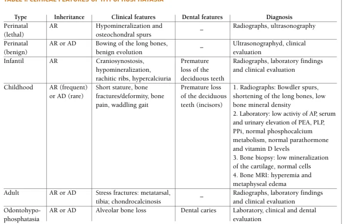
perina-tal (lethal or benign), infantile, childhood, adult and odontohypophosphatasia (Table I)1-4.
The childhood form is characterised by low bone mineral density and unexplained fractures, premature loss of deciduous teeth (beginning with incisors), short stature and delay in walking, bone pain (bone oema), joint pain (condrocalcinosis and osteoarthritis de-velop with aging), pseu dofracture in the lateral cortex of the femoral diaphysis (Looser zones), and sometimes a waddling gait2-4. Specific diagnostic clues are shown
in Table I, but screening for mutations in ALPL is es-sential to confirm the diagnosis4. Childhood form does
the differential diagnosis with rickets, osteogenesis imper fecta, dentinogenesis imperfecta, cleidocranial
Childhood hypophosphatasia with myopathy:
clinical report with recent update
ACTA REUMATOL PORT. 2012;37:92-96
AbstrAct
Hypophosphatasia is a rare genetic disease with low tis-sue nonspeficic alkaline phosphatase activity (TNSALP), due to ALPL gene mutation. There are 6 clinical forms. Childhood form is caractheri zed by short stature, premature loss of decidous teeth and diffuse bone pain associated with a pathological bone fracture in the past. Laboratory findings present low serum le -vel of alkaline phosphatase and high le-vels of serum and urinary extracelular metabolytes. It is described a case report of a 34 years old woman with previous diag -nosis of childhood hypophosphatasia, caryotype 46,XX, and molecular screening for the gene ALPL with a c.1426>A p.E476K mutation, who complained of proximal muscular weakness intensified with the cold weather, exercise, and a waddling gait. The elec-tromyography was compatible with myopathy but the muscle biopsy was normal. The serum crea tine kinase levels were normal, as well as the others muscle en-zymes. Clinical and laboratory/ /imaging dissociation is frequent in other metabo lic bone diseases as osteoma-lacia. The rarity of this case of childhood hypophos-phatasia with “de novo” non-progressive myopathy of the lower limbs, justified a case report with literature re-vision.
Keywords: Hypophosphatasia; Myopathy; Alkaline
Phosphatase; Osteomalacia.
IntroductIon
Biomineralization is a process by which hydroxyapatite is deposited in the extracellular matrix (first within ma-trix vesicles that bud from the surface membrane of
tAble I. clInIcAl feAtures of hypophosphAtAsIA
Type Inheritance Clinical features Dental features Diagnosis
Perinatal AR Hypomineralization and – Radiographs, ultrasonography (lethal) osteochondral spurs
Perinatal AR or AD Bowing of the long bones, – Ultrasonographyd, clinical
(benign) benign evolution evaluation
Infantil AR Craniosynostosis, Premature Radiographs, laboratory findings hypomineralization, loss of the and clinical evaluation
rachitic ribs, hypercalciuria deciduous teeth
Childhood AR (frequent) Short stature, bone Premature loss 1. Radiographs: Bowdler spurs, or AD (rare) fractures/deformity, bone of the deciduous shortening of the long bones, low
pain, waddling gait teeth (incisors) bone mineral density
2. Laboratory: low activiy of AP, serum and urinary elevation of PEA, PLP, PPi, normal phosphocalcium metabolism, normal parathormone and vitamin D levels
3. Bone biopsy: low mineralization of the cartilage, normal cells 4. Bone MRI: hyperemia and metaphyseal edema
Adult AR or AD Stress fractures: metatarsal, – Radiographs, laboratory findings tibia; chondrocalcinosis and clinical evaluation
Odontohypo- AR or AD Alveolar bone loss Dental caries Laboratory, clinical and dental
phosphatasia evaluation
Legend: AR= autosomal recessive; AD= autosomal dominant; AP= alkaline phosphatase; PEA = phophoethanolamine; PLP = pyridoxal-5-phosphate; PPi = inorganic pyrophosphate; MRI = magnetic resonance imaging.
dysostosis, Cole-Carpenter syndrome, idiopatic juve-nil osteoporosis and renal osteodystrophy2. Milder
forms of hypophosphatasia pose a pro blem for an ac-curate diagnosis7. Symptomatic treatment is applied,
with orthopedic management1-5,8. Positive response to
teriparatide as been reported and enzyme replacement therapy (ERT) looks promi sing7.
It is presented a case report of hypophosphatasia with myopathy and a literature revision.
cAse report
A 34-year-old woman was referred to our clinic with di-fuse bone pain and myalgias of the lower limbs and of the pelvic girdle that lasted for 10 years. Two years be-fore she started with mild proxi mal muscle weakness in her lower limbs and pelvic girdle, with difficulty in walking and inability to climb stairs. She also com-plained of morning muscle stiffness for more than an
hour. Her complaints intensified with physical de-manding activities and with cold weather. She pre-sented previous history of early delivery at 30 weeks, growth retardation, dental caries, pathologic coccyx fracture and the diagnosis of childhood hypophos-phatasia (caryo type 46,XX; autosomal recessive; mu-tation c.1426G>A p.E476K heterozygote – Institut für Humangenetik, Universitatsklinikum Schleswig--Hols tein, Kiel, Germany) when she was 20 years old.
The physical examination revealed short stature (weight 48 Kg; height 140 cm) (Figure 1), shorte ning of the 4th metatarsals, mild (grade 4/5) proximal mus-cle weakness in the lower limbs with difficulty in get-ting off the floor and in climbing stairs and a waddling gait. Deep tendon reflexes were normal and the mus-cles were not tender.
Serum AP showed low levels (28 U/L) without other abnormality in the muscle enzymes. Serum, uri-nary and immunologic analysis were normal (PPi, PLP and PEA were not performed). Radiologi cal
examina-tion showed widening of the bone structures in the knee joint, shortening of the 4th metatarsals (Figure 2) and acetabular dysplasia (Figure 3). Electromyogra-phy of the lower limbs showed spontaneous fibrilla-tions, polyphasic and low amplitude motor action po-tentials. Muscle biopsy specimens of the left quadri-ceps were exa mined by light microscopy and histo-chemistry, showing freezing artefacts, periferic nucleus, type I muscular fibres predominance, with normal dia meter. The image was not available by the patholo gist and the patient refused another biopsy for the muscular enzymatic study and the electron mi-croscopy analysis. Mucopolysaccharidoses were ex-cluded by lysosomic studies. Bone densitometry study
showed cortical osteoporosis (T-score: -2,5) and lum-bar vertebral magnetic resonance imaging was normal. The patient was diagnosed with myopathy associa -ted with metabolic bone diseases. She star -ted on sympto matic treatment with vigilance of stress frac-tures, pseudofractures and dental care. The cortical os-teoporosis was treated with teriparatide.
dIscussIon
We describe a case report of a woman with the previ-ous diagnosis of childhood hypophosphatasia who complains of myalgias and severe diffuse bone pain of the lower limbs for 10 years . Symptons of “de novo” myopathy of the lower limbs and pelvic girdle began 2 years ago. The radiologic and biochemical findings were characteristic of hypo phosphatasia. The elec-tromyography of the lower limbs showed myopathy, but the biopsy did not showed pathognomonic alte -rations. The bone densitometry study showed cortical osteoporosis. We explored the myopathy etiology in this patient. Adults with myopathy usually present proximal and symmetric muscle weakness, myalgias at rest, exercise intolerance, serum elevation of creatine kinase (CK) or myoglobinuria. The patient had typi-cal symptons of myopathy: weakness, myalgia and stiffness. Her symptons worsened with demanding metabolic exercises but sometimes persisted even in rest periods.
First it was excluded non-neuromuscular episodic or persistent causes of weakness (hypotension, hy-poxia, hyperventilation, hypoglycemia, anemia, infec-tion, malignancy, malnutriinfec-tion, hyperthyroidism, hy-perparathyroidism, hypophosphatemia)9.
fi gu re 1.Short stature (weight 48 Kg and height 140 cm)
fi gu re 2.Feet radiograph with short 4th metatarsals
Investigation for causes of proximal muscle weak-ness (inflammatory, drug and toxin-induced, infec-tions, metabolic, muscular dystrophies, muscle chan-nelopathies, neoplasm) were negative9.
Polymyositis, dermatomyositis, inclusion body myositis and connective tissue diseases were exclu ded by the clinical, biochemical and hystological findings. She had no sarcoidosis or Behçet clini cal features. She had no history of drugs with the potential of inducing the symptons and no history of previous infection.
Although the enzymatic muscle study was not been performed, her symptons were not compati ble with glycogen storage diseases (e.g. rabdomyo lisis with exer cise, relieve at rest, normal electromyography, posi tive PAS byopsy and low serum lactate after exer-cise), with lipid storage diseases (e.g. rabdomyolisis, byopsy stained by Sudan red and high levels of serum lactate after exercise and in fasting) or mitochondrial myo pathies (e.g. abnormalities of the oxydative meta -bo lism, myoglobinuria, systemic symptons, palpebral ptosis, seizures, retinopathy, ophthalmoplegia, cardyo -myopathy)9,10. Endocrine, nutritional and electrolyte
disorders were excluded. Neuropatic conditions typi-cally manifest with asymmetric weakness, distal in-volvement, sensorial alterations or abnormalities in the cranial nerves function. Muscular dystrophies were less likely by the biopsy, electromyography and the normal neurologic exa mination in facial, palpebral and upper limbs muscular strengh9.
Finally it was selected the only characteristic that could associate the patient complains with specific diag nosis: myopathy associated with rest pain. In this
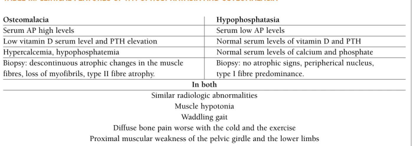
case we considered childhood dermatomyositis, hypo -thyroid myopathy, drug-induced myopathy, infectious myopathy, myo pathies associated with metabolic bone diseases and carnitine palmitoyl transferase deficien-cy. From the previous discussion the most likely cause is myopathy associated with metabolic bone di seases. The most similar metabolic bone disease with re-ported myopathy is osteomalacia. Although the bio-chemical and biopsy findings are different between hy-pophosphatasia and osteomalacia, clini cal symptons, radiologic findings and myopathy caractheristics are similar (Table II). Muscle fibres depend on 4 metabo -lic processes to replenish adenosine triphosphate stores during exercise: the CK reaction, glycogenoly-sis, oxidative phosphorilation and adenylate kinase and myoadenylate deaminase reactions9. Considering
the osteomalacea myopathy9,11,12and concerning that
this patient with hypophosphatasia has the same com-plains it was formulated 2 hypothesis for its etiolo gy: low intramuscular levels of phosphate would difficult glycolysis during demanding metabolic situations and/or a possible low intracellular calcium intake by the sarcoplasmatic reticulum proba bly by low activity of vitamin D in the myo cytes. Even with normal levels of serum vitamin D it must be assured that its actions in the myocytes is complete or is deficient.
Hypophosphatasia treatment is symptomatic. Anti--inflammatory non steroid drugs (AINEs) appear bene ficial in bone pain, blocking pros ta glandins (PGs) synthesis for a long period of time and suppressing the bone oedema8. Girschick et al, supports that PGs
elevation in HP might be explained by the impaired clea
-Legend: AP = alkaline phosphatase; PTH = parathormone
tAble III. clInIcAl feAtures of hypophosphAtAsIA And osteomAlAcIA
Osteomalacia Hypophosphatasia
Serum AP high levels Serum low AP levels
Low vitamin D serum level and PTH elevation Normal serum levels of vitamin D and PTH Hypercalcemia, hypophosphatemia Normal serum levels of calcium and phosphate Biopsy: descontinuous atrophic changes in the muscle Biopsy: no atrophic signs, peripherical nucleus, fibres, loss of myofibrils, type II fibre atrophy. type I fibre predominance.
In both
Similar radiologic abnormalities Muscle hypotonia
Waddling gait
Diffuse bone pain worse with the cold and the exercise Proximal muscular weakness of the pelvic girdle and the lower limbs
rance of calcium pyrophosphate resulting from alka-line phosphatase deficiency. PGs can reduce physical activity in these patients, thus AINEs may improve exer cise tolerance and bone mineralization8. The
cor-tical osteoporosis was treated with teriparatide (during 18 months) with improvement of the pain complains, together with calcium 1g/d and vitamin D 800 UI/d (increases the number of type II fibres with gait stabi-lization and prevention against falls).
Vigilance of serum and urinary calcium levels must be done once parathormone and vitamin D levels are normal. In these patients the bisphosphonates are avoidable as they have a similar conformation to PPi and inhibit bone mineralization. Teriparatide seems to upregulate wildtype ALPL gene expression, to reduce bone pain, to increase AP serum activity and to im-prove biochemical markers of bone turnover. Published data support our decision13,14.
Glucosamine sulfate 1500mg/d may prevent secon -da ry osteoarthritis13. Low impact exercises are impor
tant to improve muscle mass and maintain the mecha -nical stimuli that avoids the acute loss of bone in the muscle paralysis. Gross TS15, published that acute res
-ponse to transient muscle paralysis is due to a RANKL mediated osteoclastic activation and that it is restric -ted to the affec-ted limb. Vigilance of stress fractures, pseudofractures and dental care are mandatory2.
ERT has been developed and tested in mouse and clinical trials soon will be available to patients2. Millan
and colleagues showed that ERT with a semisynthe -tic bone-targeted form of TNSALP can prevent bone abnormalities of HP in the mouse7. Allogeneic
trans-plant of human mesenchymal stem cells in the child (allogeneic cultured osteoblasts and bone fragments from crushed iliac bone; allogeneic HLA-matched T-cell-depleted marrow; allogeneic heterogeneous popu lation of marrow cells and bone fragments) by intrave nous administration resulted in clinical and radiolo gic evidence of bone mineralization, some pa-tients began to walk or run, and one child turn its phe-notype to mild HP3.
In childhood HP the physician must perform a regu -lar vigilance of the bone fractures, dental caries, serum and urinary calcium levels, vitamin D serum levels and general clinical examination. Foot orthotics may help in the management of the tarsal fractures in adults2. This
form of HP presents a better prognosis than HP perina-tal and infantil1-5.
This patient improved her symptons and keeps a regu lar follow-up in the rheumatology department.
This report shows that a non-progressive proxi mal myopathy with muscle pain and stiffness may be an early sign of an osteomalacic syndrome.
correspondence to
Inês Maria Crispim Gomes da Silva Serviço Reumatologia,
Centro Hospitalar de Lisboa Ocidental, Hospital de Egas Moniz Rua da Junqueira, 126, 1300 Lisboa
Tel: +351965790032
E-mail: inescrispin@hotmail.com
references
1. Orimo H. The mechanism of mineralization and the role of al-kaline phosphatase in the health and di sease. J Nippon Med Sch 2010; 77:4-12.
2. Mornet E, Nunes ME. Hypophosphatasia. Gene Reviews – NCBI Bookshelf 2010. 1-16.
3. Undale AH, Westendorf JJ, Yaszemski MJ et al. Me senchymal stem cells for bone repair and metabolic bone diseases. Mayo Clin Proc 2009; 84: 893-902.
4. Mornet E. Hypophosphatasia. Orphanet Journal of Rare Di-seases 2007; 2: 1-8.
5. Henthorn PS, Raducha M, Fedde KN, et al. Different missence mutations at the tissue-nonspecific alkaline phosphatase gene locus in autosomal recessively inherited forms of mild and se-vere hypophosphatasia. Proc. Natl. Acad. Sci. USA 1992; 89:9924-9928.
6. Taillandier A, Sallinen SL, Brun-Heath I et al. Childhood hy-pophosphatasia due to a de novo missense mutation in the tis-sue-nonspecific alkaline phosphatase gene. The Journal of Cli-nical Endocrinology and Metabolism 2005; 90: 2436-2439. 7. Cole DEC. Hypophosphatasia update: recent advances in
diag-nosis and treatment. Clin Genet 2008; 73: 232-235. 8. Girschick HJ, Schneider P, Haubitz I, et al. Effective NSAID
treatment indicates that hyperprostaglandinism is affecting the clinical severity of childhood hypophosphatasia. Orphanet Journal of rare diseases 2006; 1:24; 1-12.
9. Baer AN, Wortmann RL. Metabolic, drug-induced and other non-inflammatory myopathies. In: Hochberg Mc,Ed. Rheu-matology,vol 2, 4th ed. Mosby Elsevier; 2008. P.1469-1478. 10. Das AM, Steuerwald U, Illsinger S. Inborn errors of energy
me-tabolism associated with myopathies. Journal of Biomedicine and Biotechnology 2010; 1-19.
11. Seshia SS, Derbyshire G, Haworth JC, et al. Myopathy with hy-pophosphatasia. Archives of Disease in Childhood 1990; 65: 130-131.
12. Schott GD, Wills MR. Muscle weakness in osteomalacia. Lan-cet 1976; i: 626-9.
13. Camacho PM, Painter S, Kadanoff R. Endocr. Pract 2008; 14(2): 204-208.
14. Whyte MP, Mumm S, Deal C. Adult hypophosphatasia treated with teriparatide. J Clin Endocrinol Metab 2007; 92: 1203--1208.
15. Gross TS, Poliachik SL, Prasad J, et al. The effect of muscle dys-function on bone mass and morphology. J Musculoskelet Neu-ronal Interact 2010; 10: 25-34.