256 Rev Odonto Cienc 2012;27(3):256-259
Received: July 21, 2012 Accepted: September 28, 2012
Conflict of Interests: The authors state that there are no financial and personal conflicts of interest that could have inappropriately influenced their work.
Copyright: © 2012 Berthold et al.; licensee EDIPUCRS. This is an Open Access article distributed under the terms of the Creative Commons Attribution-Noncommercial-No Derivative Works 3.0 Unported License.
Case Report
Treatment of Class II division 1 malocclusion
in a patient with traumatized central incisors:
A case report
Tratamento de maloclusão Classe II divisão 1 em um paciente
com incisivos centrais traumatizados: relato de caso
Telmo Bandeira Berthold a,d Roger Corrêa de Barros Berthold b Claiton Heitz c
Rosana Kalaoun b
a Department of Orthodontics, Pontifical Catholic
University of Rio Grande do Sul, Porto Alegre, RS, Brazil
b Graduate Program in Dentistry / Oral and
Maxillofacial Surgery, Pontifical Catholic University of Rio Grande do Sul, Porto Alegre, RS, Brazil
c Department of Oral and Maxillofacial Surgery,
Pontifical Catholic University of Rio Grande do Sul, Porto Alegre, RS, Brazil
d Department of Orthodontics, Federal University
of Rio Grande do Sul, Porto Alegre, RS, Brazil
Correspondence:
Telmo B. Berthold
Pontifical Catholic University of Rio Grande do Sul (PUCRS)
School of Dentistry
Av. Ipiranga 6681 – Building 6 Porto Alegre, RS – Brazil 90616-900
E-mail: [email protected]
Abstract
Purpose: The objective of this paper is to report the clinical case of a patient with a Class II division 1 malocclusion with traumatized central incisors and the treatment option of extracting them, followed by space closure with lateral incisors replacement.
Case description: A female patient aged 10 years old reported a previous facial trauma due to a bike accident resulting in avulsion and reimplantation of both central incisors when she was 9 years old. The treatment plan included extraction of central incisors, pulling the lateral incisors to the central incisor position and restoring them to improve esthetics.
Conclusions: Extraction of maxillary central incisors is not a usual treatment protocol. However, for patients with traumatized central incisors with internal and external root reabsorption and Class II division 1 malocclusion it might be a good alternative for correction of the increased overjet.
Key words: Dental trauma; orthodontic treatment; tooth avulsion
Resumo
Objetivo: O objetivo deste trabalho é relatar o caso clínico de uma paciente portadora de maloclusão Classe II divisão 1 com incisivos centrais superiores traumatizados com reabsorção interna e externa e a opção de tratamento de extraí-los, seguido de fechamento do espaço com a mesialização dos incisivos laterais superiores.
Descrição do caso: Paciente do sexo feminino com 10 anos de idade relatou trauma facial, após queda de bicicleta, e avulsão e reimplante dos incisivos centrais superiores aos 9 anos de idade. Optou-se pela extração dos incisivos centrais superiores mesializando os incisivos laterais para a posição dos centrais, restaurando-os para ficar esteticamente aceitável. Conclusão: A extração dos incisivos centrais superiores não é um protocolo de tratamento comum. Porém, para pacientes portadores de maloclusão Classe II divisão 1 com incisivos centrais traumatizados e com reabsorção radicular externa e interna pode ser uma boa solução para a correção do sobrepasse aumentado.
Rev Odonto Cienc 2012;27(3):256-259 257
Berthold et al.
Introduction
Dental avulsion injuries are mostly seen in young patients in the anterior region of the maxilla (1). The main
physical characteristics outlined as signiicant risk factors
for dentoalveolar trauma are poor lip coverage and an increased overjet, which are both present in Class II division 1 malocclusion (2). If teeth are reimplanted there is a serious
risk for permanent ankylosis and internal and/or external root resorption (3). Ankylosis of the central incisors is a
challenge for any orthodontist.
In cases of Class II division 1 malocclusion with
traumatized and ankylosed central incisors, extraction and
space closure might be a solution to treat the increased overjet. However, this treatment option requires the lateral incisors to assume the role of central incisors and the canines to assume the role of the lateral incisors.
The irst premolars take the place of the canines, doing
the excursive movements with restorative treatment for
camoulage the positional changes. Therefore, the objective
of this article was to demonstrate this procedure in a young patient.
Case description
A female patient aged 10 years old sought orthodontic treatment at a private clinic in Porto Alegre, RS, Brazil. Her chief complaint was poor esthetics and incisor protrusion. She and her parents reported a previous facial trauma due
to a bike accident with avulsion and reimplantation of both
central incisors when she was 9 years old. The patient and her parents did not recall the real conditions and procedure of the teeth reimplantation.
The clinical examination showed both central incisors with color change of the crowns (Fig. 1). The left central incisor was extensively restored due to crown fracture in the trauma episode. The radiographs showed both central incisors with internal and external root resorption. The left central incisor showed a root fracture in the cervical region and the right central incisor had an extensive external root resorption. The left lateral incisor presented endodontic treatment (Fig. 2).
She was diagnosed with a Class II division 1 malocclusion with lip incompetence. The main objectives of the orthodontic treatment plan were to eliminate protrusion of the superior teeth and establish stable occlusion with acceptable anterior dental esthetics.
Two treatment options were considered. The irst option was to extract two maxillary premolars and both ankylosed central incisors replacing them by a ixed or removable
prosthesis until her facial growth was completed. Then a restorative treatment with implants would be considered.
The case would inish in Angle Class II and the young
patient would use a temporary prosthesis in an extremely
esthetic region until her skeletal growth has ended. The
second option was to extract the central incisors only and pulling the lateral incisors to the central incisor position,
restoring them to improve esthetics, and the treatment
would inish in Angle Class II. This option seemed to be
the most acceptable for the orthodontist, the patient and her parents.
The treatment started by installing a removable appliance in the superior arch (Fig. 3). This device had the following purposes:
1. Preserve the patient aesthetics after the extractions of
the central incisors (two artiicial teeth were added to the appliance) (Fig. 3). There was a signiicant gain in
patient self-esteem considering that her natural teeth
were projected and darkened due to endodontic
treat-ment.
2. Improve the overbite, incorporating a stop bite into the removable orthodontic appliance.
3. Start the approach of lateral incisors with caution by means of springs added in the removable appliance.
The artiicial teeth were gradually reduced proximally
allowing movement of the lateral incisors without harming the aesthetics of the patient.
A ixed appliance was installed in the mandibular arch
initially for Tandem mechanics in molars. We opted for this mechanics to increase space in the mandibular arch and simultaneously to lose maxillary anchorage through
the use of elastic Class III. After this stage brackets on
incisors, canines and mandibular premolars were bonded and treatment was accomplished with conventional mechanics. Fixed appliance was installed in the maxillary arch. The
inal space closure was performed with intramaxillary elastic
chain and with a sequence of arch wires leaving a well
distributed interproximal space to be illed by composite
restorations.
During all treatment the patient underwent radiographic monitoring to verify parallelism and pulp vitality of the lateral incisors. The right lateral incisors needed endodontic treatment during orthodontic treatment (Fig. 2).
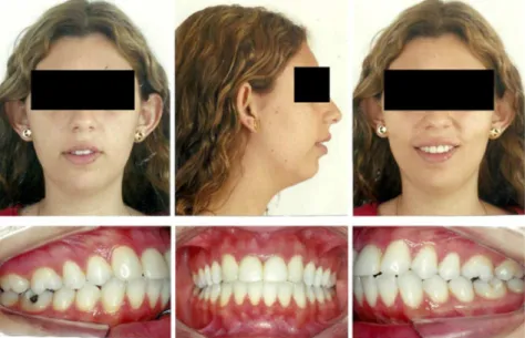
After orthodontic treatment the appliances were removed and the patient was referred to restorative treatment. Direct composite restorations were made in the lateral incisors to mimic central incisors. Enamelplasty was done in the canines to transform them in lateral incisors. The patient and her parents were pleased with the treatment outcome (Fig. 4).
258 Rev Odonto Cienc 2012;27(3):256-259
Class II division 1 malocclusion
Fig. 4. Post treatment facial and
intraoral photographs.
Fig 1. Pretreatment facial and
intraoral photographs.
Fig 2. Initial radiograph (A); During orthodontic treatment
radiograph (B); Post treatment radiograph: lateral incisors with temporary composite restorations (C).
Fig 3. Post central incisors extraction aspect (A); Removable
Rev Odonto Cienc 2012;27(3):256-259 259
Berthold et al.
Discussion
Dental injuries confront dental practitioners quite often (1-4). The incisors are the most common injured teeth and bicycle accidents are often involved (2). Children with a
6 mm or higher overjet had four times higher risk of suffering
dentoalveolar trauma, compared with those with lower overjet (5). An Angle class II malocclusion is traditionally treated orthodontically with extractions of premolars. However this patient also had protruded, traumatized and
ankylosed central incisors, which compromised esthetics
and function. In this case we opted for extraction of central incisors and pulled the lateral incisors to the extraction sites.
The treatment of a young patient with a dental prosthesis
in aesthetic region until inalization of growth may not be
the best option because of the bad impact of dental injuries on quality of life, especially for young girls (6,7). There were no extractions in the mandibular arch because this
could increase the convex proile of the patient. The dental
crowding was corrected by means of dental interproximal stripping and Tandem mechanics. Although she had lateral incisors with a large mesiodistal distance (8 mm), we opted for the restorative treatment to improve esthetics and hide the discoloration of the lateral incisors crowns due to
endodontic treatment. This unconventional treatment plan
requires speciic teeth placement so they could be restored
and reshaped (4,8). Extensive dental grinding can be
performed without signiicant discomfort, and with minor or
no pulp and dentin reactions (9). The canines were trimmed in the palatal side to avoid interference with the mandibular
incisors. The labial side was lattened and the cuspids were
reduced 2 mm in both canines.
Canine-protected occlusion is not feasible when the canine is replaced by the premolar (8). As a result, forces generated through lateral excursive movements are placed
on the smaller and thinner roots of the irst premolar (10).
However long-term periodontal and occlusal studies on congenitally missing lateral incisors have shown that orthodontic space closure with premolar substitution of canines was periodontally preferable to prosthetic replacement of the missing lateral incisor (8,11).
Extraction of the maxillary central incisors is not a usual treatment protocol. However, in patients with traumatized
and ankylosed central incisors and Class II division 1
malocclusion, this might be a good alternative to preserve tooth structure and eliminate patient’s dependence of a permanent prosthesis. Nevertheless, additional cosmetic
inishing might be necessary.
1. Skaare AB, Jacobsen I. Dental injuries in Norwegians aged 7-18 years. Dent Traumatol 2003;19:67-71.
2. Kahabuka FK, Mugonzibwa EA. Risk factors for injuries to maxillary permanent incisors and upper lip among schoolchildren in Dares Salaam, Tanzania. Int J Paediatr Dent 2009;19:148-54.
3. Hecova H, Tzigkounakis V, Merglova V, Netolicky J. A retrospective study of 889 injured permanent teeth. Dent Traumatol 2010;26:466-75.
4. Sabri R.Treatment of a Class I crowded malocclusion with an akylosed maxillary central incisor. Am J orthod dentofacacial Orthop 2002;122:557-65.
5. Schatz JP, Hakeberg M, Ostini E, Kiliaridis S. Prevalence of traumatic injuries to permanent dentition and its association with overjet in a Swiss child population. Dent Traumatol 2012, May 24. doi: 10.1111/j.1600-9657.2012.01150.x.
6. Aldrigui JM, Abanto J, Carvalho TS, Mendes FM, Wanderley MT, Bönecker M, et al. Impact of traumatic dental injuries and malocclusions on quality of life of young children. Health Qual Life Outcomes 2011;24,9:78.
7. Traebert J, de Lacerda JT, Foster Page LA, Thomson WM, Bortoluzzi MC. Impact of traumatic dental injurieson the quality of life of schoolchildren. Dental Traumatology 2012; doi: 10.1111/j.1600-9657.2012.01114.x
8. Janson G, Valarelli DP, Valarelli FP, Freitas MR, Pinzan A. Atypical extraction of maxillary central incisors. Am J Orthod Dentofacial Orthop 2010;138:510-7.
9. Zachrisson BU, Mjor IA. Remodeling of teeth by grinding. Am J Orthod Dentofacial Orthop 1975;68:545-53.
10. Tuverson DL. Orthodontic treatment using canines in place of missing maxillary lateral incisors. Am J Orthod Dentofacial Orthop 1970; 58:109-27.
11. Senty El. The maxillary cuspid and missing lateral incisors: esthetics and occlusion. Angle Orthod 1976;46:365-71.