(Annals of the Brazilian Academy of Sciences)
Printed version ISSN 0001-3765 / Online version ISSN 1678-2690 www.scielo.br/aabc
Growth and hematology of juvenile pacu
Piaractus mesopotamicus
(Holmberg 1887)
fed with increasing levels of vitamin E (DL-α-tocopheryl acetate)
RICARDO Y. SADO1, ÁLVARO J.A. BICUDO2 and JOSÉ E.P. CYRINO3 1Universidade Tecnológica Federal do Paraná, Campus de Dois Vizinhos,
Estrada para Boa Esperança, Km 04, 85660-000 Dois Vizinhos, PR, Brasil 2
Universidade Federal Rural de Pernambuco, Unidade Acadêmica de Garanhuns, Avenida Bom Pastor, s/n, 55296-901 Garanhuns, PE, Brasil
3Escola Superior de Agricultura “Luiz de Queiroz”, Universidade de São Paulo,
Caixa Postal 9, 13418-900 Piracicaba, SP, Brasil
ABSTRACT
Intensive fish production systems are characterized by 100% artificial feeding, so any dietary imbalances or deficiencies may lead to diseases outbreaks and economic losses. This study was set out to determine
the effects of increasing levels of dietary vitamin E on growth and hematology of juvenile pacu. Fishes
were fed for 90 days, twice a day until apparent satiation with semi-purified diets containing 0.0; 25; 50;
150; 300 or 600 mg.kg-1 diet DL-α-tocopheryl acetate in a completely randomized design trial (n=4); biometrical and hematological data were collected and analyzed. Fishes fed with vit E diet (150 mg.kg-1)
showed higher (p<0.05) weight gain and specific growth. Hematocrit, erythroblast number and total plasma protein were increased (p<0.05) in fishes fed diet with no vit E diet. Vitamin E supplementation in artificial
diets for pacu is essential for growth and maintenance of normal erythropoiesis.
Key words: fish nutrition, hematology, Piaractus mesopotamicus, vitamin E.
Corresponding to: Ricardo Yuji Sado email: [email protected]
INTRODUCTION
Global fish consumption has doubled since the
1970s and it is still growing (Naylor and Burke
2005). Following this trend, Latin America has
shown increased aquaculture production and
per capita
consumption (FAO 2009), as well
as intensification of fish production systems,
characterized by high biomass and use of balanced,
pelletized or extruded diets to meet fish nutritional
requirements. Therefore, imbalanced artificial diets
would lead to significant economical losses due to
nutritional deficiencies or diseases outbreaks.
Provided by exogenous source (i.e., diet),
vitamins are essential for the metabolism in fishes
playing an important role on biochemical reactions
related to growth and health (Halver 2002, NRC
2011, Tocher et al. 2003). Within the fat-soluble
vitamins (A, D, E, and K), vitamin E is one of the
most studied in fish dietetics.
activity (Chen et al. 2004, Pulsford et al. 1995, Wise et
al. 1993). Effects of vitamin E have been determined
for several, economically important fish species such
as gilthead seabream
Sparus aurata
(Montero et al.
1999), grouper
Epinephelus malabaricus
(Lin and
Shiau 2005), rainbow trout
Oncorhynchus mykiss
(Kiron et al. 2004, Pearce et al. 2003, Trenzato et
al. 2007), Atlantic salmon
Salmo salar
(Hamre et al.
1997, 2004), red drum
Sciaenops ocellatus
(Peng
and Gatlin 2009) and channel catfish
Ictalurus
punctatus
(Wise et al. 1993). Analysis of blood
components may provide important information
regarding 64 general condition and possible effects
of vitamin E on fish.
Pacu (
Piaractus mesopotamicus
) is a neotropical
freshwater Characin native of Parana, Paraguay
and Uruguay basins. Because of its herbivorous/
omnivorous habits, high growth rates, good meat
quality, consumer acceptance and suitability for
sports fishery, the specie is widely used in aquaculture
(Jomori et al. 2005, Urbinati and Gonçalves 2005).
Few studies regarding the effect of dietary vitamin E
for pacu are available (Belo et al. 2005a, b, Garcia
et al. 2007). This study was set out to evaluate the
effects of increasing levels of dietary vitamin E on
growth and hematology of pacu juveniles.
MATERIALS AND METHODS
EXPERIMENTAL DESIGN AND ANIMALS
Trials were set up in a closed water recirculation
system, with supplemental aeration and emergency
oxygenation systems. Water quality parameters such
as pH (7.60 ± 0.20), dissolved oxygen (5.8 ± 0.30
mg.L-1), ammonia (≤ 0.5 mg.L
-1) and temperature
(30.3 ± 1.8 °C) remained within acceptable values
for the specie (Urbinati and Gonçalves 2005). A
12h light/12h dark photoperiod was maintained.
Juvenile pacu (7.83 ± 0.04 g) obtained from
commercial hatchery were acclimatized to the
experimental conditions for seven days feeding on
a 40% crude protein (CP) commercial diet.
EXPERIMENTAL DIETS
The basal, experimental semi-purified diets were
made according 88 to the species’ requirements
(Table I). The vitamin and mineral mix did not
contain vitamin E; dietary vitamin levels were
set according to vitamin E activity of the dietary
source (ROVIMX E 50 Adsorbate Roche
®; 50%
vitamin E activity). Ingredients were weighed,
homogenized and mixed, moistened with distilled
water (25-30%) and pelleted (2.0 mm) in a
mincer. Prepared rations were dried in a forced
ventilation oven at 45 °C for 24h; the dried pellets
were packed in black plastic bags and stored at
-4 °C until use. Diets were analyzed for vitamin
E contents at a commercial laboratory (CBO
Assessoria & Analises; Campinas, Sao Paulo,
Brazil). The concentrations of vitamin E of the
experimental diets are presented in Table II.
TABLE I
Composition of basal semi-purified diet for P. mesopotamicus.
Ingredient %
Starch 34.24
Albumin 30.86
Cellulose 13.0
Gelatin 7.0
Dextrin 5.0
Bicalcium phosphate 4.0
Soy oil 3.87
Vitamin and mineral mix1 (vit E free) 2.0
BHT (antioxidant) 0.02
Calculated composition %
Crude protein 30.1
Dry matter 92.66
Ether extract 3.2
Ash 5.66
1
ROUTINE PROCEDURES
After acclimation period, fishes randomly assigned
to 60 L cages (20 fish per cage) were fed a vitamin
E-free diet for 15 days to zero vitamin E reserves
and as well as diets containing 0.0; 25; 50; 150;
300 and 600 mg.kg
-1vit E until apparent satiation,
twice a day (07:00 and 16:00 h) for 90 days, in
a completely randomized experimental design
(n=4). At the end of experimental period, fish
were fasted for 24h, anesthetized with alcoholic
solution of benzocaine at 50 mg.L
-1, weighted,
measured, and had blood samples drawn for
analysis. Growth parameters were evaluated
according to Tacon 107 (1990) as follows:
• Weight gain (WG)
•
WG = FW - IW
• Feed conversion ratio (FCR)
FCR =
feed consumptionweight gain• Daily feed consumption (FC)
FCR =
feed consumptiont• Specific growth rate (SGR)
SGR = 100×
(lnFW lnIW)twhere: FW = final weight (g); IW= initial weight
(g); t = experimental time (days).
Blood samples were drawn from the caudal
vein using sterilized syringes and 10% EDTA-coated
needles. Red blood cells (RBC) count was performed
in Neubauer chamber using the Natt and Herrick
(1952) diluent; hematocrit evaluation followed
the microhematocrit method of Goldenfarb et al.
(1971); hemoglobin concentration was performed
following the cyanometahemoglobin method
(Blaxhall and Daisley 1973). Hematimetric indexes
calculated were mean corpuscular volume (MCV),
mean corpuscular hemoglobin (MCH), and mean
corpuscular hemoglobin concentration (MCHC)
(Wintrobe 1934). Total plasma protein concentrations
were determined using a portable refractometer
(WZ-301/Protein 0.0-12 g.dL
-1) after blood centrifugation
and plasma collection (Sado et al. 2008). Plasma
glucose was determined by the enzymatic method
using a standard kit (GLICOSE GOD-PAP Liquid
Stable Mono Reagente, LABORLAB
®; Guarulhos,
Sao Paulo, Brazil).
Blood smears from individual fish were stained
with May-Grünwald-Giemsa (Rosenfeld 1947)
and examined under light microscopy using an
oil immersion objective for differential leukocyte
count, and white blood cell (WBC), thrombocyte and
erythroblast counts. White blood cell (WBC),
throm-bocyte and erythroblast count were performed by
indirect method (Garcia et al. 2007, Sado et al. 2010)
as follow: WBC (μL
-1) = (leukocytes number in blood
smear x erythrocyte number. μL
-1) ÷ 2,000 erythrocytes
counted in the blood smear; Thrombocytes (μL
-1) =
(thrombocytes number in blood smear x erythrocyte
number.μL
-1) ÷ 2,000 erythrocytes counted in the
blood smear; Erythroblast (μL
-1) = (erythroblast
number in blood smear x erythrocyte number.μL
-1)
÷ 2,000 erythrocytes counted in the blood smear.
Data were submitted to ANOVA. Significant
differences between treatment means were
further compared by Tukey test (α=0.05) (Steel
and Torrie 1980).
Treatments vit E* expected level (mg.kg-1)
vit E* detected level (mg.kg-1)
I 0.0 Not detected**
II 25 32.79
III 50 50.33
IV 150 136.40
V 300 246.17
VI 600 457.46
* Vitamin E source: DL-α-tocopheryl acetate (50% activity) –
ROVIMIX E 50 adsorbate - Roche®
** Under quantification limit: 2.0 mg.kg-1
TABLE II
RESULTS AND DISCUSSION
Fishes performance and survival data are
summa-rized in Table III. The acceptability of experimental
diets was adequate in all treatments. Weight gain
and specific growth rate were affected by dietary
vitamin E levels (p<0.05). Fishes fed diet with 136.4
mg.kg
-1of vitamin E diet showed better weight gain
when compared to fishes fed with no vitamin E.
Specific growth rate showed higher values in fishes
fed on diet containing 136.4; 246.17 and 457.46 mg
vit E per kg when compared to no vit E treatment.
Dietary vitamin E levels influenced (p<0.05)
hematological parameters and blood biochemistry
(Table IV). Hematocrit values were higher in fishes
fed with no vitamin E (32.6%) and 50.33 mg.kg
-1vitamin E diet (32.3%) when compared to values
recorded for fishes fed with 457.46 mg.kg
-1vitamin
E diet (30.1%). Total plasma protein concentrations
were higher (5.5 g.dL
-1; p<0.05) for fishes fed with
no vitamin E diets than fishes fed with diets of
32.79; 136.4; 246.17 and 457.46 mg vit E per kg.
Significantly higher number of erythroblasts
was registered for fish fed with diet devoid of
Vit. E* (mg.kg-1 diet)
Growth parameters
WG (g) FC (g) FCR SGR (%) Survival (%)
0.0 64.17±6.0a 75.02±7.0 1.17±0.08 3.25±0.12a 93.3±6.6
32.79 73.96±6.7ab 83.55±4.6 1.13±0.07 3.44±0.11ab 90.0±3.8
50.33 73.63±8.0ab 82.84±7.5 1.08±0.05 3.49±0.13ab 91.6±6.3
136.4 86.50±12.5b 95.34±14.1 1.10±0.10 3.65±0.18b 84.4±16.7
246.17 85.36±12.2ab 91.47±11.9 1.07±0.08 3.63±0.19b 91.1±3.8
457.46 84.77±0.9ab 91.84±2.2 1.08±0.03 3.63±0.00b 88.8±13.8
Variables Vitamin E* (mg.kg
-1 diet)
0.0 32.79 50.33 136.4 246.17 457.46
RBC 106.μL-1 1.46±0.2 1.43±0.3 1.36±0.2 1.35±0.2 1.31±0.2 1.31±0.2
Htc % 32.6±1.1a 31.5±1.7ab 32.3±1.9a 31.5±1.8ab 314±17ab 30.1±1.2b
Hb g.dL-1 10.4±0.7 10.2±0.6 10.3±0.7 10.2±0.9 9.7±0.7 10.0±0.8
MCV ƒL 229.5±42.5 226.3±41.0 239.6±27.0 239.5±32.3 245.5±43.7 237.7±41.5
MHC pg.cell-1 72.7±12.0 73.8±14.5 76.3±9.8 76.9±8.9 77.8±14.4 78.5±11.8
MCHC g.dL-1 31.8±1.5 32.6±2.3 31.8±1.9 32.2±1.6 31.8±3.1 33.2±2.0
Prtn g.dL-1 5.5±0.4a 5.0±0.3bc 5.3±0.3ab 5.0±0.4bc 4.9±0.4bc 4.6±0.4c
Gluc mg.dL-1 71.9±11.5 76.8±8.8 79.6±14.6 67.8±10.1 84.0±22.8 84.9±21.1 TABLE III
Means and standard deviation (SD) of individual weight gain (WG), feed consumption (FC), feed conversion rate (FCR), specific growth rate (SGR) and survival rate (SR) of juvenile pacu P. mesopotamicus fed increasing levels of dietary vitamin E.
TABLE IV
Hematological parameters (μ ± SD) of juvenile pacu P. mesopotamicus supplemented with increasing levels of dietary vitamin E.
Different letters superscript at same columns denote differences by Tukey test (α=0.05). * Vitamin E source: DL-α-tocopheryl acetate (50% activity) – ROVIMIX E 50
adsorbate - Roche®
Different letters superscript at same lines denote differences by Tukey test (α=0.05). * Vitamin E source: DL-α-tocopheryl acetate (50% activity) – ROVIMIX E 50
adsorbate - Roche®, RBC: Red Blood Cell, Htc: hematocrit, Hb: hemoglobin concentration, MCV: mean corpuscular volume, MHC: mean hemoglobin concentration, MCHC: mean corpuscular hemoglobin concentration, Ptrn: total plasma protein concentration
vitamin E in comparison to fish fed with diets
containing increasing levels of vitamin E (Fig. 1).
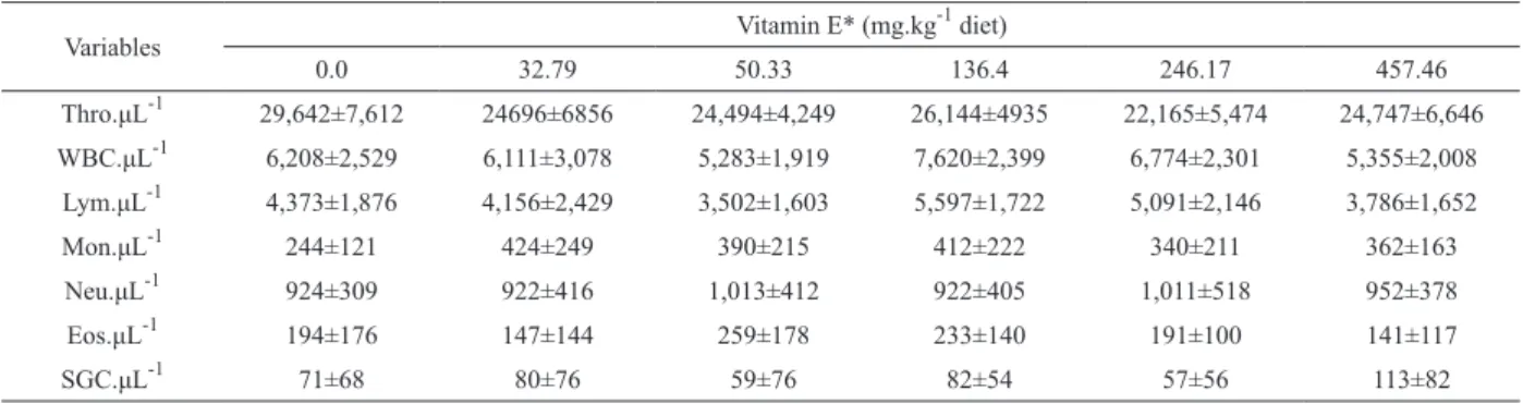
No effect of dietary vitamin E supplementation 159
was found on WBC and thrombocyte count, as well
as on differential leukocyte count (Table V).
Diet and nutrition can influence growth and
disease resistance of domestic and farm animals
(Alcorn et al. 2003, Blazer 1991, Landolt 1989,
Sitja-Bobadilla and Pérez-Sanchez 1999). In this study,
fishes fed with a vitamin E-free diet had poor growth
Variables Vitamin E* (mg.kg
-1 diet)
0.0 32.79 50.33 136.4 246.17 457.46
Thro.μL-1
29,642±7,612 24696±6856 24,494±4,249 26,144±4935 22,165±5,474 24,747±6,646
WBC.μL-1
6,208±2,529 6,111±3,078 5,283±1,919 7,620±2,399 6,774±2,301 5,355±2,008
Lym.μL-1
4,373±1,876 4,156±2,429 3,502±1,603 5,597±1,722 5,091±2,146 3,786±1,652
Mon.μL-1
244±121 424±249 390±215 412±222 340±211 362±163
Neu.μL-1
924±309 922±416 1,013±412 922±405 1,011±518 952±378
Eos.μL-1
194±176 147±144 259±178 233±140 191±100 141±117
SGC.μL-1
71±68 80±76 59±76 82±54 57±56 113±82
TABLE V
Hematological parameters (μ ± SD) of juvenile pacu P. mesopotamicus supplemented with increasing levels of dietary vitamin E.
* Vitamin E source: DL-α-tocopheryl acetate (50% activity) – ROVIMIX E 50
adsorbate - Roche®, Thro: thrombocytes, WBC: White blood cell, Lym: lymphocytes, Mon: monocytes, Neu: neutrophils, Eos: eosinophils, SGC: special granulocytic cells
and weight gain in comparison to fishes fed with diets
supplemented with vitamin E. The effect of dietary
vitamin E on fish growth still steers controversy.
Significant positive effects of increased dietary
contents of vitamin E on fish growth have been
described by several authors for different species, such
as Atlantic salmon (Hamre et al. 1997), rainbow-trout
(Pearce et al. 2003, Trenzato et al. 2007), Chinook
salmon,
Oncorhynchus tshawytscha
(Thorarinsson
et al. 1994), grouper (Lin and Shiau 2005) and rohu
(Sau et al. 2004), and poor growth performance was
also registered for fishes fed with vitamin E-deficient
diets, that is, vitamin E plays an important role in fish
dietetics. Like any other animal, fish cannot synthesize
vitamin E (Peng and Gatlin 2009).
However, many authors reported no effects of
vitamin E supplementation on growth in some fish
species, such as the golden shiner,
Notemigonus
crysoleucas
(Chen et al. 2004), channel catfish
(Gaylord et al. 1998, Wise et al. 1993), gilthead
seabream (Montero et al. 1999), Nile tilapia (Lim
et al. 2010), rainbow-trout (Clerton et al. 2001,
Kiron et al. 2004), Atlantic salmon (Hardie et
al. 1990, Poston et al. 1976) and hybrid striped
bass (Trushenski and Kohler 2008). Belo et al.
(2005b) also did not report effects of vitamin E
supplementation on growth performance of pacu,
possibly as a consequence of fish size – initial
average weight 7.8 g in the current study against
96.4 g in Belo et al. (2005b) experiment. As a matter
of fact, vitamin E is a fat-soluble nutrient that can
Fig. 1 – Erythroblast number (μ ± SD) of juvenile pacu P.mesopotamicus fed increasing levels of dietary vitamin E. Different
be stored in liver tissue (Halver 2002). Therefore;
larger fish may stand longer periods feeding on
diets with low or zero vitamin E contents, relying
and using vitamin E deposited in body lipids for
growth and maintenance of body functions.
In exception of poor growth in fishes fed
with the vitamin E-deficient diet, no clinical signs
of vitamin deficiency were recorded. Vitamin
E deficiency signs in fishes are characterized
by darkened skin (Chen et al. 2004) or lack of
pigmentation (Hamre et al. 1997), dystrophy and
necrosis of epaxial muscles (Chen et al. 2004), and
hematological disorders (Chen et al. 2004, Hamre et
al. 1997, Pearce et al. 2003, Poston et al. 1976, Wise et
al. 1993). Although vitamin E concentrations on fish
tissues were not determined, the authors presume that a
15-day withdrawal period in addition to experimental
time was not sufficient to decrease vitamin E reserves
of pacu; since the same was reported by Hardie et
al. (1990) for Atlantic salmon. Belo et al. (2005b)
and Garcia et al. (2007) also did not report clinical
signs of vitamin deficiency in pacus running
90-day feeding trials, and Clerton et al. (2001) also did
not report clinical signs on rainbow trout feeding on
vitamin E-free diets for 80 days.
Similar to data herein reported no effects of
dietary vitamin E on number and proportion of
leukocytes was reported by Garcia et al. (2007)
for pacu, Lim et al. (2010) for Nile tilapia, and
Hardie et al. (1990) for Atlantic salmon. However,
increasing and decreasing number of defense cells
was reported for fishes fed with vitamin E-deficient
diets by Chen et al. (2004) for golden shiner and Lin
and Shiau (2005) for the grouper. Although Chen et
al. (2004) reported increasing number of leukocytes
and thrombocytes, when differential leukocyte
count was considered, a decreasing number of
lymphocyte was observed. Decreasing lymphocyte
number could be a consequence of membrane
fragility and cell lysis and/or lymphocyte migration
to degenerated muscular tissue, ordinarily observed
in fishes showing vitamin E deficiency signs.
No evaluation of the immunological status of
fishes' immunological status was herein performed.
However, significant effects of vitamin E on their
immune systems of fish have been already soundly
demonstrated (Chen et al. 2004, Hardie et al. 1990,
Kiron et al. 2004, Lin and Shiau 2005, Lygren et al.
2008, Montero et al. 1999, Pearce et al. 2003, Wise
et al. 1993). Even thought there were no significant
differences in phagocyte numbers, possibly as a
result of the high variation, values of monocytes
count increased in absolute values in fishes fed with
diet of increasing vitamin E contents, regarded a
sign of immunity stimulation, given that in
in vivo
fish inflammatory response studies, macrophages
derived from blood circulating monocytes have
been reported to differentiate into multinucleated
giant cells (Sado and Matushima 2008). The
macrophage recruitment and giant cell formation in
pacu seems to be strongly related to dietary vitamin
E supplementation (Belo et al. 2005b).
Vitamin E protects cell membranes against
lipid peroxidation. Dietary vitamin E deficiency
in fish increases deformities of membranes and
fragility of erythrocytes, easing hemolysis (Halver
2002), and reducing cell survival time, leading
to hematological disturbs such as decreased
hematocrit values and hemoglobin concentrations
(Chen et al. 2004, Pearce et al. 2003, Poston et
al. 1976, Thorarinsson et al. 1995). However, no
differences on hemoglobin concentrations and
RBC were observed between treatments, despite
the higher values for RBC found in literature
regarding pacu (Martins et al. 1995, Ranzani-Paiva
et al. 1998/1999, Tavares-Dias et al. 1999, 2002,
Tavares-Dias and Mataqueiro 2004). Elevated
hematocrit values were herein reported for fishes
fed with vitamin E-deficient diets; similar results
were reported by Garcia et al. (2007).
under light microscopy (Esteban et al. 2000).
In some pathological conditions the number of
immature cells can be elevated, as observed in this
study and also reported by Poston et al. (1976) and
Garcia et al. (2007). A compensatory effect can be
seen as a decrease on erythrocytes life time and
the consequent release of more immature cells to
the blood stream. The red cell maturation process
involves chromatin condensation, increase on
hemoglobin concentrations and decrease on nuclear
and cell size (Esteban et al. 1989). Erythroblasts,
i.e. young erythrocytes, are larger than mature
red cells. Therefore, an increase in numbers of
those cells in fish blood circulation would explain
the high hematocrit values found in the current
experiment and by Garcia et al. (2007) in fishes fed
with vitamin E-deficient diets.
Physiological status can be determined through
hematological and biochemical parameters.
Experimental procedures and rearing conditions
can be deemed adequate since no differences on
plasma glucose concentrations were found. A close
relationship can be found between total plasma
protein concentrations and both protein metabolism
and nutritional status (Coles 1984). Elevated values
found in fishes fed on diets with no vitamin E
supplementation are similar to results from Poston
et al. (1976), suggesting cellular protein release
through erythrocyte hemolysis.
Conflicting results demonstrate that, in fish,
ideal dietary vitamin E concentration for growth
and health maintenance depend on several factors,
such as vitamin type and source (Norouzitallab et
al. 2009, Trushenski and Kohler 2008), production
system (Gaylord et al. 1998), and nutrient
interactions (Chaiyapechara et al. 2003, Chen et
al. 2004, Garcia et al. 2007, Hamre et al. 1997,
Jaramillo et al. 2009, Kiron et al. 2004, Lin and
Shiau 2005, Montero et al. 1999, Thorarinsson
254 et al. 1994). 255 Vitamin E is a key nutrient
for growth and erythropoiesis of pacu. The
compensatory effect demands more energy and
protein to the formation of new blood cells which
could impair fish growth. Studies about vitamin
E supplementation effects in pacu are scarce, do
not reflect its economic importance on neotropical
aquaculture and shall be fostered.
ACKNOWLEDGMENTS
Authors are indebted to FINEP for financial support
(FINEP-FUSP 01.06.0407.00) and FAPESP for
doctoral scholarships granted to RY Sado (Proc.
05/51967-2) and AJA Bicudo (Proc. 05/51968-9).
JEP Cyrino is a CNPq research scholar.
RESUMO
Sistemas intensivos de produção utilizam 100% de dietas artificiais sendo que, qualquer imbalanço ou deficiência
de algum nutriente pode ocasionar surtos de doenças e perdas econômicas. O presente estudo determinou o efeito de níveis crescentes de vitamina E na dieta sobre o desempenho e hematologia de juvenis de pacu. Os peixes foram alimentados por 90 dias até aparente
saciedade com dietas semi purificadas contendo 0,0; 25;
50; 150; 300 ou 600 mg.kg-1 de DL-α-tocoferil-acetato,
rações em um delineamento experimental inteiramente
casualizado (n=4). Parâmetros de desempenho e hematológicos foram coletados e analisados. Peixes alimentados com ração de 150 mg.kg-1 de vit E apresentaram maior ganho de peso (p<0,05) e taxa
de crescimento específico. Hematócrito, número de
eritroblastos e proteina total plasmática foram maiores (p<0,05) nos peixes alimentados com a dieta insenta de vitamina E. O suplemento de vitamina E em dietas
artificiais é essencial para o crescimento e a manutenção
da eritropoiese nos valores normais para a espécie.
Palavras-chave: nutrição de peixes, hematologia,
Piaractus mesopotamicus, vitamina E.
REFERENCES
BELO MAA, FENERICK JrJ, SOARES VE AND MORAES FR. 2005a. Suplementação com DL-α acetate de tocoferila e para sitismo por Anacanthorus penilabiatus (Monogea: Dactylogyridae) em Piaractus mesopotamicus (Osteichthyes: Characidae). Acta Sci Anim Sci 27(1): 73-79.
BELO MAA, SCHALCH SHC, MORAES FR, SOARES VE, OTOBONI AMMB AND MORAES JER. 2005b. Effect of Dietary Supplementation with Vitamin E and Stocking Density on Macrophage Recruitment and Giant Cell Formation in the Teleost Fish, Piaractus mesopotamicus. J Comp Pathol 133: 146-154.
BLAXHALL PC AND DAISLEY KW. 1973. Routine
hematolo-gical methods for use with fish blood. J Fish Biol 5: 771-781.
BLAZER VS. 1991. Piscine Macrophage Function and
Nutri-tional Influences: A Review. J Aquat Anim Health 3(2):
77-86.
CHAIYAPECHARA S, CASTEN MT, HARDY RW AND DONG FM. 2003. Fish performance, fillet characteristics, and health assesment index of rainbow trout (Oncorhynchus mykiss) fed diets containing adequate and high concentrations of lipid and vitamin E. Aquaculture 219: 715-738.
CHEN R, LOCHMANN R, GOODWIN A, PRAVEEN K, DABROWSKI K AND LEE KJ. 2004. Effects of dietary vitamins C and E on alternative complement activity, hematology, tissue composition, vitamin concentrations and response to heat stress in juvenile golden shiner (Notemigonus crysoleucas). Aquaculture 242: 553-569.
CLERTON P, TROUTAUD D, VERLHAC V, GABAUDAN J AND DESCHAUX P. 2001. Dietary vitamin E and rainbow trout (Oncorhynchus mykiss) phagocytic functions: effect on
gut and on head kidney leucocytes. Fish Shellfish Immun
11: 1-13.
COLES EH. 1984. Função hepática. In: COLES HE (Ed), Patologia Clínica Veterinária, 3a ed., Ed. Manole, São Paulo, SP, Brasil, p. 185-219.
ESTEBAN MA, MESEGUER J, AYALA AG AND AGULLEIRO B. 1989. Erytropoiesis and Thrombopoiesis in the Head-Kidney of Sea Bass (Dicentrarchus labrax L.): An Ultrastructural Study. Arch Histol Cytol 52(4): 407- 419.
ESTEBAN MA, MUÑOZ J AND MESEGUER J. 2000. Blood Cells of Sea Bass (Dicentrarchus labrax L.). Flow Cytometric and Microscopic Studies. Anat Rec 258: 80-89.
FAO. 2009. The State of World Fisheries and Aquaculture. FAO Fisheries Department, Rome, Italy, 196 p.
GARCIA F, PILARSKI F, ONAKA EM, MORAES FR AND MARTINS ML. 2007. Hematology of Piaractus mesopo-tamicus fed diets supplemented with vitamins C and E, challenged by Aeromonas hydrophila. Aquaculture 271: 39-46.
GAYLORD TG, RAWLES SD AND GATLIN IIIDM. 1998. Re-evaluation of vitamin E supplementation of pratical diets
for channel catfish, Ictalurus punctatus, production. Aquacult Nutr 4: 109-114.
GOLDENFARB PB, BOWYER FP, HALL E AND BROSIOUS E. 1971. Reproducibility in the hematology laboratory: The microhematocrit determination. Am J Clin Pathol 56: 35-39.
HALVER JE. 2002. The Vitamins. In: HALVER JE AND HARDY RW (Eds), Fish Nutrition, 3rd ed., Academic Press Inc, San Diego, CA, USA, p. 62-141.
HAMRE K, CHRISTIANSEN R, WAAGBO R, MAAGE A, TORSTENSEN BE, LYGREN B, LIE O, WATHNE E AND ALBREKTSEN S. 2004. Antioxidant vitamins, minerals and lipid levels in diets for Atlantic salmon (Salmo salar, L.):
effects on growth performance and fillet quality. Aquacult
Nutr 10: 113-123.
HAMRE K, WAAGBO R, BERGE RK AND LIE O. 1997. Vitamins C and E Interact in juvenile Atlantic salmon (Salmo salar, L.). Free Radical Biology & Medicine 22(1/2): 137-149. HARDIE LJ, FLETCHER TC AND SECOMBES CJ. 1990. The
Effect of Viamin E on the Immune Response of the Atlantic Salmon (Salmo salar L.). Aquaculture 87: 1-13. JARAMILLO Jr F, PENG L AND GATLIN III DM. 2009.
Selenium nutrition of hybrid striped bass (Morone chrysops
x M. saxatilis) bioavailability, toxicity and interaction with vitamin E. Aquacult Nutr 15: 160-165.
JOMORI RK, CARNEIRO DJ, MARTINS MIEG AND PORTELLA MC. 2005. Economic evaluation of Piaractus mesopo-tamicus juvenile production in different rearing systems. Aquaculture 243: 175-183.
KIRON V, PUANGKAEW J, ISHIZAKA K, SATOH S AND WATANABE T. 2004. Antioxidant status and nonspecific immune responses in rainbow trout (Oncorhynchus mykiss) fed two levels of vitamin E along with three lipid sources. Aquaculture 234: 361-379.
LANDOLT ML. 1989. The Relationship Between Diet and the Immune Response of Fish. Aquaculture 79: 193-206. LIM C, YILDIRIM-AKSOY M, WELKER T AND KLESIUS PH.
2010. Growth Performance, Immune Response, and Resistance to Streptococcus iniae of Nile Tilapia,
Oreochromis niloticus, Fed Diets Containing Various Levels of Vitamins C and E. J World Aquacult Soc 41(1): 35-48. LIN YH AND SHIAU SY. 2005. Dietary vitamin E
requi-rement of grouper, Epinephelus malabaricus, at two lipid levels, and their effects on immune responses. Aquaculture 248: 235-244.
LYGREN B, HJELTNES B AND WAAGBO R. 2001. Immune response and disease resistance in Atlantic salmon (Salmo salar L.) fed three levels of dietary vitamin E and the effect of vaccination on the liver status of antioxidant vitamins. Aquacult Int 9: 401-411.
MARTINS ML, CASTAGNOLLI N, ZUIM SMF AND URBINATI EC. 1995. Influência de diferentes níveis de vitamina C na ração sobre parâmetros hematológicos de alevinos de Piaractus mesopotamicus Holmberg, 1887 (Osteichthyes: Characidae). R Bras Zootec 12(3): 609-618.
MONTERO D, MARRERO M, IZQUIERDO MS, ROBAINA L, VERGARA JM AND TORT L. 1999. Effects of vitamin E and C dietary supplementation on some immune parameters of gilthead seabream (Sparus aurata) juveniles subjected to crowding stress. Aquaculture 171: 269-278.
NAYLOR R AND BURKE M. 2005. Aquaculture and Ocean Resources: Raising Tigers of the Sea. Ann Rev Environ Resour 30:185-218.
NOROUZITALLAB P, FARHANGI M, BABAPOUR M, RAHIMI R, SINHA AK AND BARUAH K. 2009. Comparing the efficacy
of dietary α-tocopherol with that of DL-α-tocopheryl
acetate, both either alone or in combination with ascorbic
acid, on growth and stress resistance of angelfish, Pterophylum scalare, juveniles. Aquacult Int 17: 207-216. NRC - NATIONAL RESEARCH COUNCIL 2011. Nutrient Requi-rement of Fish and Shrimp. Washington: National Academic Press, Washington, 105 p.
PEARCE J, HARRIS JE AND DAVIES SJ. 2003. The effect of vitamin E on the serum complement activity of rainbow trout, Onchorynchus mykiss (Walbaum). Aquacult Nutr 9: 337-340.
PENG LI AND GATLIN III DM. 2009. Dietary vitamin E requirement of the red drum Sciaenops ocellatus. Aquacult Nutr 15: 313-319.
POSTON HA, COMBS JrGF AND LEIBOVITZ L. 1976. Vitamin E and Selenium Interrelation in the Diet of Atlantic Salmon (Salmo salar): Gross, Histological and Biochemical
Deficiency Signs. J Nutr 106: 892-904.
PULSFORD AL, CRAMPE M, LANGSTON A AND GLYNN PJ. 1995. Modulatory effects of disease, stress, copper, TBT and
vitamin E on the immune system of flatfish. Fish Shellfish
Immun 5: 631-643.
RANZANI-PAIVA MJT, SALLES FA, EIRAS JC, EIRAS AC, ISHIKAWA CM AND ALEXANDRINO AC. 1998/1999. Análises hematológicas de curimbatá (Prochilodus scrofa), pacu (Piaractus mesopotamicus) e tambaqui (Colossoma macropomum) das estações de piscicultura do Instituto de
Pesca, Estado de São Paulo. Bol Inst Pesca 25: 77-83. ROSENFELD G. 1947. Corante pancrômico para hematologia e
citologia clínica. Nova combinação dos componentes do May-Grünwald e do Giemsa num só corante de emprego rápido. Mem Inst Butantan 20: 329-334.
SADO RY, BICUDO AJA AND CYRINO JEP. 2008. Feeding Dietary Mannan Oligosaccharides to Juvenile Nile Tilapia
Oreochromis niloticus, Has No Effect on Hematological Parameters and Showed Decreased Feed Consumption. J World Aquacult Soc 39(6): 821-826.
SADO RY, BICUDO AJA AND CYRINO JEP. 2010. Dietary
Levamisole Influenced Hematological Parameters of
Juvenile Pacu, Piaractus mesopotamicus (Holmberg 1887). J World Aquacult Soc 41(S1): 66-75.
SADO RY AND MATUSHIMA ER. 2008. Histopathological, Immunohistochemical and Ultraestructural Evaluation
of Inflammatory Response in Arius genus Fish under Experimental Inoculation of BCG. Braz Arch Biol Technol 51(5): 929-935.
SAU SK, PAUL BN, MOHANTA KN AND MOHANTY SN. 2004.
Dietary vitamin E requirement, fish performance and
carcass composition of rohu (Labeo rohita) fry. Aquacult 240: 359-368.
SITJà-BOBADILLA A AND PéREZ-SáNCHEZ J. 1999. Diet
related changes in non specific immune response of
European sea bass (Dicentrarchus labrax L.). Fish
Shellfish Immun 9: 637-640.
STEEL RGD AND TORRIE JH. 1980. Principles and Procedures of Statistics: A biometrical approach. 2nd ed., McGraw-Hill, New York, NY, USA, 569 p.
TACON AGJ.1990. Standard Methods for the Nutrition of Farmed Fish and Shrimp. Argent Laboratories Press, Seattle, WA, USA, 208 p.
TAVARES-DIAS M, MARTINS ML, SCHALCH SHC, ONAKA EM, QUINTANA CIF, MORAES JRE AND MORAES FR. 2002.
Alterações hematológicas e histopatológicas em pacu, Piaractus mesopotamicus Holmberg, 1887 (Osteichthyes: Characidae), tratado com sulfato de cobre (CuSO4). Acta Sci 24(2): 574-554.
TAVARES-DIAS M AND MATAQUEIRO MI. 2004. Características hematológicas, bioquímicas e biométricas de Piaractus mesopotamicus Holmberg, 1887 (Osteichthyes: Characidae) oriundos de cultivo intensivo. Acta Sci Biol Sci 26(2): 157-162.
TAVARES-DIAS M, TENANI RA, GIOLI LD AND FAUSTINO CD. 1999. Caracterísitcas hematológicas de teleósteos brasileiros. II. Parâmetros sangüíneos do Piaractus mesopotamicus
Holmberg, 1887 (Osteichthyes: Characidae) em policultivo intensivo. Rev Bras Zool 16: 423-431.
THORARINSSON R, LANDOLT MA, ELLIOTT DG, PASCHO RJ AND HARDY RW. 1994. Effect of dietary vitamin E and selenium on growth, survival and the prevalence of
Renibacterium salmoninarum infection in chinook salmon (Oncorhynchus tshawytscha) Aquaculture 121: 343-358. TOCHER DR, MOURENTE G, VAN DER EECHEN A, EVJEMO JO,
DIAZ E, WILLE M, BELL JG AND OLSEN Y. 2003. Comparative study of antioxidant defence mechanisms in
marine fish fed variable levels of oxidized oil and vitamin
E. Aquacult Int 11: 195-216.
TRENZATO CE, HIGUERA M AND MORALES AE. 2007.
Influence of dietary vitamins E and C and HUFA on
rainbow trout (Oncorhynchus mykiss) performance under crowding conditions. Aquaculture 236: 249-258.
TRUSHENSKI JT AND KOHLER CC. 2008. Influence of Vitamin E Source and Dietary Supplementation Level on Production Performance of Sunshine Bass, Morone chrysops♀ x Morone saxatilis♂, Fillet Tocopherol Content, and Immunocompetency during Stress and Bacterial Challenge. J World Aquacult Soc 34(4): 454-466.
URBINATI EC AND GONÇALVES FD. 2005. Pacu (Piaractus mesopotamicus). In: BALDISSEROTTO B AND GOMES LC (Eds), Espécies nativas para piscicultura no Brasil. Editora UFSM, Santa Maria, RS, Brasil, p. 225-255. WINTROBE MM. 1934. Variations on the size and hemoglobin
content of erythrocytes in the blood of various vertebrates. Folia Haematol 51: 32-49.
WISE DJ, TOMASSO JR, SCHWEDLER TE, BLAZER VS AND GATLIN IIIDM. 1993. Effect of vitamin E on the immune