Custom-made root analogue zirconia implants
Universidade Fernando Pessoa Faculdade de Ciências da Saúde
Miguel Romero Alves Pessanha de Andrade
Custom-made root analogue zirconia implants
Trabalho apresentado à Universidade Fernando Pessoa como parte dos requisites para obtenção
Resumo
O objectivo deste estudo foi conduzir uma revisão literária sobre os potenciais benefícios do uso de implantes dentários de zircónia customizados e análogos a estruturas radiculares. Uma pesquisa bibliográfica foi efectuada nas bases de dados Pub-Med e Science Direct desde 1969 a 2017. Foram explorados os seguintes itens de pesquisa: “zirconia” e “custom-made” e “dental implants”, “zirconia” e “root
-analogue” e “dental implants”, “zirconia” e “anatomical” e “dental implants”,
“zirconia” e “finite element” and “dental implants”, “zirconia” e ”customized” e
“dental implants”, “zirconia” e “mechanical properties” e “dental implants”,
“zirconia” e“biomechanical” e“dental implants”.
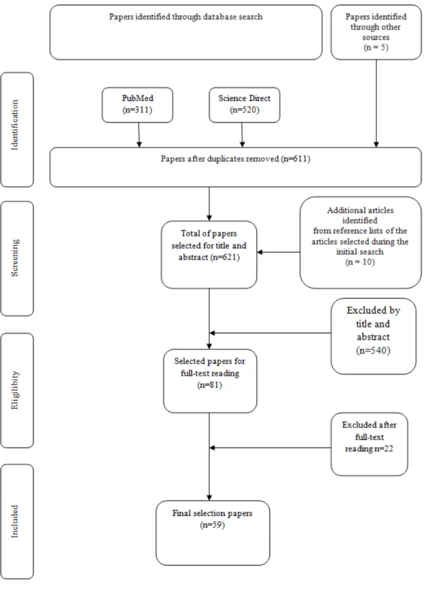
Um total de 611 trabalhos foram selecionados a partir das bases de dados eletrónicas, sendo que um total de 81 foram preliminarmente selecionados para leitura completa. Um total final de 59 estudos foram selecionados para este estudo. Foi verificado pelos trabalhos selecionados que o uso de materiais à base de zirconia tem aumentado recentemente devido às suas propriedades estéticas e sucesso biológico. Além disso, os implantes análogos a estruturas radiculares estão-se a tornar uma solução viável para ultrapassar limitações relacionadas com distribuição de tensões ao osso peri-implantar, estética e peri-implantite por acúmulo de biofilme. Além disso, os avanços tecnológicos recentes têm levado a novas estratégias para melhorar a morfologia e superfícies dos implantes de zircónia. Entretanto, poucos estudos ainda são encontrados para os implantes análogos a estruturas radiculares e ainda torna-se difícil a comparação de resultados dentre inúmeras geometrias e condições intrínsecas dos pacientes.
Abstract
The aim of this study was to conduct a literature review on the potential benefits of custom-made root analogue zirconia implants. A PubMed and ScienceDirect bibliographical search was carried out from 1969 to 2017. The following search items were explored: “zirconia” and “custom-made” and “dental implants”, “zirconia” and
“root-analogue” and “dental implants”, “zirconia” and “anatomical” and “dental
implants”, “zirconia” and “finite element” and “dental implants”, “zirconia” and “customized” and “dental implants”, “zirconia” and “mechanical properties” and
“dental implants”, “zirconia” and “biomechanical” and “dental implants”.
The increased interest in zirconia-based dental structures linked to aesthetic and biological outcomes have been reported in literature. Also, custom-made root analogue implants have become a viable alternative to overcome limitations concerning stress distribution, aesthetics and peri-implantitis induced by biofilms. Recent technological advances have focused on novel strategies to modify zirconia-based surfaces to accelerate osseointegration. However, only a few studies revealed mechanical and biological benefits of zirconia custom-made root analogue implants and therefore further studies should investigate the influence of different geometries and surface modification on the performance of such implants.
Dedicatória
Dedico aos meus pais toda a paciência, esforço, força e dedicação por nunca
duvidarem de mim e por me apoiarem desde o primeiro momento que decidi tomar o
Agradecimentos
Aos meus pais, por sempre acreditarem em mim, sem eles nada disto teria sido possível. Foram eles que através de muito esforço e dedicação me deram a possibilidade de me formar como Médico Dentista. Obrigado por me terem dado essa possibilidade e de me transmitirem também os valores certos.
Ao meu amigo, Luís Beltrão, pelo os últimos dois anos de percurso clínico como binómio, estando presente e apoiando nos bons e maus momentos.
Um agradecimento muito especial ao meu orientador Prof. Doutor Júlio Souza, por ter acreditado em mim e num tema diferente e bastante recente. Por toda a sua atenção e dedicação na realização desta dissertação, também pela sua paciência, apoio interminável e motivação ao longo do ano que tornou este projeto numa realidade. Espero que este não seja o último mas o primeiro de outros projetos desta índole a envergar no futuro.
Uma palavra também à Prof. Doutora Sandra Gavinha que ao longo destes últimos dois anos foi uma pessoa presente em momentos muito importantes na minha trajectória, pelas suas palavras, conselhos mas acima de tudo pelo esforço que dedica a este curso, alunos e Universidade.
A todos os professores, com quem tive o gosto de me cruzar neste percurso e que contribuíram de certa forma para a minha formação académica e pessoal.
General index
Resumo ...iv
Abstract ... v
Dedicatória ...vi
Agradecimentos ... vii
General index ... viii
Figure index ... ix
Table index ... x
List of abbreviations and acronyms ... xi
I. Introduction ... 1
1.Search strategy ... 2
II. Theorical Foundations ... 3
2.1. Immediate custom-made root analogue implants ... 3
2.2. Manufacturing and placement of root analogue implant ... 5
2.3. Mechanical and biomechanical properties of materials ... 6
2.4. Zirconia surfaces for custom made root analogue implants ... 9
III. Results ... 12
IV. Discussion ... 13
IV. Conclusions ... 13
V.References ... 15
Figure index
Figure 1: Search strategy used in this study………20
Table index
Table 1– Summary of relevant studies on bone to implant contact……….23
List of abbreviations and acronyms
A
AAD - advanced abutment design
B
BIC- Bone in contact
C
CAD/CAM - Computer Aided Design / Computer Aided Manufacturing technique
Cp Ti – Commercial pure Titanium CT – Computer Tomography
E
EBM–Electron Beam Melting
F
FEA – Finite Element Analysis FEM – Finite Element Modelling FGP - functionally graded porosity
T
Ti -Titanium
Y
I. Introduction
Dental implants are currently used to replace missing teeth in oral rehabilitation (Nam and Tokutomi, 2014), concerning a long-term success due to the osseointegration of titanium surfaces (Dhima et al., 2013). Long-term success rates have been reported for titanium-based dental implants ranging at around 90%, within a follow up over 10 years (Pirker and Kocher, 2008; Pirker et al., 2011; Pirker and Kocher, 2009). However, anterior tooth replacement with titanium based implants has shown major concerns related to esthetics and release of degradation products to peri-implant tissues (Apaza-Bedoya et al., 2017; Broggini et al., 2006).
Following technological developments in dentistry, patients have desired more esthetic oral rehabilitations that has led to the improvement of metal-free structures for implant-supported prostheses (Navar et al., 2015). Ceramic materials with tooth-like color (Manicone et al., 2007) and high biocompatibility (Navar et al., 2015) has had an increased demand on all-ceramic structures of more than 12% per year (Chevalier, 2006). It is noteworthy that the dental community has seen the aesthetical and mechanical benefits of zirconia-based materials, which have made it a potential to replace titanium implants (Chahine et al., 2011). Zirconia-based implants have become a solution for certain cases considering morphological aspects of peri-implant soft tissues and patient phenotype (Gungor and Yilmaz, 2016; Mobilio, 2013). The color of yttria-stabilized tetragonal zirconia polycrystalline (YTZP) can vary depending on the oxide content to mimic the color of natural teeth. That is a significant outcome to overcome aesthetic issues of implant system related to the use of titanium (Mangano, 2012). Considering mechanical properties YTZP has a Young´s modulus around 210-240 GPa associated with a three-bending-strength ranging from 900 to 1200 MPa (Langhoff, 2008; Piconi and Maccauro, 1999). Additionally, YTZP is a biologically inert material possessing a high biocompatibility that can provide osseointegration (Pirker and Kocher, 2008; Pirker et al., 2011; Pirker and Kocher, 2009).
peri-implant response and lesser biofilm accumulation (Prithviraj et al., 2012). Limited data are available on the stress distribution on YTZP implants and surrounding tissues (Gungor and Yilmaz, 2016). The design of the custom-made zirconia or titanium implant can maintain the stress distribution pattern in the surrounding bone, due to the design mimicking of the alveolar region (Chahine et al., 2011).
1.Search strategy
The aim of this study was to conduct a literature review on the potential benefits of the use of custom-made root analogue zirconia implants. It was hypothesized that zirconia custom-made root analogue implants can provide proper stress distribution, biocompatibility and healthy state peri-implant when compared to standard implants.
A PubMed and ScienceDirect bibliographical search was carried out from 1969 to 2017. The following search items were explored: “zirconia” and “custom-made” and “dental implants”, “zirconia” and “root-analogue” and “dental implants”, “zirconia” and “anatomical” and “dental implants”, “zirconia” and “finite element” and “dental implants”, “zirconia” and ”customized” and “dental implants”, “zirconia” and “mechanical properties” and “dental implants”, “zirconia” and “biomechanical” and “dental implants”. The eligibility inclusion criteria used for article search were: Meta-analysis; randomized controlled trials; prospective cohort studies; retrospective cohort studies; as well as articles and reviews written in English.
II. Theorical Foundations
2.1. Immediate custom-made root analogue implants
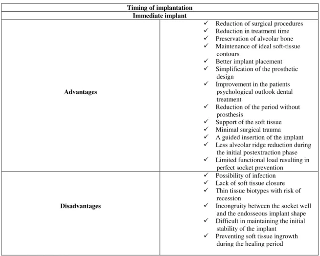
After the removal of a tooth, there is an alveolar bone resorption within loss of bone (buccal plate) followed by the loss of the supported soft tissue contours. The physiological morphology of the inter-dental papillae is changed due to such bone loss (Jivrai and Chee, 2006). That often leads to two distinct problems: firstly, the manufacture of conventional or implant-supported prosthesis results in an aesthetic issue; secondly, the bone volume can decrease decreasing the possibility to place an endosseous implant. Thus, it is crucial to preserve the alveolar process dimensions in extraction areas (Camargo, 2000). The immediate placement of dental implants can prevent a loss of alveolar bone volume leading to an improved esthetic and functional prosthodontic result (Schropp et al., 2003; Beagle, 2006). Thus, immediate implant placements have some advantages and disadvantages as seen in Table 2.
The extraction site is often larger than the implant diameter that can result in gap between bone and implant. That misfit with the extraction site requires the use of a barrier membrane or biomaterials for bone augmentation to prevent down-growth of connective tissue or epithelium between the implant and socket (Pirker and Kocher, 2011). Botticelli et al., (2003), reported the healing that occurred adjacent to implants placed in recipient sites with a wide marginal defect. In these cases, the new bone formation in the test sites resulted not only in the elimination of the gap but also in the establishment of a high degree of bone-to-implant contact or osseointegration. For instance, the amount of mineralized bone found in the test sites (70.3-75.6 %) was similar to that found in the control sites (74.1%), although the quality of the bone that filled the gap was markedly different. In the test site within the gap, most the bone grown was immature. Accordingly, a gap of 0.5 mm between the bone and the implant can decrease the success rate of acceptable bone-to-implant contact.
degree of predictability. The study reported that the incongruence (gaps) between the bony walls of the tooth socket and the root analogue implants should be avoided. Therefore, a good curettage of the periodontal ligament remains should be done, in order to secure osseointegration of the analogue implant. Gaps located in the marginal area, lead to the possibility of down-growth of supracrestal connective tissue, so a well fit root analogue implant with controlled techniques it is necessary to result in a bone-to-implant contact osseointegration.
Pirker and Kocher, (2008) reported a successful clinical use of a modified root-analogue zirconia implant for immediate single tooth replacement. A right maxillary premolar in 64-year-old patient was removed and a custom-made, root-analogue, roughened zirconia implant with macro-retentions in the interdental space was produced and placed into the extraction socket 4 days later (Figure 2 in annex). In general, the authors concluded that a good fit between implant and host bed by additional retentions was an important factor to decrease bone resorption and therefore that zirconia has excellent biocompatibility and improved esthetic (Table 1 in annex). Pirker and Kocher, (2009) also reported two novel approaches for dental root replacement in humans and evaluated the use of root analogue zirconia implants prospectively in 18 patients. The clinical trial indicated that immediate implantation of a root analogue replica allows instantaneous support of soft tissue and limited functional load, resulting in perfect socket preservation with minimal bone loss. Also, confirmed the need of macro-retentions and implant diameter reduction next to the cortical bone, that primary stability and excellent osseointegration of immediate root-analogue zirconium can be achieved.
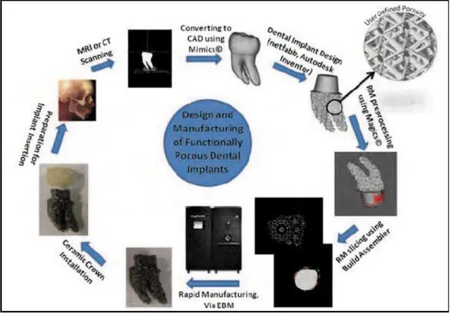
pervious natural tooth that was developed by computer aided designing (CAD) as illustrated in Figure 3 (see annex).
2.2. Manufacturing and placement of root analogue implant
The new concept of custom-made root-analogue zirconia implants can be achieved by using a Computer Aided Design/Computer Aided Manufacturing technique (CAD/CAM), as illustrated in Figure 3. It is possible to manufacture precise custom-made, root-analogue implants by combining the use of cone beam computed tomography 3D data and CAD/CAM technology (Mangano, 2012). The manufacturing of custom-made root analogue implants has been reported before and after extraction. On both cases, the root-form of the extraction surgical site is preserved by minimizing trauma, that can lead to a faster healing of the surrounding bone (Misch et al., 2005; Pirker and Kocher, 2011).
In the cases that the fabrication of the custom-made root analogue implant happens after extraction, the surgical site is cleaned by curettage followed by saliva irrigation and then a iodonform-soacked cottom gauze is placed in the wound (Figure 2). The root can be laser-scanned after extraction and then, the root analogue implant is milled from a medical-grade YTZP block (Pirker and Kocher, 2008). The root analogue implant is cleaned in an ultrasonic bath containing 96% ethanol for 10 min, packaged and sterilized in a steam sterilizer before placement in the surgical site (Pirker and Kocher, 2008) (Figure 2). After 1-8 days extraction the iodoform cotton gauze is removed, and the alveolar socket curetted and flushed with sterile physiologic saline solution (Pirker and Kocher, 2008; Patankar et al., 2016; Pirker and Kocher, 2009; Pirker et al., 2011; Pirker and Kocher, 2011). The custom-made root-analogue implant can be placed into the socket by using finger pressure, following gentle tapping with a hammer and a mallet. Palpation and percussion is used to check primary stability (Pirker and Kocher, A. 2008; Patankar et al., 2016; Pirker and Kocher, 2009; Pirker et al., 2011; Pirker and Kocher, 2011) (Figure 2).
design features: functionally graded porosity (FGP) and advanced abutment design (AAD) (Chahine et al., 2011). Finally, the design is prepared to be manufactured by a computer-aided manufacturing (CAM) via electron beam melting (EBM) or by other additive manufacturing technique and the tooth can be three-dimensionally produced
(Figure 3). The implant undergoes post-manufacturing processing steps before being sent to the dentist office before extraction (Chahine 2009;Chahine et al., 2010). From a clinical point of view, the implantation is accomplished in one dental visit. The implant can be ready upon the initial visit of the patient where the dentist can carefully remove the damaged tooth and insert the implant with minimal to no site preparation (Chahine, et al., 2011).
2.3. Mechanical and biomechanical properties of materials
A success-rate of a dental implant is determined by several aspects related to the implant, surgery, prosthetic and patient conditions. The type of material used is also a key factor to the implant osseointegration and clinical success-rate. Commercially pure titanium (cp Ti) is the most common material used in the last 20 years, but zirconia (YTZP) has growingly become a potential material in implant dentistry (Choi et al., 2012).Zirconia-based materials have appeared in dentistry for metal-free structures due to an excellent biocompatibility, improved esthetic results, high flexural strength, fracture toughness, and high chemical resistance (Pirker and Kocher, 2009; Gahlert et al., 2010; Pirker et al., 2011).
The Finite Element Analysis (FEA) has become an increasingly useful tool for the prediction of the effects of stress on implants and the surrounding bone (Caglar et al., 2011; Cheng et al., 2017). TheFinite Element Method (FEM) generates an accurate analytical model of a dental implant essential to produce realistic solutions using appropriate engineering software. FEM can simulate the stress distribution around implants and determine a proper design to dissipate the stresses from occlusal forces (Van Staden, et al., 2006).
tissues can result in patient discomfort, pressure necrosis, and the eventual failure of the implant system (Choi et al., 2012).
Choi et al., (2012) evaluated the biomechanical behaviour of Ti6Al4V and PS-ZrO2 dental implants inserted into the human mandible during clenching using a three-dimensional anatomically realistic finite element model. Ti6Al4V and PS-ZrO2 dental implants were modeled as cylindrical structure with a diameter of 5.26 mm and length of 12 mm and placed in the first molar region on the right hemi-mandible. On Ti6Al4V dental implants, the maximum tensile stress, compressive stress and Von Mises stress values recorded were at 11.02, -12.39 and 11.37 MPa respectively. On PS-ZrO2, the maximum tensile stress, compressive stress and Von Mises stress values recorded were at 14, -15.3 and 14.2 MPa, respectively. The results revealed an increase of 2-3% in the tensile and compressive stress mean values while Von Mises stress increased in 8% in the bone-implant interface when PS-ZrO2 dental implant was used instead of Ti-6Al-4V dental implant (Table 1).
In a study conducted by Siegele and Soltesz, (1989), the stress distribution generated was assessed in jaw bone by FEA considering 5 different types of dental implants designs (cylindrical, conical, stepped, screw-shaped and hollow cylindrical). The results showed that the different implant shapes led to significant variations in stress distribution surrounding peri-implant bone. In conclusion, the lowest von mises stresses were noticed by the cylindrical and implant fixtures. The stepped and hollow-cylindrical implants led to maximum compressive stresses at 5 MPa. In maximum stresses, the results were significantly different, the conical implants revealed the highest (25 MPa) compared with the other shapes. The most favorable were the cylindrical (7.5 MPA) and screw- designed (6 MPA). Fuh et al., (2013) investigated the effects of different thread designs on the bone around YTZP and cp Ti implants. A total of 18 finite element models comprising two implant materials (YTZP or titanium), three thread designs, and three interface conditions were assessed considering the stress distribution on bone tissue. In the immediately loaded implant, the stress was highly concentrated at one site of the peri-implant bone. Also, zirconia implant can reduce the bone stress in the crestal cortical region.
lithium dissilicate or YTZP. That study concluded that stress distribution was lower around YTZP implants than that around cpTi under horizontal loading although similar stress values for YTZP and cpTi were reported for oblique loading. In a study performed by Himmlova et al., (2004), a mathematical simulation of stress distribution was used to determine influence of length and diameter to dissipate stress. A FEA was carried out to simulate masticatory forces. The results were that a decrease in stress of 31.5% was noted for implants when the diameter implant from 3.6 mm up to 4.2 mm. Stress for the 5.0 mm diameter implant was only at 16.4%. This finite element study showed that increased implant diameter better dissipated the simulated masticatory force and decreased the stress around the implant neck, in conclusion an increase in the implant diameter decreased the maximum von Mises equivalent stress around the implant neck more than an increase in the implant length, as a result of a more favorable distribution of the simulated masticatory forces applied in this study.
Mobilio, (2013) compared the stress in bone around zirconia and titanium implants under loading. A one-piece YTZP implant and a replica of the same implant made of commercially pure titanium were embedded in two self-curing acrylic resin blocks. Loads of 50, 100, and 150 N, with orientations of 30, 45 and 60 degrees with respect to the implant axis were applied on the implant. Strain under all loading conditions on both samples was measured. Three-dimensional virtual replicas of both the implants were reproduced using the FEM and inserted into a virtual acrylic resin block. The two implants were assessed by FEA and revealed different biomechanical behavior. Titanium implant revealed higher stress values on the cortical bone while the YTZP implant showed higher stress values on the trabecular bone. The stress magnitude were similar in bone of the two implants in all cases even if the stiffness of YTZP was twice that of titanium. On mechanical point of view, YTZP was thus a feasible substitute for dental implants.
and lingual regions than on the proximal ones. Stress values were ranged from 1.3-2 MPa and 4-5 MPa at the cortical and critical/cancellous transition regions. The Re-implant root analogue structures showed a better stress distribution at the bone-to-implant junction when compared to standard bone-to-implants (Table 1). In a clinical study, Moin et al., (2016), selected a right upper human canine in a patient with 64 years old, to create a 3D surface model of a root analogue implant. Based on the standard triangulation language, five different (targeted) press-fit design root were built: non-modified standard, target press-fit prism, targeted press-fit fins, targeted press-fit plug and targeted press-fit bulbs. Two different loadings were applied to stimulate anterior bite forces. The stress mean levels caused by oblique loading were higher when compared to vertical loading. The study concluded that the optimization of standard root design, preferably fins or bulbs, will have a positive effect on stress distribution and lower stress concentration on peri-implant bone.
2.4. Zirconia surfaces for custom made root analogue implants
A main factor that strongly influences wound healing at the implantation site is the morphology of the implant surface, which subsequently affects osseointegration (Albrektsson et al., 1981). Several chemical and physical methods have been used to modify implant surfaces considering increase in roughness, wet ability and bioactivity (Depprich et al., 2009; Mangano 2012). Indeed, studies have shown that implants with rough surface have a higher resistance to removal torque when compared to smooth surface implants (Chahine, et al., 2011). Smooth surface implants are not generally used since such implants reveal a lower contact area of interaction with bone tissue (Puleo and Thomas, 2006). Consequently, the clinical use of YTZP dental implants is limited due to the manufacturing process of YTZP structures involving a morphological enhancement of the surface as found on titanium implants (Zkurt and Kazazog, 2011).
electrochemical methods to chemically modify the surface in a specific environment containing bioactive materials. Hanawa, (2010) has reported the functionalization of titanium surfaces by using ceramics or polymeric materials.
In a study performed by Langhoff, (2008), two main approaches for surface modifications were assessed. At first, micro-roughness was achieved by gritblasting and acid-etching and, then bioactive coating composed of calcium phosphate, bisphonate and collagen type 1 was applied on the rough surface. Six different types of dental implants within a core based on YTZP or titanium were tested for osseointegration. After two weeks, zirconia-based implants showed a higher bone-implant interface (BIC at 77%) compared to the BIC on titanium-based implants (BIC around 57-61%) as seen in Table 1. An increase in BIC was detected on the pharmacologically and chemically modified titanium implants. In a study carried out by Kohal, (2004), 12 custom-made titanium (control group) and 12 custom-made YTZP implants (test group) were placed in the extraction sites in six monkeys after five months of tooth extraction. Titanium implant surfaces were grit-blasted with Al2O3 and subsequently acid-etched with H2O2/HF. YTZP implants were only grit-blasted once YTZP cannot be modified by acid etching. After 5 months of loading, BIC was recorded around 72.9 % for titanium implants and 67.4% for YTZP implants (Table 1). In conclusion, the custom-made zirconia implants osseointegrated to the same extent as custom-made titanium control implants.
58.6% ± 9.5 was recorded for titanium implans. After 12 weeks, the BIC percentage was higher for YTZP at 71.4 ± 7.8% while titanium surfaces revealed a BIC at 82.9 ± 10.7%, as seen in Table 1. Those BIC results are quite similar to the results recorded in another study in minipigs (Schliephakeet al., (2010).
Han, et al., (2016) evaluated the biomechanical and histological of a ceria-stabilized zirconia-alumina nano composite (NanoZr) in comparison with that of YTZP in Sprague-Dawley rats. The average BIC percentage within the bone marrow area for YTZP was at 25.26% while NanoZrO showed a BIC at 31.51% after 2 weeks. After 4 weeks, BIC for YTZP was at 38% while NanoZrO was at 46.78%. Finally, BIC was recorded at 47.88% for YTZP and at 56.81% for NanoZrO after 8 weeks. On cortical bone, the mean BIC percentage values within the cortical area were 38.86% and 58.42% at 2 weeks, 66.82% and 57.74% at 4 weeks, and 79.91% and 78.97% at 8 weeks, respectively. Animal studies have shown that 3Y-TZP and pure titanium have similar BIC (Table 2).
Two rough surfaces of titanium and YTZP were also assessed after placement in Sprague-Dawley rat femur model (Kohal et al., (2009). Four groups of implants were tested: machined YTZP implants (m-YTZP), rough YTZP implants (r-YTZP), machined titanium implants (m-Ti), and electrochemically roughned titanium (Ti Unite) surface. For 14 days of bone healing, the BIC percentage was at 23.2% for m-Ti, 30.9% for m-YTZP, 36.4% for TiUnite group and 45.3% for r-YTZP. After 28 days, the BIC percentage increased for m-Ti in 39.4%, 46.6% for m-YTZP, 55.2% for TiUnite and 59.4% for r-YTZP group. No significant differences could be found within the groups after 28 days of healing.
A good number of studies confirmed the osseointegration of zirconia to be like or even better than that of titanium. The results show potential in using zirconia implants for dental application in the future. Such studies also showed that zirconia performance can be highly dependent on surface preparation and that every new surface modification should be tested regarding aging and fatigue before clinical use (Sanon et al., 2015; Prithviraj et al., 2012; Fuh et al., 2013). Roughening the surface of machined zirconia implants enhances bone apposition and enhances the ability to withstand shear stress. On balance, the osseointegration of zirconia implants is promising considering addictive manufacturing techniques (Gahlert et al., 2010). Functionality graded YTZP has been developed by additive manufacturing including pores in the outer region (Chahine et al., 2011). Such development has been introduced in YTZP implants ranging from a micro- to a nano-scale level to enhance osseointegration.
III. Results
A total of 611 papers were selected by the search on the electronica database, a total of 81 were preliminarily selected for full-test reading and then 59 were evaluated as most relevant regarding the purpose of the present study. The selection of studies is illustrated in Figure 1 (see annex). Within the most relevant articles found in the present bibliographical search, the main significant factors that should be highlighted on custom made root analogue implants are described along this section and in Table 1. Such key factors linked to the placement, manufacturing, mechanical behavior and surface conditions can be drawn as follow:
The time of dental implant placement is determinant on the alveolar bone remodeling. Immediate root analogue implant can prevent a loss of alveolar bone volume within maintenance of peri-implant soft tissues leading to an improved esthetic and functional prosthodontic result (Tables 1 and 2);
The manufacturing of custom-made root analogue implants can be achieved by CAD/CAM as well as on additive manufacturing technology combined with cone beam computed tomography focusing on the tooth which should be removed (Figures 2 and 3);
implant-supported prosthesis. Clinical conditions vary from different cases that can be assessed preliminary by finite element analysis considering different conditions such as materials, design, loading and maxillofacial positioning of implants. Root analogue implants can promote a proper distribution of stresses trough the materials towards to the bone that can decrease the early peri-implant bone loss;
Titanium-based implant surfaces can me modified by different methods resulting in well-known micro- and nano-scale topography. However, zirconia-based surfaces are currently studied to achieve morphological features that enhance the adsorption of proteins and then the migration of osteogenic cells. The major concern is based on the high chemical resistance of zirconia-based surfaces which cannot be modified by usual acid etching procedures. Additive manufacturing approaches are the first choice to improve the morphology of zirconia surfaces.
IV. Discussion
The present study assessed the relevant studies published in literature on the biomechanical and biological benefits of zirconia custom made implants. The results found in the selected articles validate the hypothesis of the present study. They showed significant enhancement in the stress distribution through custom made root analogue implants associated to desirable aesthetic outcomes and low risks of peri-implantitis induced by bacterial accumulation. Basically, a good fit between an implant and the bone provided by a root analogue implant is an important factor to immediate implant success and will raise dental implantology to a new level of truly anatomic implants. The main outcomes from the selected studies in the present review are discussed as follow.
IV. Conclusions
Zirconia-based materials have shown increased interest to replace titanium-based structures considering aesthetics and biological success. The optical properties of zirconia-based structures can mimic the color of natural teeth while their chemical composition enhances the biocompatibility to soft and bone tissues;
The surgical protocol of custom-made zirconia immediate implants is slightly invasive and atraumatic when compared to that for standard titanium implants. In fact, the immediate implantation consists the standard protocol in placement of custom-made zirconia implants;
The design of the custom-made zirconia can maintain the stress distribution in the surrounding bone, due to the geometrical mimicking of the alveolar region. That can decrease the bone resorption due to occlusal overloading associated to peri-implant inflammatory reactions;
V.References
Albrektsson, T. et alli. (1981). Osseointegrated titanium implants. Requirements for ensuring a long-lasting, direct bone-to-implant anchorage in man. Acta Odontologica Scandinava, 52(2), pp:155-70.
Apaza-Bedoya, K. et alli. (2017). Synergistic interactions between corrosion and wear a titanium-based dental implant connections: A scoping review, Journal of Periodontal Research, In press.
Beagle, J.R. (2006). The immediate placement of endosseous dental implants in fresh extraction sites,
Dental Clinics of North America. 50(3). pp: 375-89.
Botticelli, D. et alli. (2003). The jumping distance revisited: An experimental study in the dog, Clinical Oral Implants Research, 14(1), pp: 35-42.
Broggini, N. et alli. (2006). Peri-implant inflammation defined by the implant-abutment interface,
Journal of Dental Research, 85(5). pp. 473.8.
Caglar, A. et alli. (2011). Three-dimensional finite element analysis of titanium and yttrium-stabilized zirconium dioxide abutments and implants, The International Journal Oral & Maxillofacial Implants, 26(5), pp: 961-9.
Camargo, P. M. (2000). Influence of bioactive glass on chances in alveolar process dimensions after exodontias, Oral Surgery, Oral Medicine, Oral Pathology, Oral Radiology, and Endodontics.90(5). pp:581-6.
Chahine, G. (2009). Design optimization of a customized dental implant manufactured via Electron Beam Melting, International Solid Freeform Fabrication Symposium.
Chahine, G., Smith, P., and Kovacevic, R.(2010). Application of structural optimization in modern rapid manufacturing, International Solid Freeform Fabrication Symposium Austin Texas.
Chahine,G. et alli. (2011). Digital Engineering of Bio-Adaptable Dental Implants. In: Turkyilmaz, I. (Ed).
Implant dentistry – A Rapidly Evolving Practice. In Tech, pp. 252-266.
Chen, S. T., Wilson, T.G. and Hammerle, C.H. (2004). Immediate or early placement of implants following tooth extraction: review of biological basis, clinical procedures, and outcomes, The international Journal of oral and Maxillofacial Implants,19. pp: 12-25.
Cheng, Y.C. et alli. (2017). Dental implant customization using numerical optimization design and 3-dimensional printing fabrication of zirconia ceramic, International Journal for Numerical Methods in Biomedical Engineering, 33(5).
Chevalier, J. (2006). What future for zirconia as a biomaterial ?, Biomaterials, 27(4). pp. 535-543.
Choi, A.H., Matinlinna, J.P. and Ben-Nissan, B. (2012). Finite element stress analysis of Ti-6Al-4V partially stabilized zirconia dental implant during clenching, Acta Odontologica Scandinava, 70(5), pp:353-61.
Depprich, R. et alli. (2008). Osseointegration of zirconia implants: an SEM observation of the bone-implant interface, Head & Face Medicine, 4(25).
Depprich, R. et alli. (2009). Osseointegration of zirconia implants compared with titanium: An in vivo study, Head & Face Medicine, 4(1), pp:30.
Dhima, M. et alli. (2013). Practice- Based Evidence from 29-year Outcome Analysis of Management of the Edentulous Jaw Using Osseointegrated Dental Implants, Journal of Prosthodontics, 23(3). pp. 173-81.
Gahlert, M. et alli. (2010). A comparison study of the osseointegration of zirconia and titanium dental implants. A biomechanical evaluation in the maxilla of pigs, Clinical Implant Dentistry and Related Research. 12(4), pp: 297-305.
Glickman, R.S., Bae, R. e Karlis, V. (1990). A model to evaluate bone substitutes for immediate implant placement, Implant Dentistry, 10(3), pp: 209-15
Gotfredsen, K., Nimb, L. and Hjorting-Hansen, E. (1994). Immediate implant placement using a biodegradable barrier, polyhydroxybutyrate-hydroxyvalerate reinforced with polyglactin 910. An experimental study in dogs, Clinical Oral Implants Research, 5(2), pp: 83-91.
Gungor, M.B. and Yilmaz, H. (2016). Evaluation of stress distributions occurring on zirconia and titanium implant-supported prostheses: A three-dimensional finite element analysis, The Journal of Prosthetic Dentistry, 116(3). pp. 346-55.
Han, J. et alli. (2016). Biomechanical and histological evaluation of the osseointegration capacity of two types of zirconia implant, International Journal of Nanomedicine, 11, 5507-6516.
Hanawa, T. (2011). A comprehensive review of techniques for biofunctionalization of titanium, Journal of Periodontal and Implant Science, 41(6), pp: 263-272.
Harris, W.H. et alli. (1983). Bony ingrowth of the acetabular component in canine hip joint arthroplasty,
Clinical Orthopaedics and Related Research,(176), pp:7-11.
Himmlova, L. et alli. (2004). Influence of implant length and diameter on stress distribution: a finite element analysis, The Journal of Prosthetic Dentistry, 91(1), pp: 20-5.
Hisbergues, M., Vendeville, S. and Vendeville, P. (2009). Zirconia: Established facts and perspectives for a biomaterial in dental implantology, Journal of Biomedical Materials Research,88(2), pp: 519-29.
Hodosh, M., Poyar, M. and Shklar, G. (1969). The dental polymer implant concept, Journal of Prosthetic Dentistry, 22, pp: 371.380.
Jivrai, S. and Chee, W. (2006). Treatment planning of implants in the aesthetic zone, British Dental Journal, 201(2). pp: 77-89.
Khandare, K.K., Jaju, S.B. and Patil, P.G. (2013). Fem analysis for stress distribution of root analogue zirconia dental implant: A review, International Journal of Innovative Research in Science, Engineering and Techonology, 2(6).
Kohal, R.J. (2004). Loaded custom-made zirconia and titanium implants show similar osseointegration: an animal experiment, Journal of Periodontology.75(9), pp:1262-8.
Kohal, R.J. et alli. (1997). Custom-made root analogue titanium implants placed into extraction sockets. An experimental study in monkeys, Clinical Oral Implants Research,8(5), pp:386-92.
Kohal, R.J. et alli. (2009). Biomechanical and histological behavior of zirconia implants: an experiment in the rat, Clinical Oral Implants Research,20(4), 333-9.
Langhoff, J. D. (2008). Comparison of chemically and pharmaceutically modified titanium and zirconia implant surfaces in dentistry: a study in a sheep, The International Journal of Oral and Maxillofacial Implants, 37(12). pp: 1125-32.
Lundgren, D. et alli. (1992). Healing-in of root analogue titanium implants placed in extraction sockets. An experimental study in the beagle dog, Clinical Oral Implants Research, 3(3), pp:136-43.
Mangano, F.G. (2012). Custom-made, root-analogue direct laser metal forming implant: a case report,
Manicone, F. P., Iommetti, P. R. and Raffaelli, L. (2007). An overview of zirconia ceramics: Basic properties and clinical applications, Journal of Dentistry, 35(11). pp. 819-826.
Mendonça, G. et alli. (2008). Advancing dental implant surface technology from micron to nanotopography, Biomaterials, 29(28), pp: 2322-35.
Misch, C. et al (2005). A positive correlation between occlusal trauma and peri-implant bone loss: Literature Support. Implant Dentistry, 14(2), 108-116.
Mobilio, N. (2013). Experimental and numeric stress analysis of titanium and zirconia one-piece dental implants, The International Journal of Oral and Maxillofacial Implants, 28(3). pp. 135-42.
Moin, D.A., Hassan, B. and Wismeijer, D.(2016). A patient specific biomechanical analysis of custom root analogue implant designs on alveolar bone stress: a finite element study, International Journal of Dentistry, Volume 2016.
Nam, J. and Tokutomi, H. (2014). Using zirconia based prosthesis in a complete-mouth reconstruction treatment for worn dentition with the altered vertical dimension of occlusion, The Journal of Prosthetic Dentistry, 113(2). pp. 81-1.
Navar, S., Aruna, U. and Bhat, W. M. (2015). Enhanced aesthetics with all ceramics restoration, Journal of Pharmacy Bioallied Sciences, 7(1). pp. 282-284.
Patankar, A. et alli. (2016). Immediate, Non Submerged Root Analog Zirconia Implant in Single Rooted Tooth Replacement: Case Report with 2 years follow up, Journal of Maxillofacial and Oral Surgery,15(2). pp: 270-3.
Piconi, C. and Maccauro, G. (1999). Zirconia as a ceramic biomaterial, Biomaterials, 20(1). pp: 1-25.
Pirker, W. and Kocher, A. (2008). Immediate, non-submerged, root-analogue zirconia implant in single tooth replacement, International Journal of Oral and Maxillofacial Surgery, 37(3). pp. 293-5.
Pirker, W. and Kocher, A. (2009). Imeddiate, non-submerged, root-analogue zirconia implants placed into single-rooted extraction sockets: 2-year follow-up of a clinical study, International Journal of Oral and Maxillofacial Surgery, 38(11). pp. 1127-32.
Pirker, W. and Kocher, A.(2011). Root analog zirconia implants: true anatomical design for molar replacement- a case report, The international Journal of Periodontics & Restorative Dentistry,31(6). pp: 663-8.
Pirker, W. et alli. (2011). Immediate, single stage, truly anatomic zirconia implant in lower molar replacement: a case report with 2.5 years follow-up, International Journal of Oral and Maxillofacial Surgery, 40(2). pp. 212-6.
Prithviraj, D.R. et alli. (2012). A systematic review of zirconia as an implant material, Indian Journal of Dental Research, 23(5). pp: 643-9 .
Puleo, D.A. and Thomas, M.V. (2006). Implant surfaces, Dental Clinics of North America, 50(3), pp:323-38.
Salem, N.A., Abo Taleb, A.L. and Aboushelib, M.N. (2013). Biomechanical and histomorphometric evaluation of osseointegration of fusion-sputtered zirconia implants, Journal of Prosthodontics, 22(4), pp:261-7.
Sanon, C. et alli. (2015). A new testing protocol for zirconia dental implants, Dental Materials, 31(1), pp: 15-25.
Schropp, L. et alli. (2003). Bone healing and soft tissue contour changes following single-tooth extraction: a clinical and radiographic 12-month prospective study, The International Journal of Periodontics & Restorative Dentistry. 23 (4). pp: 313-23.
Siegele, D. and Soltesz, U. (1989). Numerical investigations of the influence of implant shape on stress distribution in the jaw bone, , The international Journal of Oral Maxillofacial Implant, 4(4), pp: 333-40.
Smeets, R. (2016). Impact of dental implant surface modifications on osseointegration, Biomedical Research International.
Van Staden, R.C., Guan, H. and Loo, Y.C. (2006). Application of the finite element method in dental implant research, Computer methods in Biomechanics and Biomedical Engineering, 9(4), pp: 257-70.
Miguel Romero Alves Pessanha de Andrade
Custom-made root analogue zirconia implants
Attachments
Universidade Fernando Pessoa Faculdade de Ciências da Saúde
Table 1– Summary of relevant studies on bone to implant contact
Authors and year of
publication Purpose
Sample size and
groups Assessment methods
Study design Follow-up Bone implant contact
(BIC)
Langhoff et al. (2008) To investigate surface modifications and materials on the same
implant geometry
Six types of dental implants 5 titanium implants
1 zirconia implant
Macroscopic, radiographic and histomorphometric
methods
2 weeks, 4 and 8 At two weeks titanium implants (57-61%) Zirconia was better (77%)
The main BIC increased between 2 and 4 weeks. There were no significant
differences in the results Kohal et al. (2004) To investigate the
histological behavior of loaded zirconia implants in an animal model and to
compare it to titanium implants
12 custom-made titanium implants (control-group) 12 custom-made zirconia implants (test
group)
Light microscope 9 months 72.9% for the titanium
implants 67.4% for the zirconia
implants
Depprich et al. (2008) To investigate the osseointegration of zirconia implants with the
modified ablative surface
24 zirconia implants with modified ablative
surfaces 24 titanium implants with modified ablative
surfaces
Scanning electron microscopy
1 week, 4 weeks or 12 weeks
After 4 week intimate contact with bone cells
both in titanium and zirconia implant surfaces After 12 weeks successful
osseointegration of the zirconia as well titanium Depprich et al. (2009) To compare the osseous
healing of zirconia implants with titanium
implants
24 screw-type zirconia implants with modified acid-etched surfaces 24 screw- type titanium
implants with acid-etched surface
Histomorphometic evaluation
1 week, 4 and 12 After 1 week of healing, the mean BIC was 35.5% +- 10.8 % for the zirconia and 47.7% +- 9.1 for the
titanium implants, respectively. After 4 weeks in situ, BIC of the
zirconia implants averaged 45.3% +- 15.7 and 58.6% +- 9.5 for the titanium implans. After 12 weeks the BIC values
were 71.4% +- 71.8 for the zirconia implants and
Authors and year of publication
Purpose Sample size and
groups
Assessment methods Study design Follow-up Bone implant contact
(BIC)
Han et al. (2016) Evaluate the biomechanical and histological behaviour Ceria-stabilized zirconia-alumina nanocomposite(NanoZr)
in comparison with that of yttria-stabilized tetragonal zirconia polycrystalline (3Y-TZP) Scanning white-light interferometry and scanning electron microscopy
In vivo(Sprague Dawley rats)
2 weeks,4, and 8 Bone marrow area: For 3Y-TZP and NanoZr
were 25.26% and 31.51% at 2 weeks, 46.78% and 38% at 4 weeks, and 47.88% and
56.81% at 8 weeks, respectively. Cortical are: 38.86% and 58.42% at 2
weeks, 66.82% and 57.74% at 4 weeks, and 79.91% and 78.97% at 8
weeks Salem et al.
(2013)
Evaluate osseointegration of fusion-sputtered zirconia
implantis in comparison with sandblasted, acid-etched titanium implants
in a biomechanical and histomorphometric
study
60 zirconia were manufactured. Half
received fusion sputtering surface treatment. Standart Ti
implants of the same shape and dimensions
served as control
Histological and histometric analyses
In vivo(30 adult New Zealand white male
rabbits)
4 weeks,8 and 12 Fusion sputtered zirconia with a bic of
69,66% at 4 weeks, 88.03% at 8 weeks and
89.09% at 12 weeks. Titanium with a BIC of
62.83% at 2 weeks, 82.94% at 8 weeks and
86.77% at 12 weeks. Control Zirconia with a
Authors and year of publication
Purpose Sample size and
groups
Assessment methods Study design Follow-up Bone implant contact
(BIC)
Kohal et al. (2008)
Evaluate the integration of zircnonia implants
4 groupos of implants were utilized: machined
zirconia implants, zirconia implants with a rough surface, machined titanium implants, and titanium implants with an electrochemically
roughened surface
Histological and histomorphometric
In vivo(rat femur model) 14 days and 28 days of healing
For 14 days bone healing, the BIC percentage was at 23.2%
for Ti, 30.9% for m-YTZ36.4% for TiUnite group and 45.3% for
r-YTZP. After 28 days, the bone to implant contact increased for all
groups. The BIC percentage for m-Ti was
Table 2 - Advantages versus Disadvantages regarding implantation timing. Source: (Gotfredsen et al., 1994; Beagle, 2006; Glickman, et al., 1990)
Timing of implantation Immediate implant
Advantages
Reduction of surgical procedures
Reduction in treatment time
Preservation of alveolar bone
Maintenance of ideal soft-tissue contours
Better implant placement
Simplification of the prosthetic design
Improvement in the patients psychological outlook dental treatment
Reduction of the period without prosthesis
Support of the soft tissue
Minimal surgical trauma
A guided insertion of the implant
Less alveolar ridge reduction during the initial postextraction phase
Limited functional load resulting in perfect socket prevention
Disadvantages
Possibility of infection
Lack of soft tissue closure
Thin tissue biotypes with risk of recession
Incongruity between the socket well and the endosseous implant shape
Difficult in maintaining the initial stability of the implant