Vol.52, n. 3: pp.549-554, May-June 2009
ISSN 1516-8913 Printed in Brazil BRAZILIAN ARCHIVES OF
BIOLOGY AND TECHNOLOGY
A N I N T E R N A T I O N A L J O U R N A L
Plant Regeneration through Somatic Embryogenesis of
Yacón [
Smallanthus sonchifolius
(Poepp. and Endl.) H.
Robinson]
Cynthia Manyra Corrêa
1, Graciele Nicolodi de Oliveira
2,
Leandro Vieira Astarita
1and
Eliane Romanato Santarém
1*1Laboratório de Biotecnologia Vegetal; Departamento de Biologia Celular e Molecular; Faculdade de Biociências;
Pontifícia Universidade Católica do Rio Grande do Sul; Av. Ipiranga, 6681; 90619-900; Porto Alegre - RS - Brasil.
2
Laboratório de Cultura de Tecidos Vegetais; Universidade de Cruz Alta; Cruz Alta - RS - Brasil
ABSTRACT
Smallanthus sonchifoliushas tuberous roots containing large amounts of fructo-oligosaccharides and its medicinal use has increased due to the hypoglycemic properties reported for this species. An efficient system for propagation via somatic embryogenesis is reported using petiole segments cultivated on MS medium supplemented with combinations of BA, kinetin and 2,4-D, under light and darkness conditions. Embryogenic callus was formed in most of the treatments; however, somatic embryogenesis was promoted by the presence of light. Clusters of somatic embryos appeared on callus surface after 50 days of culture. The highest number of embryos was produced on 0.45 µM BA and 4.5 µM 2,4-D. Embryogenic calli were maintained on MS medium containing 4.5 µM BA and 0.045 µM
2,4-D. Embryos converted on hormone-free half-strength MS medium with 2 g.L-1 activated charcoal and plantlets
were transferred to non-sterile conditions for acclimatization, showing 100% of survival.
Key words: Medicinal plant, tissue culture, Polymnia sonchifolia, micropropagation
* Author for correspondence: [email protected]
INTRODUCTION
Yacón [Smallanthus sonchifolius (Poepp. and
Endl.) H. Robinson], a member of Asteraceae originally cultivated in the mountain regions of South America, has recently been introduced for cultivation in Brazil. It is a perennial herb, 2 to 2.5 m tall with a root system composed of 4 to 20 edible fleshy tuberous storage roots (Zardini, 1991). This species has received attention because of the high content of fructo-oligosaccharides in its roots. Considered by early Andean inhabitants as a “fruit”, it has a relatively low energy value despite
Yacón is propagated from offset, stem cutting and tuber division. However, virus infections are frequent as result of asexual propagation (Grau and Rea, 1997). As alternative for multiplication and maintenance of germplasm of this species,
efficient in vitro techniques have become
necessary. Direct organogenesis from stem cuttings and leaf segments has been reported as a tool for germoplasm conservation (Estrella and Lazart, 1994; Niwa, 2002). In spite of the procedures described for organogenesis, somatic embryogenesis has not been reported for this species. In this work, somatic embryogenesis in yacón from vegetative parts of the plant was studied.
MATERIAL AND METHODS
Somatic embryo induction
Mature leaves of yacón [Smallanthus sonchifolius (Poepp. and Endl.) H. Robinson] were collected from one-year-old plants and surface disinfected by immersion in 20% (v/v) commercial sodium hypoclorite solution (0.4% NaClO) for 10 min. Under aseptic conditions, leaves were rinsed in sterile distilled water and petiole segments (5 mm) were excised. The basal culture medium contained MS salts and vitamins (Murashige and Skoog, 1962), 30 g.L-1 sucrose and 6 g.L-1 agar. The pH was adjusted to 5.8 before autoclaving. The effect of different growth regulators on embryogenesis induction was tested by adding benzyladenine (BA) and kinetin (Kin) at 0.045,
0.45 and 4.5 µM to the basal medium in
combination with 0.45 or 4.5µM of
2,4-dichlorophenoxyacetic acid (2,4-D). Hormone-free medium was used as control. Subcultures into fresh medium were carried out every 25 days. Cultures were maintained at 26 ± 2 ºC under light (16 h photoperiod at 30 µmol m-2 s-1 of irradiance) or darkness. Somatic embryo proliferation was
tested under light condition on medium
supplemented with 4.5 µM BA and either 0.045 or 0.45 µM 2,4-D.
Twenty explants were used per treatment. The experiment was repeated twice. The embryogenic efficiency was calculated as the number of embryogenic callus per total number of explants after 50 days of culture initiation. Callus was considered embryogenic when presented at least one somatic embryo. The number of somatic
embryos induced per explant was also evaluated at
50 days. Data were normalized using
transformation x’= log (x+1) (Fowler and Cohen, 1990) and analyzed by analysis of variance. Means were separated using the Tukey’s test, α=0.05.
Embryo conversion
Somatic embryos induced on callus were separated and transferred to half-strength MS medium (½ MS) containing 20 g.L-1 sucrose, 2 g.L-1 activated charcoal and 6 g.L-1 agar and cultivated under light conditions. Regenerated plants were recorded after 30 days. In vitro plantlets with well-developed roots were removed from the culture medium and roots were gently washed under running tap water. Plantlets were transferred to plastic bags containing a moistened mixture of soil and vermiculite (2:1) under non-sterile conditions. Potted plants were covered with a clear plastic bag and maintained inside the plant growth chamber at 26 ± 2 ºC and 16 h photoperiod for 7 days. Plants were acclimated to ambient humidity levels by gradually removing the plastic bag during the following 5 days. Acclimated plants were then transferred to a greenhouse and the number of surviving plants was recorded after 10 days.
Histological analysis
For histological studies, the embryogenic and non embryogenic calli were collected during the induction process. Samples were fixed in glutaraldehyde, dehydrated through an ethanol series, infiltrated with Historesin Plus (Leica) and further sectioned (5 µm thickness) using a rotatory microtome (Leica). The sections were stained with 0.05% toluidine blue and observed under light microscope (Zeiss).
RESULTS AND DISCUSSION
were cultured either on medium devoid of any growth regulator or in the presence of BA or Kin
individually, although proliferation of
undifferentiated cell masses was obtained (Table 1). Callus proliferation from tissues of most
dicotyledonous plants usually requires the
presence of both an auxin and a cytokinin in the growth medium. The addition of cytokinin (0.1 to 1 µM BA or Kin) to the medium tends to increase the growth rate of embryogenic callus (George,
1993). Nevertheless, although there was a beneficial effect resulting from the combination of auxin and cytokinin, the presence of 2,4-D alone also promoted the embryogenic callus formation (Table 1).
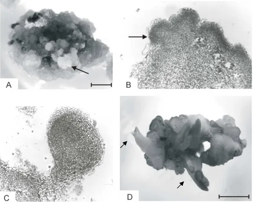
Clusters of somatic embryos were recorded on the
callus surface after 50 days of culture
(Fig.1A).Induction of somatic embryos was affected by culture medium composition and light conditions.
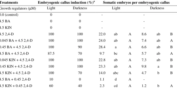
Table 1 - Effect of different types and concentrations of growth regulators on induction of embryogenic callus and somatic embryos of yacón (Smallanthus sonchifolius) in the presence or absence of light.
Treatments Embryogenic callus induction (%)a Somatic embryos per embryogenic callus
Growth regulators (µM) Light Darkness Light Darkness
0.0 (control) 0 0 - -
4.5 BA 0 0 - -
4.5 KIN 0 0 - -
4.5 2,4-D 100 100 22.0 ab A 8.6 ab B
0.045 BA + 4.5 2,4-D 100 100 24.0 ab A 7.4 ab A
0.45 BA + 4.5 2,4-D 100 90 28.4 a A 6.6 ab B
4.5 BA + 4.5 2,4-D 87.5 70 9.7 bc A 5.7 ab A
0.045 KIN + 4.5 2,4-D 100 100 22.8 ab A 7.3 ab B
0.45 KIN + 4.5 2,4-D 100 100 23.3 ab A 9.8 a B
4.5 KIN + 4.5 2,4-D 100 70 14.0 abc A 4.7 b B
4.5 BA + 0.45 2,4-D 10 0 1.1 d A -
4.5 KIN + 0.45 2,4-D 60 40 2.3 cd A 1.2 b A
a
Frequency of embryogenic callus induction and average number of somatic embryos per embryogenic callus from two independent experiments after 50 days of cultivation.
Means followed by different small letters in each column and capital letters in each row are significantly different according to
Tukey’s Test at P≤ 0.05.
The presence of light promoted somatic
embryogenesis on callus induced under various combinations of growth regulators, reaching an average of 28.4 embryos per callus, while under darkness only 9.8 somatic embryos were observed (Table 1). Requirement of light during in vitro morphogenetic processes has been proved to depend on the species tested. Somatic embryos were induced when explants were cultured either in the presence of light (Fernando et al., 2002; Martin et al., 2000; Martin, 2003) or under dark conditions (Fernando et al., 2001; Gogate et al.,
2003). Medium composition affected the
embryogenic response of yacón as well. The highest number of embryos was observed on callus induced on medium supplemented with low BA
and Kin along with 4.5 µM 2,4-D under light. In
Anacardium occidentale, BA was the most
effective growth regulator for somatic
embryogenesis (Martin, 2003), while Kin
revealed that embryogenic calli were more friable and yellowish in appearance than the non embryogenic calli. Regions with densely staining cells were observed at the peripheral layer of friable callus (Fig. 1B). Somatic embryos were observed to arise from the outermost cell layer of the callus and no vascular connection between the callus and the developing embryos was observed (Fig. 1C). Similarly, somatic embryos of Carica
papaya L. were originated from embryogenic
superficial cells located on peripheral cell layers of callus (Fernando et al., 2001). Somatic embryos induced on different treatments were transferred to
proliferation medium with 4.5 µM BA and 0.045 or 0.45 µM 2,4-D. Though the media evaluated did not result on embryo proliferation, the addition
of 0.045 µM 2,4-D led somatic embryos to
develop through globular to cotyledonary stage within 30 days (Fig. 1D). Since no embryo germination into complete plants was obtained on this medium, developed embryos were transferred to hormone-free conversion medium, containing
activated charcoal. Embryo conversion or
germination was defined as the development of embryos into plantlets with well-defined root and shoot systems
.
Figure 1 - Somatic embryogenesis and plant regeneration in yacón (Smallanthus sonchifolius). (A) Embryogenic callus induced on 0.45 µM BA + 4.5 µM 2,4-D after 50 days of culture. Arrow indicates a cluster of somatic embryos. Bar represents 2 mm. (B) Longitudinal section of embryogenic callus showing densely stained cells at peripheral layer (arrow) (100×). (C) Longitudinal section of a somatic embryo (400×) (D) Developed somatic embryo (arrows). Bar represents 3 mm.
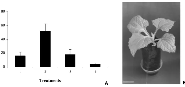
Embryos that were induced in the presence of either BA or Kin (0.045 µM and 0.45 µM) and 4.5 µM 2,4-D were able to convert into plantlets (Fig. 2A). However, the highest percentage of conversion (52%) was obtained from embryos induced on 0.45 µM BA and 4.5 µM 2,4-D and cultivated on hormone-free ½ MS conversion medium (Fig. 2A). Conversion of somatic
Figure 2 - (A) Conversion of somatic embryos of yacón (Smallanthus sonchifolius) on hormone-free MS medium supplemented with 2 g.L-1activated charcoal. Treatments refer to the culture media used for somatic embryos induction, under light conditions: (1) 0.045 µM BA + 4.5 µM 2,4-D; (2) 0.45 µM BA + 4.5 µM 2,4-D; (3) 0.045 µM KIN + 4.5 µM 2,4-D; (4) 0.45 µM KIN + 4.5 µM 2,4-D. Bars represent standard deviation. Concentration of growth regulators is expressed in µM. (B) Plant regenerated from somatic embryo and acclimated under environment conditions. Bar represents 14 cm.
Micropropagation of yacón through organogenesis has previously been reported. From stem cuttings, an average of 7.6 shoots per explant was obtained using 4.4 µM BA and 0.49 µM IBA (Estrella and Lazart, 1994). Shoot regeneration was also reported through callus induced from leaf segments (Niwa, 2002). Thus, the procedure for somatic embryogenesis allowed a pottentially higher number of regenerated plants, showing the efficiency of micropropagation.
This paper reports an efficient procedure for somatic embryo induction and plant regeneration in yacón. Somatic embryogenesis and plant regeneration were dependent on auxin and cytokinin concentration during the embryo induction phase. Future research shall refine this technique for achieving higher frequencies of embryo proliferation to allow long term culture maintenance.
ACKNOWLEDGEMENTS
Research support was provided by Fundação de Amparo à Pesquisa do Estado do Rio Grande do Sul/FAPERGS. Authors are grateful to Dr. Júlio
César Bicca-Marques for editing suggestions in the preparation of the manuscript.
RESUMO
Smallanthus sonchifolius apresenta raízes
tuberosas, que contêm grandes quantidades de frutoligosacarídeos e seu uso medicinal tem aumentado devido à propriedade hipoglicemiante relatada para esta espécie. Um sistema eficiente para propagação via embriogênese somática é
descrito, utilizando segmentos peciolares
cultivados em meio MS suplementado com diversas combinações de BA, cinetina e 2,4-D, sob condições de luz ou escuro. A maioria dos tratamentos resultou na formação de calos embriogênicos; no entanto, a embriogênese somática foi promovida em presença de luz e
agregados de embriões somáticos foram
observados na superfície dos calos após 50 dias de cultivo. O maior número de embriões foi obtido em presença de 0,45 µM BA e 4,5 µM 2,4-D. Os calos embriogênicos foram mantidos em meio MS com adição de 4,5 µM BA e 0,045 µM 2,4-D. A conversão dos embriões somáticos foi obtida em
A B
0 20 40 60 80
1 2 3 4
E
m
b
r
y
o
c
o
n
v
e
r
si
o
n
(%
)
meio MS, com a concentração de sais reduzida à metade, 2 g.L-1 de carvão ativado, sem reguladores de crescimento. As plantas regeneradas foram aclimatadas em condições ambientais, com sobrevivência de 100 %.
REFERENCES
Aybar, M. J.; Riera, A. N.S.; Grau, A. and Sanchez, S.S. (2001), Hypoglycemic effect of the water extract
of Smallanthus sonchifolius(yacón) leaves in normal
and diabetic rats. J. Ethnopharmacol., 74, 125-132. Castillo, P.; Márquez, J.; Rubluo, A.; Hernandez, G.
and Lara, M. (2000), Plant regeneration from callus and suspension cultures of Valeriana edulis ssp.
procera via simultaneous organogenesis and somatic
embryogenesis. Plant Sci., 151, 115-119.
Estrella, J. E. and Lazart, J. E. (1994), In vitro
propagation of jicama (Polymnia sonchifolius Poepp. and Endlicher): A neglected Andean crop.
HortScience, 29, 331.
Grau, A. and Rea, J. (1997), Yacon, Smallanthus
sonchifolius (Poepp. and Endl.) H. Robinson. In:
Andean roots and tubers: Ahipa, arracacha, maca yacon. Promoting the conservation and use of
underutilized and neglected crops, eds. M. Hermann
and, J. Heller. Gatersieben/International Plant Genetic Resources Institute. Rome, pp. 199-242. Available at: http://www.cipotato.org/artc/yacon.asp Accessed at Apr, 27th, 2007.
Fernando, J.A.; Melo, M.; Soares, M.K.M. and Appezzato-da-Glória, B. (2001), Anatomy of somatic embryogenesis in Carica papaya L. Braz. arch. biol.
technol., 44, 247-255.
Fernando, J.A.; Vieira, M.L.C.; Gerardi, I.O. and Appezzato-da-Glória, B. (2002), Anatomical study of somatic embryogenesis in Glycine max (L.) Merrill.
Braz. arch. biol. technol., 45, 277-286.
Fowler, J. and Cohen, L. (1990), Practical statistics for
field biology. Open University Press, Buckingham.
George, E.F. (1993), Plant propagation by tissue
culture. Part 1. The Technology. 2ed. Exegetics
Limited, England.
Gogate, S.S. and Nadgauda, R.S. (2003), Direct induction of somatic embryogenesis from immatue zygotic embryo of cashewnut (Anacardim
occidentale L.). Sci. Hort., 97, 75-82.
Martin, K.P. (2003), Plant regeneration through direct somatic embryogenesis on seed coat explants of cashew (Anacardium occidentale L.). Sci. Hort., 98, 299-304.
Martin, A. B.; Cuadrado, Y.; Guerra, H.; Gallego, P.; Hita, O.; Martin, L.; Dorado, A. and Villalobos, N. (2000), Differences in the contents of total sugars, reducing sugars, starch and sucrose in embryogenic and non-embryogenic calli from Medicago arborea
L. Plant Sci., 154, 143-151.
Murashige, T. and Skoog, F. (1962), A revised medium for rapid growth and bioassays with tobacco tissue culture. Physiol. Plant., 15, 473-497.
Niwa, M. (2002), Plant regeneration through leaf culture of yacon. J. Japan. Soc. Hort. Sci., 71, 561-567.
Rout, G.R.; Samantaray, S. and Das, P. (2000), In vitro
somatic embryogenesis from callus cultures of
Cephaelis ipecacuanha A. Richard. Sci. Hort., 86,
71-79.
Valentova, K.; Moncion, A.; de Waziers, I. and Ulrichova, J (2005), The effect of Smallanthus
sonchifolius leaf extracts on rat hepatic metabolism.
Cell Biol. Toxicol., 20, 109-120.
Te-chato, S. and Rungnoi, O. (2000), Induction of somatic embryogenesis from leaves of Sadao Chang
(Azadirachta excelsa (Jack) Jacobs). Sci. Hort., 86,
311-321.
Zardini, E. (1991), Ethnobotanical notes on yacón,
Polymnia sonchifolia (Asteraceae). Econ. Bot., 45,
72-85.