AIMS Microbiology, 3(4): 960-975. DOI: 10.3934/microbiol.2017.4.960 Received: 02 October 2017
Accepted: 05 December 2017 Published: 14 December 2017 http://www.aimspress.com/journal/microbiology
Research article
Assessment of occupational exposure to azole resistant fungi in 10
Portuguese bakeries
Liliana Aranha Caetano 1,2,*, Tiago Faria 1, Ana Crespo Batista 1, Susana Viegas 1,3, and Carla Viegas 1,3
1
Environment and Health Research Group (GIAS) Escola Superior de Tecnologia da Saúde de Lisboa, ESTeSL, Instituto Politécnico de Lisboa, Lisbon, Portugal
2
Research Institute for Medicines (iMed.ULisboa), Faculty of Pharmacy, University of Lisbon, Lisbon, Portugal
3
Centro de Investigação em Saúde Pública Escola Nacional de Saúde Pública, Universidade Nova de Lisboa, Lisbon, Portugal
* Correspondence: Email: liliana.caetano@estesl.ipl.pt; Tel: +351-218-980-458.
AIMS Microbiology Volume 3, Issue 4, 960-975.
to mycobiota, and highlights the importance of studying the prevalence of azole-resistant strains in specific occupational environments.
Keywords: azole-resistance; occupational exposure; bakeries; fungi; Aspergillus; Mucorales
1. Introduction
Handling of flour dust and raw materials in the food industry can be associated with health problems as raw materials may be associated with allergen sensitization and fungal colonization [1–6]. Several reports on the relation between fungi levels in diverse occupational environments and health effects [7–15] corroborate that fungi are potential occupational health hazards that should be taken into account in risk assessment strategies in occupational settings, including bakeries.
Exposure to flour dust and related bioaerosols in the bakery industry is described to occur mainly during grinding, sifting and mixing operations [16]. When mixing occurs, abundant organic dust particles originating from flour dust disperse into the air and are suspended for a long time before deposited on the floor due to gravitational sedimentation. Consequently, high spreading of fungi and their spores and metabolites, such as volatile organic compounds and mycotoxins, will also probably be suspended [17].
Raw materials used in bakeries consists of finely milled cereal or grains (e.g., wheat, rye, barley, oats, rice, malt, carob, corn) and additional non-cereal ingredients (e.g., enzymes, antioxidants, flavorings and spices, baker’s yeast, sugar powder) that are used for dough improvement [16]. Some of these raw materials are ideal microbial growth substrates and can generate elevated levels of bioaerosols [18,19]. The genus Aspergillus, including A. fumigatus, is ubiquitous in nature and one of the most prevalent in crops and cereals such as corn, wheat, barley, oat, rice, and sorghum [20].
Aspergillus disease affects a broad patient population, from patients with asthma to immunocompromised patients [21]. Invasive fungal diseases, such as aspergillosis, are still a life-threatening complication for immunocompromised patients [22]. Azole drugs are critical in long-term therapy for chronic pulmonary aspergillosis, as they are the only anti-Aspergillus agents orally available. This class includes itraconazole (available for clinical use since 1997), voriconazole (since 2002), posaconazole (since 2006), and, most recently, isavuconazole [23,24]. However, azole resistance has been increasingly reported in both clinical and environmental Aspergillus strains [25–29].
Until now, no data regarding exposure to bioaerosols nor azole-resistance distribution in bakeries have been reported for Portugal, and this omission has delayed the application of preventive measures for the protection of workers health. Therefore, the aim of this study was to assess fungal contamination in ten bakeries in Portugal and to determine the prevalence of azole-resistant fungal species in this occupational setting.
2. Materials and Method
This study was conducted between May 2016 and June 2017 in 10 Portuguese bakeries located in the Lisbon district and is part of an enlarged exploratory study with financial support from the Portuguese Authority for Working Conditions aiming to characterize occupational exposure to fungi and particles on Portuguese bakeries. While being part of a larger study in which additional environmental characterization was carried out, this paper presents the preliminary results regarding environmental samples collected by passive methods in which azole-resistance monitoring was performed.
2.1. Bakeries characteristics
Most bakeries were organized in three different areas: Production—where kneading machines and ovens were located and where dough shaping was performed; Raw material warehouse—where workers collected the raw materials for dough preparation for several times during process; Store—where final product was sold (bread or pastry). In one bakery with no store a distinct area was characterized: Expedition—where distribution of final product for other units occurs. One bakery was dedicated to pastry. The sampling sites and collection periods for each bakery were determined based on the high amount of time spent by workers on those places or dislocation frequency during their occupational activity. In these settings environmental samples (settled dust and electrostatic dust cloth) and several raw materials were collected for the assessment of fungal burden and screening of azole resistance.
2.2. Environmental and raw material samples
In total, 34 environmental samples and 26 raw material samples were collected and analysed by culture-based methods (Table 1).
One settled dust sample in each bakery unit (7/10) was collected, by collecting the floor dust into a sterilized bag. After sampling, 4.4 g of the collected floor settled dust were weighted and extracted with 40 mL of distilled water for 20 minutes at 200 rpm, as previously described [40–43].
AIMS Microbiology Volume 3, Issue 4, 960-975.
10 EDCs weighted before sampling was subtracted. Dust from EDC cloths was extracted with 20 mL 0.9% NaCl with 0.05% Tween80™ by orbital shaking (250 rpm, 60 minutes, at room temperature [40].
Table 1. Type and number of samples collected in ten bakeries.
Bakery Settled dust EDC Raw material
1 NA 2 NA
2 NA 3 NA
3 1 2 NA
4 1 2 7
5 1 3 5
6 1 3 5
7 1 3 4
8 1 3 4
9 1 3 NA
10 NA 3 NA
n = 7 n = 27 n = 26 NA: not applicable.
Twenty six samples of bread/pastry raw material, including different types of flour, sugar and/or spices, were collected (4 to 7 samples per unit) from half of the bakeries evaluated in this study (5 out of 10 units) and prepared as follows: 4.4 g of raw material was weighted and washed with 40 mL of distilled water for 20 minutes at 200 rpm [40–43].
2.3. Culture-based methods and screening of azole-resistance
The fungal burden was determined in environmental and raw material samples through the inoculation of 150 µL of the wash suspension on 2% malt extract agar (MEA) supplemented with chloramphenicol (0.05%) and dichloran glycerol (DG18). DG18 was used due to its ability to restrict the colony size of fast-growing genera [47], allowing a more complete characterization of fungal growth in complex matrices such as environmental and substrate samples. The prevalence of azole-resistance was determined in all the collected samples using azole-supplemented media by seeding 150 µL of the wash suspension on Saboraud agar supplemented with 4 mg/L itraconazole, 1 mg/L voriconazole, or 0.5 mg/L posaconazole, according to the EUCAST guidelines [48]. All of the collected samples were incubated at 27 °C for 5–7 days, in order to allow the growth of all fungal species present in the samples.
2.4. Fungal contamination characterization
lactophenol cotton blue mount procedures. Morphological identification was achieved through macro and microscopic characteristics as noted by De Hoog et al. [49].
2.5. Data analysis
The data analysis was performed using univariate descriptive statistics using frequency (n; %), median and graphical representations appropriate to the nature of the data.
3. Results
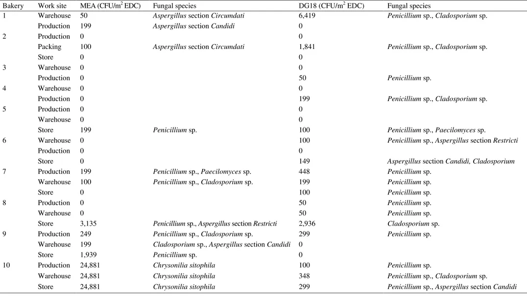
Seven different fungal species were detected in EDC samples from all analyzed bakery units (Figure 1). Considering MEA and DG18 combined (Table 2), Chrysonilia sitophila was the predominant species (79.3%), followed by Penicillium sp. (12.1%) and Cladosporium sp.(7.8%). In addition, Aspergillus sp. (0.8%) and Paecilomyces sp. (0.1%) were also isolated. Among Aspergillus genera, four different species were isolated belonging to three different sections, namely, Candidi (0.5%), Circumdati (0.2%) and Restricti (0.1%) (Table 3). Regarding fungal load distribution among work areas (Table 3), higher fungal counts (CFU/m2) were determined either in store/expedition (units nº 5, 6, 8 and 9), production (units nº 3, 4 and 7), or warehouse/packing areas (units nº 1, 2 and 10), with Penicillium sp. and Cladosporium sp. being the most prevalent species, with one exception (unit nº 10, presenting countless CFU/m2 of Chrysonilia sitophila at all sampling sites). No fungal growth was detected in settled dust samples.
Table 2. Fungal distribution in EDC and raw material samples (fungal count for MEA and DG18 combined).
Fungal species EDC (CFU/m2 EDC) (n; %)
Fungal species Raw material (CFU/g) (n; %)
Chrysonilia sitophila 74,642; 79.3 Penicillium sp. 14; 63.6
Penicillium sp. 11,346; 12.1 Aspergillus sp. 6; 27.3
Cladosporium sp. 7,315; 7.8 Mucoralesorder 2; 9.1
Aspergillus sp. 746; 0.8
Paecilomyces sp. 100; 0.1
CFU were calculated as follows: (n) = (CFU in MEA + CFU in DG18); (%) = (CFU in MEA + CFU in DG18)/(total CFU in MEA + total CFU in DG18) × 100
AIMS Microbiology Volume 3, Issue 4, 960-975.
Table 3.Fungal distribution in different work areas assessed by EDC.
Bakery Work site MEA (CFU/m2 EDC) Fungal species DG18 (CFU/m2 EDC) Fungal species
1 Warehouse 50 Aspergillus section Circumdati 6,419 Penicillium sp., Cladosporium sp. Production 199 Aspergillus section Candidi 0
2 Production 0 0
Packing 100 Aspergillus section Circumdati 1,841 Penicillium sp., Cladosporium sp.
Store 0 0
3 Warehouse 0 0
Production 0 50 Penicillium sp.
4 Warehouse 0 0
Production 0 199 Penicillium sp., Cladosporium sp.
5 Production 0 0
Warehouse 0 0
Store 199 Penicillium sp. 100 Penicillium sp., Paecilomyces sp.
6 Warehouse 0 100 Penicillium sp., Aspergillus section Restricti
Production 0 0
Store 0 149 Aspergillus section Candidi, Cladosporium
7 Production 199 Penicillium sp., Paecilomyces sp. 448 Penicillium sp. Warehouse 100 Penicillium sp., Cladosporium sp. 199 Penicillium sp.
Store 0 100 Penicillium sp.
8 Production 0 50 Penicillium sp.
Warehouse 0 50 Penicillium sp.
Store 3,135 Penicillium sp., Aspergillus section Restricti 2,936 Cladosporium sp. 9 Production 249 Penicillium sp., Cladosporium sp. 299 Penicillium sp.
Warehouse 199 Cladosporium sp., Aspergillus section Candidi 0
Store 1,939 Penicillium sp. 0
10 Production 24,881 Chrysonilia sitophila 100 Penicillium sp.
Warehouse 24,881 Chrysonilia sitophila 348 Penicillium sp., Cladosporium sp.
Figure 1. Fungal load in EDC after inoculation onto MEA and DG18 media.
Table 4. Azole-resistant fungal species distribution after EDC inoculation onto azole-supplemented Saboraud media.
EDC (CFU/m2 EDC) (n; %)
Fungal species 4 mg/L ITC 1 mg/L VRC 0.05 mg/L PSC Total
Chrysonilia sitophila 0; 0 49,761; 65.8 0; 0 49,761; 64.8
Rhizopus sp. 0; 0 24,930; 33.0 0; 0 24,930; 32.5
Cladosporium sp. 498; 71.4 249; 0.3 249; 55.6 995; 1.3
Penicillium sp. 100; 14.3 398; 0.5 149; 33.3 647; 0.8
Chrysosporium sp. 0; 0 100; 0.1 50; 11.1 149; 0.2
Aureobasidium sp. 50; 7.1 50; 0.1 0; 0 100; 0.1
Aspergillus section Circumdati 0; 0 50; 0.1 0; 0 50; 0.1
Paecilomyces sp. 50; 7.1 0; 0 0; 0 50; 0.1
Chrysonilia sp. 0; 0 50; 0.1 0; 0 50; 0.1
Alternaria sp. 0; 0 50; 0.1 0; 0 50; 0.1
ITC, itraconazole; VRC, voriconazole; PSC, posaconazole; N, number of species isolates; %, number of species isolates per total of resistant isolates.
0 20000 40000 60000 80000 C. sitophila
Penicillium sp. Aspergillus section Candidi Cladosporium sp. Aspergillus section Circumdati Aspergillus section Restricti Paecilomyces sp.
CFU/m2
MEA
0 2000 4000 6000 8000
Cladosporium sp. Penicillium sp. Aspergillus section Candidi Aspergillus section Restricti Paecilomyces sp.
CFU/m2
AIMS Microbiology Volume 3, Issue 4, 960-975.
Regarding raw material samples, six different groups of fungal species were isolated (Figure 2). Considering MEA and DG18 combined (Table 2), Penicillium sp. was the most prevalent genera (63.6%), followed by Aspergillus sp. (27.3%) and Mucorales group (9.1%). This fungal contamination was present in 27% (7/26) of the raw material samples collected in four of the five assessed bakeries (Table 5). Among Aspergillus genera, four different species were isolated belonging to the sections Versicolores (18.2%), Candidi (4.5%) and Circumdati (4.5%). Among Mucorales, the isolated species were Mucor sp. (4.5%), and Syncephalastrum racemosum (4.5%). Two azole-resistant fungal species were identified in two distinct raw materials, namely Chrysosporium sp. (1 CFU/g) not susceptible to 4 mg/L itraconazole, and Mucor sp. (1 CFU/g) not susceptible to 1 mg/L voriconazole (Table 6). No azole-resistant Aspergillus species were identified in raw material samples.
Table 5. Fungal distribution in raw materials (n = 26) collected at five bakeries (CFU/g).
Raw material ID* (Code)
MEA (CFU/g)
Fungal species DG18 (CFU/g)
Fungal species
4A 0 0
4B 0 0
4C 0 0
4D 1 Aspergillus section Versicolores 0
4E 0 0
4F 0 0
4G 0 0
5E 0 0
5B 0 0
5G 1 Aspergillus section Versicolores 0
5C 0 0
5F 3 Aspergillus section Versicolores,
Mucor sp.
1 Penicillium sp.
6H 0 0
6I 0 13 Penicillium sp.
6J 0 0
6K 0 1 Syncephalastrum racemosum
6L 0 1 Aspergillus section Candidi
7M 0 0
7N 0 1 Aspergillus section Circumdati
7O 0 0
7P 0 0
8B 0 0
8F 0 0
8Q 0 0
8G 0 0
8E 0 0
Figure 2. Fungal load in raw material after inoculation onto MEA and DG18 media.
Table 6. Azole-resistant fungal species distribution after raw material inoculation onto azole-supplemented Saboraud media.
Raw material (CFU/g) (n; %)
Fungal species 4 mg/L ITC 1 mg/L VRC 0.05 mg/L PSC Total
Chrysosporium sp. 1; 100 1; 50
Mucor sp. 1; 100 1; 50
ITC, itraconazole; VRC, voriconazole; PSC, posaconazole.
4. Discussion
This is the first study in Portugal determining the fungal load and prevalence of antifungal-resistant species in bakeries. Overall, with the exception of Chrysonilia sitophila for one bakery, the most prevalent fungi isolated in both media were Cladosporium sp. and Penicillium sp.. Other species with recognized toxigenic potential belonging to the genus Aspergillus were also isolated both in the environment and in raw materials.
Exposure to bioaerosols in bakeries may potentially place workers at higher health risk, since exposure to high levels of flour dust potentiates the exposure to airborne microorganisms, which may reach infectious levels within a confined space more readily [3,7,16]. In our study, the highest fungal load was found in the production area (1,793 CFU/m2 EDC, in MEA and DG18), followed by the warehouse (1,052 CFU/m2 EDC, in MEA and DG18) and the store/packing area (957 CFU/m2 EDC, in MEA and DG18). Previous studies identified being near the kneading machines during ingredients mixing as the task with higher values for the smallest particles [15,16]. One possible explanation for
0 1 2 3 4 5
Aspergillus section Versicolores
Mucor sp.
CFU/g
MEA
0 5 10 15
Penicillium sp. Aspergillus section Candidi Aspergillus section
Circumdati Syncephalastrum
racemosum
CFU/g
AIMS Microbiology Volume 3, Issue 4, 960-975.
this might be related with the use of open machines for mixing without localized exhaustion [15], which is of most importance when, as was the case in the present study, none of the workers used respiratory protection devices.
In the assessment of EDC fungal contamination, different results were obtained using different culture media regarding both fungal load and mycobiota diversity. As an example, the isolates from Chrysonilia sitophila were only identified on MEA media. This is due to the ability of DG18 to restrict the colony size of fast-growing genera, such as Chrysonilia sitophila, allowing a different and a more complete characterization of fungal contamination. Noteworthy, the use of both media allowed to identify toxigenic genus Penicillium and Aspergillus (sections Candidi, Circumdati and Versicolores) in the analyzed samples, unveiling a common scenario of potential co-exposure to more than one risk factor—mycobiota and mycotoxins (e.g., aflatoxin) in the baking industry.
Concerning the resistance prevalence in the assessed bakeries, eleven different azole-resistant species, including isolates identified as Aspergillus sp. and Mucor sp., were detected in the environment and in raw materials. Of note, the number of azole-resistant isolates belonging to the genus Aspergillus may be underestimated, in general and in our study, due to the dominance of other genera in the azole-supplemented media with faster growth rates [50]. Aspergillus growth restriction can be circumvented by using higher incubation temperatures, as most Aspergilli are highly thermotolerant. However, since the evaluation of occupational exposure aims to characterize the complete bioburden, not only Aspergillus genera, conventional incubation temperatures were used. The identification of Mucor sp. in raw materials is also of concern because invasive fungal diseases due to Mucorales are increasing [34,35,36]. Belonging to the order Mucorales, Mucor is one of the most commonly identified human pathogenic genera in Europe [51]. Mucorales are not susceptible to voriconazole, the first-line antifungal drug for invasive aspergillosis. The dominant and fast growth of these species in voriconazole screening media may hinder the presence of Aspergillus and other species [52].
Although most isolates were not susceptible to 1 mg/L voriconazole only, in four EDC from distinct bakery units three other genus (Penicilliumsp., Cladosporium sp., Aureobasidium sp.) were identified as not susceptible to more than one azole. Azole-resistant Penicillium and Cladosporium spp. were previously reported in clinical isolates for itraconazole and voriconazole [53]. High MICs of voriconazole and isavuconazole were also reported in vitro for Aureobasidium pullulans in both clinical and environmental isolates [54]. Intrinsic resistance to available antifungals reported in some
fungi such as Fusarium, Rhizopus, Rhizomucor and Scedosporium spp. has been pointed out as a
major issue by Alhanout and colleagues [55]. However, little is known regarding intrinsic
azole-resistance of Penicillium sp., Cladosporium sp., and Aureobasidium sp. In fact, data on intrinsic resistance to azoles are still very limited for non-A. fumigatus fungal species. One known example is the intrinsic resistance of Aspergillus section Terrei to itraconazole [56]. The fact that overall reported MIC-distributions include only a limited number of clinical isolates for most non-A. section Fumigati species compared to A. section Fumigati hinders our ability to distinguish in vitro susceptibility at species level. Therefore, molecular identification remains important to gain more insight into the efficacy of antifungal agents [55].
administration in animals [58]. Also Aspergillus section Circumdati, and Penicillium species can produce ochratoxin A, an hazard for human health due to its carcinogenic, nephrotoxic, hepatotoxic, immunotoxic, and teratogenic effects in animals [59]. One possible measure to reduce fungal burden in this setting would be the use of cleaning products containing fungicides. However, several antifungal substances used as pesticides have been described as potential inducers of azole resistance in environmental Aspergillus section Fumigati species. This is due to the fact that fungicides present similar structures to the molecules of clinical azoles [60,61]. Therefore, it is important to characterize the setting in relation to the prevalence of antifungal resistant species in order to determine which specific biocidals can be used [39,62].
While the emergence of drug-resistant bacteria such as methicillin-resistant Staphylococcus aureus and extensively drug-resistant Mycobacterium tuberculosis are already under surveillance policies, it was not until recently that the global problem of antifungal resistance has been recognized as an issue [39]. The increasing occurrence of cryptic species, often drug resistant, as well as of emerging species that are resistant to all antifungal classes [63] illustrates the importance of molecular biology techniques in association with culture-based methodologies [64] for the assessment of occupational exposure to mycobiota [65,66,67], as well as for the correct identification of Aspergillus species of the section Fumigati [68]. This study also corroborates the importance of passive methods (EDC, settled dust and raw material) to complement the exposure assessment. The use of EDC adds information regarding the cumulative presence of bioaerosols in the environment (as they are placed at 1.5 m height and stay in place for 15 days), needing, however, an integrated analysis from the obtained data. It should be pointed out that the main advantage from passive methods is that they can collect contamination from a larger period of time (weeks to several months), whereas air samples can only reflect the load from a shorter period of time (mostly minutes) [69]. Further molecular analyses will be performed in future studies to Aspergillus isolates to support a wider project aiming to characterize the prevalence and distribution of Aspergillus genera and Mucorales order in different Portuguese occupational environments.
Global warming is increasing the prevalence of crop fungal pathogens, and may also increase the prevalence of fungal disease in humans as fungi adapt to survive in warmer temperatures [70]. It is, therefore, of the outmost importance to perform surveillance studies both in clinical settings and in the environment, including the characterization of azole-resistance prevalence in specific environment compartments (water, soil) and in occupational settings where high fungal load and azole pressure might be expected [67,71,72]. International and collaborative efforts are required to understand how resistance develops in the environment to allow effective measures to be implemented aimed at retaining the use of azoles both for food production and human medicine.
5. Conclusion
AIMS Microbiology Volume 3, Issue 4, 960-975.
and screening methods. In order to improve the assessment of occupational exposure to mycobiota and antifungal resistance, both culture-based and molecular methods should be used.
Acknowledgments
The authors are grateful to Portuguese Authority for Working Conditions for funding the Project ―Occupational exposure assessment to particulate matter and fungi and health effects of workers from Portuguese Bakeries‖ (005DBB/12) and also to Occupational Health Services from the Bakeries engaged in this study.
Conflict of Interest
All authors declare no conflicts of interest in this paper.
References
1. Blancocarmona JG, Picón SJ, Sotillos MG (1991) Occupational asthma in bakeries caused by sensitivity to alpha-amylase. Allergy 46: 274–276.
2. Burdorf A, Lillienberg L, Brisman J (1994) Characterization of exposure to inhalable flour dust in Swedish bakeries. Ann Occup Hyg 38: 67–78.
3. Lilienberg L, Brisman J (1996) Peak exposure concentrations of dust in bakeries, In: Proceeding of the 2nd International Symposium on Modern Principles of Air Monitoring, Feb 5–8, 1996, Salen, Sweden: 47.
4. Sander I, Flagge A, Mergret R, et al. (2001) Identification of wheat flout allergens by means of 2-dimensional immunoblotting. Allergy Clin Immunol 107: 907–913.
5. Bush RK, Portnoy JM, Saxon A, et al. (2006) The medical effects of mold exposure. J Allergy Clin Immunol 117: 326–333.
6. Salcedo G, Quirce S, Diaz-Perales A (2011) Wheat allergens associated with baker’s asthma. J Invest Allergy Clin 21: 81–92.
7. Cullinan P, Cook A, Nieuwenhuijsen MJ, et al. (2001) Allergen and dust exposure as determinants of work-related symptoms and sensitization in a cohort of flour-exposed workers; a case-control analysis. Ann Occup Hyg 45: 97–103.
8. Brandl H (2011) Bioaerosols in indoor environment—A review with special reference to residential and occupational locations. Open Environ Biol Monit J 4: 83–96.
9. Sahlberg B, Gunnbjörnsdottir M, Soon A, et al. (2013) Airborne molds and bacteria, microbial volatile organic compounds (MVOC), plasticizers and formaldehyde in dwellings in three North European cities in relation to sick building syndrome (SBS). Sci Total Environ 444: 433–440. 10.Viegas C, Carolino E, Sabino R, et al. (2013) Fungal contamination in swine: A potential
occupational health threat. J Toxicol Env Heal A 76: 272–280.
11.Viegas C, Faria T, dos Santos M, et al. (2015) Fungal burden in waste industry: an occupational risk to be solved. Environ Monit Assess 187: 199.
13.Viegas C, Faria T, Meneses M, et al. (2016) Analysis of surfaces for characterization of fungal burden—Does it matter? Int J Occup Med Environ Health 29: 623–632.
14.Viegas S, Caetano L, Korkalainen M, et al. (2017) Cytotoxic and inflammatory potential of air samples from occupational settings with exposure to organic dust. Toxics 5: 8.
15.Viegas S, Faria T, Viegas C (2017) Bakers exposure to flour dust—a exploratory study in a Portuguese Bakery. International Symposium on Occupational Safety and Hygiene SHO 2017. 16.Stobnicka A, Górny RL (2015) Exposure to flour dust in the occupational environment. Int J
Occup Saf Ergon 21: 241–249.
17.Tsapko V, Chudnovets A, Sterenbogen M, et al. (2011) Exposure to bioaerosols in the selected agricultural facilities of the Ukraine and Poland—A review. Ann Agr Env Med 18: 19–27.
18.Tiikkainen U, Louhelainen K, Nordman H (1996) The Nordic expert group for criteria documentation of health risks from chemicals 120. Flour Dust. Arbete Hälsa 27.
19.Mohammadien HA, Hussein MT, El-Sokkary RT (2013) Effects of exposure to flour dust on respiratory symptoms and pulmonary function of mill workers. Egypt J Chest Dis Tuberc 62: 745–753.
20.Gisi U (2013) Assessment of selection and resistance risk for DMI fungicides in Aspergillus fumigatus in agriculture and medicine: a critical review. Pest Manag Sci 70: 352–364.
21.Vermeulen E, Maertens J, De Bel A, et al. (2015) Nationwide surveillance of azole resistance in Aspergillus diseases. Antimicrob Agents Ch 59: 4569–4576.
22.Meletiadis J, Roilides E (2013) Rare invasive fungal infections: Epidemiology, diagnosis and management. Curr Fungal Infect Rep 7: 351–360.
23.Walsh TJ, Anaissie EJ, Denning DW, et al. (2008) Treatment of aspergillosis: Clinical practice guidelines of the infectious diseases society of America. Clin Infect Dis 46: 327–360.
24.Kontoyiannis DP (2012) Invasive mycoses: Strategies for effective management. Am J Med 125: S25–S38.
25.Howard SJ, Webster I, Moore CB, et al. (2006) Multi-azole resistance in Aspergillus fumigatus. Int J Antimicrob Agents 28: 450–453.
26.Lelièvre L, Groh M, Angebault C, et al. (2013) Azole resistant Aspergillus fumigatus: An emerging problem. Med Maladies Infect 43: 139–145.
27.Abdolrasouli A, Rhodes J, Beale M, et al (2015) Genomic context of Azole-resistance mutations in Aspergillus fumigatus using whole-genome sequencing. Mbio 6: 1–11.
28.Verweij PE, Chowdhary A, Melchers WJG, et al. (2016) Azole resistance in Aspergillus fumigatus: Can we retain the clinical use of mold-active antifungal Azoles? Clin Infect Dis 62: 362–368.
29.Zhang J, Snelders E, Zwaan BJ, et al. (2017) A novel environmental Azole resistance mutation in Aspergillus fumigatus and a possible role of sexual reproduction in its emergence. Mbio 8: e00791-17.
30.Verweij PE, Snelders E, Kema GH, et al. (2009) Azole resistance in Aspergillus fumigatus: a side-effect of environmental fungicide use? Lancet Infect Dis 9: 789–795.
31.Howard SJ, Cerar D, Anderson MJ, et al. (2009) Frequency and evolution of Azole resistance in Aspergillus fumigatus associated with treatment failure. Emerg Infect Dis 15: 1068.
AIMS Microbiology Volume 3, Issue 4, 960-975.
33.Arendrup MC (2014) Update on antifungal resistance in Aspergillus and Candida. Clin Microbiol Infect Suppl 6: 42–48.
34.Kontoyiannis DP, Lionakis MS, Lewis RE, et al. (2005) Zygomycosis in a tertiary-care cancer center in the era of Aspergillus-active antifungal therapy: a case-control observational study of 27 recent cases. J Infect Dis 191: 1350–1360.
35.Bitar D, Van Cauteren D, Lanternier F, et al. (2009) Increasing incidence of zygomycosis (mucormycosis), France, 1997–2006. Emerg Infect Dis 15: 1395–1401.
36.Auberger J, Lass-Florl C, Aigner M, et al. (2012) Invasive fungal breakthrough infections, fungal colonization and emergence of resistant strains in high-risk patients receiving antifungal prophylaxis with posaconazole: real-life data from a single-centre institutional retrospective observational study. J Antimicrob Chemoth 67: 2268–2273.
37.Odds FC, Brown AJ, Gow NA (2003) Antifungal agents: mechanisms of action. Trends Microbiol 11: 272–279.
38.Springer J, Goldenberger D, Schmidt F, et al. (2016) Development and application of two independent real-time PCR assays to detect clinically relevant Mucorales species. J Med Microbiol 65: 227–234.
39.Perlin DS, Rautemaa-Richardson R, Alastruey-Izquierdo A (2017) The global problem of antifungal resistance: Prevalence, mechanisms, and management. Lancet Infect Dis.
40.Madsen AM, Matthiesen CB, Frederiksen MW, et al. (2012) Sampling, extraction and measurement of bacteria, endotoxin, fungi and inflammatory potential of settling indoor dust. J Environ Monitor 14: 3230–3239.
41.Viegas C, Faria T, Caetano LA, et al. (2017) Fungal contamination in green coffee beans samples: a public health concern. J Toxicol Env Health A 80: 719–728.
42.ISO (2008) Microbiology of food and animal feeding stuffs: Horizontal method for the enumeration of yeasts and moulds. Part 1: Colony count technique in products with water activity greater than 0.95.
43.ISO (2008) Microbiology of food and animal feeding stuffs: Horizontal method for the enumeration of yeasts and moulds. Part 2: Colony count technique in products with water activity less than or equal to 0.95.
44.Cozen W, Avol E, Diaz-Sanchez D, et al. (2008) Use of an electrostatic dust cloth for self-administered home allergen collection. Twin Res Hum Genet 11: 150–155.
45.Viegas C, Ramalho I, Alves M, et al. (2017) Electrostatic dust cloth: A new sampling method for occupational exposure to bioaerosols. In: Arezes P, editor, International Symposium on Occupational Safety and Hygiene, Guimarães: SPOSHO, 39–41.
46.Kilburg-Basnyat B, Metwali N, Thorne PS (2016) Performance of electrostatic dust collectors (EDCs) for endotoxin assessment in homes: effect of mailing, placement, heating and electrostatic charge. J Occup Environ Hyg 13: 85–93.
47.Bergwall C, Stehn B (2002) Comparison of selective mycological agar media for the isolation and enumeration of xerophilic moulds and osmotolerant yeasts in granulated white sugar. Zuckerindustrie 127: 259–264.
49.Hoog C, Guarro J, Gené G, et al. (2000) Atlas of clinical fungi, Centraalbureau voor Schimmelcultures, Utrecht, the Netherlands.
50.Degois J, Clerc F, Simon X, et al. (2017) First metagenomic survey of the microbial diversity in bioaerosols emitted in waste sorting plants. Ann Work Expo Health 61: 1076–1086.
51.Lanternier F, Dannaoui E, Morizot G, et al. (2012) A global analysis of mucormycosis in France: the RetroZygo Study (2005–2007). Clin Infect Dis 54: S35–S43.
52.Springer J, Lackner M, Ensinger C, et al. (2016) Clinical evaluation of Mucorales-specific real-time PCR assay in tissue and serum samples. J Med Microbiol 65: 1414–1421.
53.Cuenca-Estrella M, Gomez-Lopez A, Mellado E, et al. (2006) Head-to-head comparison of the activities of currently available antifungal agents against 3,378 Spanish clinical isolates of yeasts and filamentous fungi. Antimicrob Agents Ch 50: 917–921.
54.Najafzadeh MJ, Sutton DA, Keisari MS, et al. (2014) In vitro activities of eight antifungal drugs against 104 environmental and clinical isolates of Aureobasidium pullulans. Antimicrob Agents Ch 58: 5629–5631.
55.Alhanout K, Brunel JM, Ranque S, et al. (2010) In vitro antifungal activity of aminosterols
against moulds isolated from cystic fibrosis patients. J Antimicrob Chemoth 65: 1307–1309.
56.Pound MW, Townsend ML, Dimondi V, et al. (2011) Overview of treatment options for invasive fungal infections. Med Mycol 49: 561–580.
57.Despot DJ, Kocsubé S, Bencsik O, et al. (2017) New sterigmatocystin-producing species of Aspergillus section Versicolores from indoor air in Croatia. Mycol Prog 16: 63–72.
58.EFSA Panel on Contaminants in the Food Chain (CONTAM) (2013) Scientific Opinion on the risk for public and animal health related to the presence of sterigmatocystin in food and feed. EFSA J 11: 3254
59.Ali N, Blaszkewicz M, Manirujjaman M, et al. (2014) Biomonitoring of ochratoxin A in blood plasma and exposure assessment of adult students in Bangladesh. Mol Nutr Food Res 58: 2219– 2225.
60.Kano R, Kohata E, Tateishi A, et al. (2014) Does farm fungicide use induce azole resistance in Aspergillus fumigatus? Med Mycol 53: 174–177.
61.Jeanvoine A, Rocchi S, Reboux G, et al. (2017) Azole-resistant Aspergillus fumigatus in sawmills of Eastern France. J Appl Microbiol 123: 172–184.
62.Snelders E, Huis In’t Veld RA, Rijs AJ, et al. (2009) Possible environmental origin of resistance of Aspergillus fumigatus to medical triazoles. Appl Environ Microb 75: 4053–4057.
63.Denning DW, Bowyer P (2013) Voriconazole resistance in Aspergillus fumigatus: Should we be concerned? Clin Infect Dis 57: 521–523.
64.Eduard W, Halstensen A (2009) Quantitative exposure assessment of organic dust. SJWEH Supplements 7: 30–35.
65.Viegas C, Malta-Vacas J, Sabino R (2012) Molecular biology versus conventional methods— complementary methodologies to understand occupational exposure to fungi. International Symposium on Occupational Safety and Hygiene SHO 2012.
AIMS Microbiology Volume 3, Issue 4, 960-975.
67.Viegas C, Faria T, Caetano LA, et al. (2017) Aspergillus spp. prevalence in different Portuguese occupational environments: what is the real scenario in high load settings? J Occup Environ Hyg 14: 771–785.
68.Lamoth F (2016) Aspergillus fumigatus–Related species in clinical practice. Front Microbiol 7: 683.
69.Viegas C, Faria T, dos Santos M, et al. (2015) Fungal burden in waste industry: an occupational risk to be solved. Environ Monit Assess 187: 199.
70.Garcia-Solache MA, Casadevall A (2010) Global warming will bring new fungal diseases for mammals. Mbio 1: e00061-10.
71.Vermeulen E, Maertens J, De Bel A, et al. (2015) Nationwide surveillance of azole resistance in Aspergillus diseases. Antimicrob Agents Ch 59: 4569–4576.
72.Fairlamb AH, Gow NA, Matthews KR, et al. (2017) Stop neglecting fungi. Nat Microbiol 2: 17120.