Full Length Research Paper
In vitro cytotoxic effects and apoptosis induction by a
methanol leaf extract of carob tree (Ceratonia siliqua L.)
Luísa Custódio
1*, Ana Luísa Escapa
1, Eliana Fernandes
1, Alba Fajardo
2, Rosa Aligué
2,
Fernando Alberício
3,4,5,
Nuno Neng
6, José Manuel Florêncio Nogueira
6and Anabela Romano
11
Institute for Biotechnology and Bioengineering, Centre of Genomics and Biotechnology (IBB/CGB), Faculty of Sciences and Technology, Ed. 8, Campus of Gambelas, Faro, Portugal.
2
University of Barcelona, School of Medicine, Department of Cell Biology, Barcelona, Spain.
3
Institute for Research in Biomedicine, Barcelona Science Park, Baldiri Reixac 10, 08028, Barcelona, Spain.
4
CIBER-BBN, Networking Centre on Bioengineering, Biomaterials and Nanomedicine, Barcelona Science Park, Baldiri Reixac 10, 08028 Barcelona, Spain.
5
University of Barcelona, Department of Organic Chemistry, Martí i Franqués 1-11, 08028 Barcelona, Spain.
6
University of Lisbon, Faculty of Sciences, Department of Chemistry and Biochemistry and Center of Chemistry and Biochemistry, Campo Grande, Ed. C8, 1749-016 Lisboa, Portugal.
Accepted 12 January 2011
This research evaluated the in vitro apoptotic inducing properties of a methanol leaf extract of carob tree (Ceratonia siliqua L.) on a human cervical adenocarcinoma cell line (HeLa). The cell viability effect on a prostate (DU-145), breast (MDA-MB-231) and colon cell line (HCT-166) was also assessed. The effect of the extract on reactive oxygen species (ROS) production by HeLa cells was studied, and a phytochemical evaluation was made by high performance liquid chromatography with diode array detection (HPLC). Total mean yield of identified phenols was 261.1 mg/g DW, and (+)-catechin was the major compound (76.8 mg/g DW). The extract decreased cell viability in a dose- and time-dependent manner, and a more pronounced effect on HeLa line was observed. In vitro cytotoxic activity was associated with apoptosis, but not to the increase of ROS production. Among the tested compounds identified in the extract, the highest activity was detected with gallic acid (GA), (+)-catechin and quercetin, with reductions in HeLa cells viability down to 8.0, 11.9 and 27.1%, respectively. This is the first report on the apoptotic activity of a leaf extract of carob tree on a human cancer cell line, suggesting that it may be a potential source of chemopreventive compounds.
Key words: Antioxidants, antiproliferation, cell cycle, flavonoids, polyphenols, reactive oxygen species (ROS).
INTRODUCTION
Carob tree (Ceratonia siliqua L.; Fabaceae) is one of the most useful trees of the Mediterranean basin. This species is mainly used for the production of locust bean gum (LBG, E410) extracted from the seeds, with a wide application in the food industry as a thickening agent and stabiliser. Leaves and pulps of carob tree are rich in phenolic compounds, and have antioxidant and
*Correspondence author. E-mail: [email protected]. Tel: +351 289 800900 ext. 7262. Fax: +351 289 818353.
antiproliferative activities (Avallone et al., 1997; Corsi et al., 2002; Haber, 2003; Kumazawa et al., 2002; Owen et al., 2003; Makris and Kefalas, 2004; Papagiannopoulos et al., 2004; Custódio et al., 2009). Leaf extracts of this species have also potential anxiolytic and sedative effects (Avallone et al., 2002).
Several studies have emphasized the relationship between apoptosis and cancer, and increasing evidence suggests that the processes of neoplastic transformation, progression, and metastasis involve alteration of the normal apoptotic pathways (Bold et al., 1997). Apoptosis, a morphologically distinct form of programmed cell death,
is an evolutionarily highly conserved phenomenon that plays an important role in the regulation of cellular activities in eukaryotes. Apoptosis is also the main response of cells to chemotherapeutic agents (Reed, 2001). As compounds displaying apoptosis inducing activity are considered to be potential antitumor agents (Frankfurt and Krishan, 2003), many efforts have been made to discover new drugs through the isolation of apoptosis inducing agents from natural products.
In a previous work, we found that a leaf polyphenol rich extract from carob tree strongly decreased the viability of a human adenocarcinoma cell line (HeLa line) (Custódio et al., 2009). In this work, we expanded upon this study by assessing the apoptotic-inducing effects of a methanol leaf extract from female cv. Mulata on HeLa cells, and its
in vitro tumor growth inhibition against other human cancer cells, namely a prostate (DU-145), breast (MDA-MB-231) and colon cell line (HCT-166). The effect of the application of the extract on ROS production by HeLa cells was also assessed, and the chemical composition of the extract was evaluated by high performance liquid chromatography(HPLC-DAD). In addition, the antiproli-ferative activity of the phenolic compounds identified in the extract was investigated on HeLa cells.
MATERIALS AND METHODS
Chemicals used for experimentation
Dulbecco’s modified eagle medium (DMEM), Ham's F10, RPMI, penicillin and streptomycin were purchased from Biological Industries (Kibbutz Beit Haemek, Israel). Fetal bovine serum (FBS) was from PAA Laboratories (Pasching, Austria, while WST-1 [2-(4-iodophenyl)-3-(4-nitrophenyl)-5-(2,4-disulfophenyl)-2H-tetrazolium, monosodium salt] was purchased from Roche (Barcelona, Spain). Folin-Ciocalteu, (-)-epicatechin, GA, catechol and methanol were from Fluka (Steinheim, Germany). Sigma Aldrich (Steinheim, Germany) supplied all the other chemicals.
Collection of plant material and preparation of the extract
In this work we used leaves sampled from a female cultivar of carob tree (Mulata). This cultivar was used because it is the most common female cultivar present in commercial orchards in the southern of Portugal. The procedure for sampling and preparation of the extract has been previously described (Custódio et al., 2009).
Phytochemical evaluation
The extract was analyzed by HPLC-DAD at a concentration of 10 mg/ml. The analysis was conducted on an Agilent 1100 Series LC system (Agilent Technologies, Germany), constituted by the following modules: Vacuum degasser (G1322A), quaternary pump (G1311A), autosampler (G1313A), thermostat column compartment (G1316A), and the diode array detector (G1315B). The data acquisition and instrumental control were performed by the software
LC3D ChemStation (version Rev.A.10.02[1757], Agilent
Technologies). Analyses were performed on a Tracer excel 120 ODS-A column, 150 mm x 4.0 mm, 5 µm particle size (Teknokroma, Spain). The mobile phase consisted of a mixture of 2.5% acetic
acid in water (A) and Methanol (B), the applied gradient was 0 to 50 min: 30 to 80% B, 50 to 55 min: 80 to 30% B and hold for 5 min and the flow rate was 0.5 ml/min. The analyses were performed at 25ºC and the injection volume was 40 µl with a draw speed of 200 µl/min. For the identification of the phenolic compounds, the retention parameters of each assay were compared with the standard controls and the peak purity with the UV-visible spectral reference data.
The quantification of the phenolic compounds in the samples was performed by the external standard methodology using calibration standard methanol solutions having concentrations ranging from 1 to 1000 mg/l for (+)-catechin and chlorogenic, GA and gentisic acid, and 1 to 100 mg/l for syringic, caffeic, cinnamic, ferulic and protocatchuic acid, and for (-)-epicatechin, myricetin, rutin, kaempferol, methyl gallate, vanillin and catechol.
Cell culture conditions
HeLa and MDA-MB-231 cells were maintained in Dulbecco’s modified eagle medium (DMEM) with 1000 mg/ml of glucose, supplemented with 10% FBS. DU-145 cells were maintained in Ham's F10 nutrient mixture supplemented with 5% FBS and HCT-116 cells were maintained in RPMI medium supplemented with 10% FBS. All the cell lines were maintained in culture medium
supplemented with L-glutamine (2 mM), sodium pyruvate (111
mg/L), penicillin (50 U/ml) and streptomicin (50 µg/ml), and were
grown in a incubator at 37ºC, 5.1% CO2 in humidified atmosphere.
Preparation of the extract and phenolic compounds for the cell experiments
For the cell experiments, the solvent of the extract was completely removed by rotary evaporation with vacuum, and the extract was ressuspended in the appropriate culture medium. For the assays with phenolic compounds, stock solutions of (+)-catechin, (-)-epicatechin, vanillin and quercetin, and gentisic acid, GA and chlorogenic acid were prepared on phosphate-buffered saline (PBS, pH 7.4), and diluted with culture medium immediately before use to concentrations corresponding to the amounts present in the crude extract.
Effect of the extract on cell lines viability
Cell viability was evaluated by the determination of the cleavage of tetrazolium salts to formazan by cellular enzymes (Berridge et al., 1996). The expansion in the number of viable cells results in an increase in the overall activity of mitochondrial dehydrogenases in the sample. This augmentation in enzyme activity leads to an increase in the amount of formazan dye formed, which directly correlates to the number of metabolically active cells in the culture.
The
4-(3-4-iodophenyl)-2-(4-nitrophenyl)-2H-5-tetrazolio)-1,3-benzenedisulfonate (WST-1) assay was used to assess cytotoxicity towards HeLa cells as previously described (Custódio et al., 2009),
while
(3-(4,5-dimethylthiazol-2-yl)-5-(3-carboxymethoxyphenyl)-2-(4-sulfophenyl)-2H-tetrazolium, (MTT) method (Mukherjee et al., 2005) was used in the other cell lines. For the MTS assay exponentially growing cells were seeded on 96-well plates at a
density of 10 × 103 cells/well. After incubation for 24 h at 37ºC, cells
were treated with 100 µl of the extract at different concentrations (25, 50, 100, 200 and 400 µg/ml) and incubated for 24 to 72 h. In HeLa cells, two additional periods of incubation (6 and 12 h) were tested. Two hours before the end of incubation, 10 µl of MTS were added to each well and further incubated for 2 h at 37ºC. Absorbance was measured on a Multiskan Ascent V1.24 spectrophotometer at 492 nm. Results were expressed in terms of
cell viability (%), determined by comparing the absorbance of the treated cells with that of the controls containing only culture
medium, half maximal inhibitory concentration (IC50, mg/ml), and
maximal degree of inhibition (%).
Effect of the phenolic compounds identified on the extract on HeLa cells viability
The effect of the main phenolic compounds identified on the extract was determined on HeLa cells viability by the MTT assay
(Mosmann, 1983). Cells (7 × 10³/well) were seeded in 96-well
plates and allowed to attach for 24 h. Culture medium was replaced by 100 µl of medium containing phenolic compounds in concentrations corresponding to the amounts contained in the extract. The phenolic compounds and concentrations used were (+)-catechin: 768 µg/ml; gentisic acid: 565 µg/ml; chlorogenic acid: 495 µg/ml; (-)-epicatechin: 492 µg/ml; GA: 269 µg/ml; quercetin 13 µg/ml and vanillin: 9 µg/ml. After 24 or 48 h of incubation culture solutions were removed and replaced by 100 µl of fresh culture medium. Ten microliters of sterile filtered MTT solution (5 mg/ml) in PBS (pH 7.4) were added to each well and cells were incubated for 2 h at 37°C. After removing the medium and the unreacted dye, 150 µl of DMSO was added to each well. Absorbance at 570 nm of the dissolved solution was measured in an Infinite M200 (Tecan) spectrophotometer, and results were expressed in terms of cell viability (%).
Apoptosis studies
Morphological observations
Apoptotic DNA fragmentation was evaluated by TUNEL assay
using DeadEndTM Fluorimetric TUNEL System according to the
manufacturer’s instructions. Briefly, HeLa cells (2 × 10⁵/well) were
plated on poly-L-lysine-coated slides into 35 mm dishes and incubated with 1 ml of complete culture medium for 24 h. Then, the cells were treated with 1 ml of the extract at the concentration of 200 µg/ml, complete DMEM (negative control) or Anisomycine (10 µg/ml, positive control), and reincubated for 72 h. Cells were washed twice with PBS, fixed with 4% (w/v) paraformaldehyde in PBS for 15 min and rinsed again with PBS. Subsequently, the cells
were permeabilized by immersion in 0.2% Triton® X-100 in PBS for
5 min, rinsed in PBS and equilibrated with the Equilibration Buffer for 10 min. Cells were labelled by incubating at 37ºC with the rTdT Incubation Buffer for 60 min. The reaction was stopped by immersing the slides in saline sodium citrate for 15 min. Finally, slides were washed with PBS and treated with Benzimidazole Hoechst 33342 (1 mg/ml in PBS) for 15 min in the dark. Observations were made by using confocal fluorescence microscopy (Leica TCS 4D).
Cell cycle analysis
The effect of the extract on cell-cycle distribution and apoptotic cells
was evaluated by flow cytometry. In brief, HeLa cells (2 × 10⁵/well)
were seeded on 35 mm dishes, allowed to attach for 24 h and treated with 1 ml of the extract at the concentrations of 200 and 400 µg/ml. After 24 h or 72 h of incubation, cells were trypsinized, washed with PBS and centrifuged (1000 rpm, 8 min, 4ºC). The pellet was resuspended in 1 ml of ice-cold PBS and fixed overnight at -20ºC with ice-cold 70% (v/v) ethanol. Cells were centrifuged (1000 rpm, 10 min, 4ºC), washed with PBS and the pellet was incubated with 10 ml of Triton X-100 (0.1%, v/v) containig 2 mg of
RNAse A (Ribonuclease A, DNAse-free). Finally, PI (1 mg/mlin
water, w/v) was added to the cell suspension. The fluorescence
intensity was measured by Fluorescence-Activated Cell Sorter (FACS) in a flow cytometer (Coulter XL). Data from 12 000 cells per sample were collected and analyzed by a Cell Fit Cell analysis program.
ROS production
The effect of the application of the extract on ROS production was measured in both treated and control HeLa cells using 2',7'-dichlorodihydro fluorescein diacetate (DCFH-DA) (Chang et al., 2001). Briefly, cells were exposed to the extract (200 and 400 µg/ml) for 24 h, trypsinized and washed with PBS. Treated and control cells were resuspended in PBS containing 10 µM DCFH-DA
at 37ºC for 30 min and then incubated with 4 mM H2O2 (as inducer
for ROS production) for 30 min at 37°C. Relative amount of intracellular ROS (%) was subjected to evaluation by Fluorescence Activated Cell Sorting (FACS) in a flow cytometer (Coulter XL) and calculated according to the following equation:
Relative amount of intracellular ROS (%) = (FI1 / FI0) x 100%,
where FI0 was the fluorescence intensity of the negative control and
FI1 the fluorescence intensity in the presence of the extract at an
excitation wavelength of 485 nm and an emission wavelength of 530 nm.
Statistical analysis
The data were subjected to analysis of variance (ANOVA) to assess treatment differences using the SPSS statistical package for Windows (release 15.0, SPSS INC). Significance between means was tested by Duncan’s New Multiple Range Test (P <0.05). The
values for the half maximal inhibitory concentration (IC50) were
calculated by sigmoidal fitting of the data in the GraphPad Prism V 4.0 microcomputer program.
RESULTS AND DISCUSSION Phytochemical evaluation
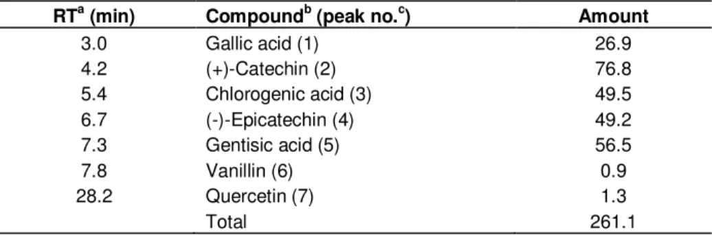
The phenolic profile of the extract determined by HLPC-DAD is represented in Table 1 and Figure 1. All standard compounds exhibited good linearity over the concentrations tested, with correlation coefficients (r2) ranging from 0.97 to 0.99 (data not shown), and the total mean yield of identified polyphenols was 261.1 mg/g. According to the literature the main phenolic compound in leaves of carob tree is GA (Corsi et al., 2002). However, in this work the major compound was (+)-catechin (76.8 mg/g), followed by gentisic acid (56.5 mg/g), chlorogenic acid (49.5 mg/g), (-)-epicatechin (49.2 mg/g), and GA (26.9 mg/g). Minor amounts of quercetin (1.3 mg/g) and vanillin (0.9 mg/g) were also identified. Myricetin, rutin, kaempferol, catechol and syringic, caffeic, cinnamic, protocatchuic and ferulic acid were investigated but not detected. This difference may be explained by the fact that phenolic compounds are secondary metabolites produced and accumulated in plant tissues, and changes in phytopathogenesis, among other factors, may result in different concentrations of these compounds in plant
Table 1. HPLC-DAD analysis of phenolic compounds contents (mg/g DW) in the leaf extract
of carob tree.
RTa (min) Compoundb (peak no.c) Amount
3.0 Gallic acid (1) 26.9 4.2 (+)-Catechin (2) 76.8 5.4 Chlorogenic acid (3) 49.5 6.7 (-)-Epicatechin (4) 49.2 7.3 Gentisic acid (5) 56.5 7.8 Vanillin (6) 0.9 28.2 Quercetin (7) 1.3 Total 261.1
Notes: aRetention times. bIdentified by comparison of the retention parameters with the standard controls and peak purity with the UV-vis spectral reference data. cCorresponding peak number in the chromatogram on Figure1.
Figure 1. HPLC-DAD analysis of phenolic compounds in the leaf extract of carob tree. Chromatogram
recorded at 260 nm. The marked peaks refer to the compounds listed on Table 1.
organs (Ferguson, 2001). In addition, the variations in polyphenol contents among extracts from the same species of different geographical origins are probably linked to genetic and geographical differences, as well as cultural conditions (Avallone et al., 1997). In fact, certain genotypes within a plant species can have widely divergent levels of inherent antioxidants as shown by cross-varietal screening tests (Lila, 2006). It should also be noted that those differences can simply be related to sample preparation and both the extraction and method of analysis.
Effect of the extract on cancer cells viability
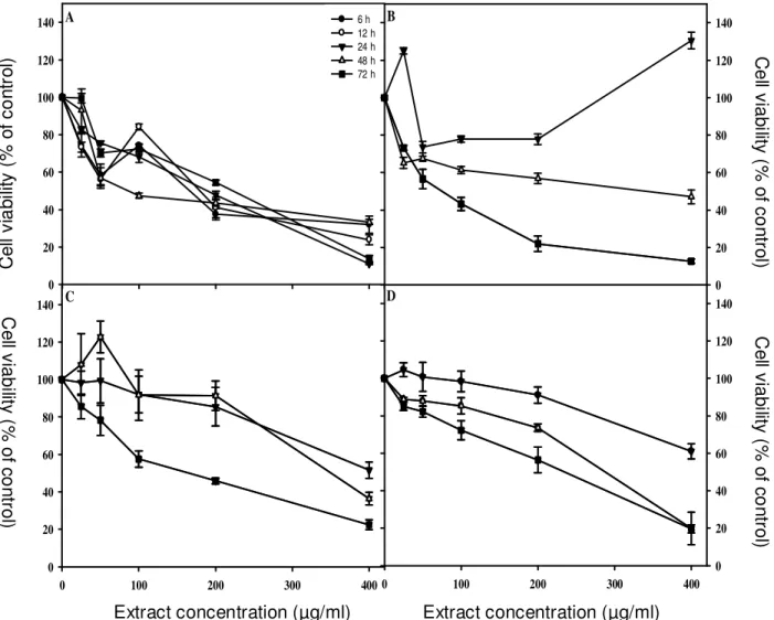
The extract had a concentration and time dependant inhibitory effect on cell viability (Figure 2). HeLa cells
were the most susceptible to the treatment, while DU-145 and HCT-116 were the least, denoted by the highest IC50
values and lower maximal degrees of inhibition (Tables 2 and 3). On HeLa cells, the inhibitory effect on cell viability was evident after 6 h of incubation (IC50 = 147.1 µg/ml),
were the treatment with 400 µg/ml produced a reduction of cell viability to 31.9%, reaching 71.1% as maximal degree of inhibition (Figure 2A; Tables 2 and 3). In this cell line the lowest IC50 value (18.6 µg/ml) was observed
after a 72 h treatment (Table 2). This a very interesting result, since extracts with an IC50 value lower than 30
µg/ml are considered promising for the search of new anticancer agents (Suffnes and Pezzuto, 1990; dos Santos Júnior et al., 2010). On breast cancer cells the extract significantly reduced cell viability after 48 and 72 h of incubation (Figure 2B). The response in terms of cell viability with increasing concentrations and periods of
Extract concentration (µg/ml) 0 100 200 300 400 C e ll v ia b il it y ( % o f co n tr o l) 0 20 40 60 80 100 120 140 C e ll v ia b il it y ( % o f c o n tr o l) 0 20 40 60 80 100 120 140 6 h 12 h 24 h 48 h 72 h X Data C e ll v ia b ili ty ( % o f c o n tr o l) 0 20 40 60 80 100 120 140 Extract concentration (µg/ml) 0 100 200 300 400 C e ll v ia b ili ty ( % o f c o n tr o l) 0 20 40 60 80 100 120 140 A B C D Extract concentration (µg/ml) Extract concentration (µg/ml) C e ll v ia b ilit y ( % o f c o n tr o l) C e ll v ia b ilit y ( % o f c o n tr o l) C e ll v ia b ilit y ( % o f c o n tr o l) C e ll v ia b ili ty ( % o f c o n tr o l)
Figure 2. Effect of the application of the leaf extract of carob tree on the viability of HeLa (A), MDA-MB-231 (B) HCT-116 (C)
and DU-145 (D) cells. Cell proliferation was measured by the WST-1 assay on HeLa cells, and by the MTS method in the other cell lines. Values represent means ± SD of 3 assessments.
Table 2. Half maximal inhibitory concentration (IC50, µg/ml) of the leaf extract of carob tree on human cancer cell lines after
different periods of incubation.
Period of incubation (h) Cell line 6 12 24 48 72 HeLa 147.1 ± 6.5b 158.1 ± 58.2b 122.3 ± 9.2bc 78.8 ± 10.8c 18.6 ± 9.6a DU-145 nd nd >400* 223.4 ± 15.3a 242.2 ± 12.6a HCT-116 nd nd >400* 230.2 ± 22.0a 221.4 ± 37.1a MDA-MB-231 nd nd >400* 144.3 ± 32.4a 74.4 ± 23.0a
Notes: Values represent means ± SD of 3 assessments. For each cell line, statistical analysis was made between periods of incubation. On each raw, values followed by different superscript are significantly different at P < 0.05 (one-way ANOVA, Duncan’s New Multiple Range Test). *Maximal dosage in this assay; nd, not determined.
incubation was similar in the other cell lines (Figures 2C and D). For the 24 h incubation period, IC50 was not
achieved, and there were no significant differences
between 48 and 72 h (Table 2). However, the highest inhibition was observed after the longest period of incubation (Table 3).
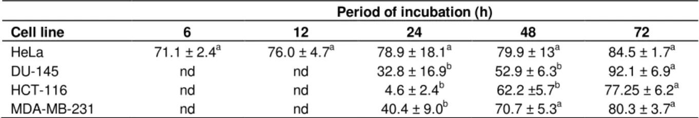
Table 3. Maximal degree of inhibition (%) of the leaf extract of carob tree on human cancer cell lines after different periods of incubation. Period of incubation (h) Cell line 6 12 24 48 72 HeLa 71.1 ± 2.4a 76.0 ± 4.7a 78.9 ± 18.1a 79.9 ± 13a 84.5 ± 1.7a DU-145 nd nd 32.8 ± 16.9b 52.9 ± 6.3b 92.1 ± 6.9a HCT-116 nd nd 4.6 ± 2.4b 62.2 ±5.7b 77.25 ± 6.2a MDA-MB-231 nd nd 40.4 ± 9.0b 70.7 ± 5.3a 80.3 ± 3.7a
Notes: Values represent means ± SD of 3 assessments. For each cell line, statistical analysis was made between periods of incubation. On each raw, values followed by different superscript are significantly different at P < 0.05 (one-way ANOVA, Duncan’s New Multiple Range Test). nd, not determined.
Period of incubation (h) 24 48 C el l v ia b il it y ( % o f co n tr o l) 0 20 40 60 80 100 120 GA (+)-catechin Chlorogenic acid (-)-epicatechin Gentisic acid Vanillin Quercetin *** *** *** *** *** *** *** ***
Figure 3. Effect of the application of the phenolic compounds detected on the leaf extract of carob tree on HeLa
cells viability after 24, and 48 h of incubation. Cell proliferation was measured by the MTT assay. Values represent means ± SD of 3 assessments. Significant differences with untreated cells: *P < 0.05, **P < 0.01, *** P < 0.001
Effect of the phenolic compounds identified in the extract on HeLa cells viability
The biological activity of carob tree leaves is believed to be the result of its phenolic content (Corsi et al., 2002). The biological activities of a crude extract can be due to a natural mixture of its components, and a single constituent may not have an activity greater than that of the total extract as a whole (Salucci et al., 2002; Búfalo et al., 2010). In order to investigate the in vitro cytotoxic activity of the extract phenolic constituents, all the phenolic compounds identified in the extract were evaluated in the viability assay at concentrations equivalent to the ones quantified in the crude extract
(Klenow et al., 2009). After 24 h of incubation, GA exhibited the strongest cytotoxic activity, reducing HeLa cells viability to 11.1% (Figure 3). For the 48 h incubation period, the highest cytotoxic activities were observed with the application of GA, (+)-catechin and quercetin, with reductions in cell viability down to 8.0, 11.9 and 27.1%, respectively (Figure 3). The basic chemical structure of phenolic compounds is a hydroxyl group (-OH) bonded directly to an aromatic hydrocarbon group. This structure allows phenolics to intercept the free radical chain of oxidation by donating hydrogen from the phenolic hydroxyl groups, thereby forming a stable end product, which does not initiate or propagate oxidation of the lipids (Pietta, 2000). Moreover, they also exhibit metal chelating
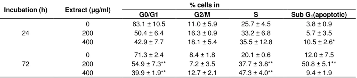
Table 4. Modulation of cell cycle progression and apoptosis in HeLa cells treated with the leaf extract of carob tree for 24 and 72 h.
Incubation (h) Extract (µg/ml) % cells in
G0/G1 G2/M S Sub G1(apoptotic) 24 0 63.1 ± 10.5 11.0 ± 5.9 25.7 ± 4.5 3.8 ± 0.9 200 50.4 ± 6.4 16.3 ± 0.9 33.2 ± 6.8 5.7 ± 3.5 400 42.9 ± 7.7 18.1 ± 5.4 35.5 ± 12.8 10.5 ± 2.6* 72 0 71.3 ± 2.4 8.4 ± 1.8 20.1 ± 0.6 12.0 ± 7.5 200 54.9 ± 7.3** 7.2 ± 3.5 37.7 ± 3.8** 50.8 ± 5.1** 400 39.9 ± 1.9** 12.7 ± 2.1 47.3 ± 4.0** 9.4 ± 1.9
Notes: Values represent means ± SD of 3 assessments. Significant difference with untreated group: * P < 0.05, ** P < 0.01, *** P < 0.001.
properties (Kähkönen et al., 1999). The antioxidant properties are the basis of the biological activities of phenolic compounds, and are associated with the protective effects of plant-based products against cardiovascular disease and certain types of cancer (Haslam, 1998). GA (3, 4, 5-trihydroxybenzoic acid) results from the hydrolysis of tannins and is ubiquitously distributed in plant material where it exists in the form of free acids, esters, catechin derivatives and hydrolysable tannins. It is one of the most biologically-active phenolic compounds of plant origin, being reported as a strong free radical scavenger and shown to reduce cellular viability and induce apoptosis in stomach, colon, melanoma and leukemia cell lines (Yoshioka et al., 2000; Lo et al., 2010).
Catechin and other related procyanidins are an interesting class of flavonoids commonly found in food. It has been shown that these compounds or extracts containing them have antitumor activity (Matito et al., 2003). Quercetin is a flavonoid with a strong growth inhibitory effect on several human cancer lines (Huang et al., 2006). The higher bioactivity observed in this work for GA, quercetin and (+)-catechin can be due to both the number of hydroxyl groups and type of bonds present in this compounds (Haslam, 1998). However, since we tested the activity of the total extract, we cannot exclude that other compounds might have contributed to the inhibition of tumoral cell viability. Moreover, the biological activity of a crude extract cannot be attributed to a single compound but also to other components present in the extract, often resulting from synergistic or additional effects among them. Since some unconfirmed compounds were also detected, the structure and cytotoxic activities of these compounds will be the focus of future studies.
Induction of apoptosis in HeLa cells by the extract Induction of apoptosis in cancer cells is one useful strategy for anticancer drug development (Hu and Kavanagh, 2003). In this respect, many compounds from plant origin have been tested for their apoptotic inducing
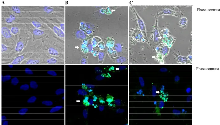
capacity (Kwon et al., 2006; Gonçalves et al., 2010; Lee and Park, 2010). As shown in Table 4, the incubation of HeLa cells with the extract at 200 or 400 µg/ml for both periods of incubation, resulted in the appearance of a large number of cells in the sub-G1 phase (apoptotic cells, Table 4). Moreover, a reduction of cells in the G0/G1 phase and cell arrest in the S phase was observed after treatment with both concentrations for 72 h (Table 4). Exposure of HeLa cells to the extract at 200 µg/ml for 24 h led to typical morphological hallmarks of apoptosis (Figure 4). Those changes included cells shrinkage, membrane blebbing, fragmented cell nuclei forming apoptotic bodies, nuclear fragmentation, cytoplasmatic membrane shrinkage and loss of contact with neighboring cells (Figure 4C). Similar observations were made on cells treated with Anysomicine (Figure 4B), whereas the exposure to the vehicle did not caused nuclear alterations as confirmed by the absence of staining (Figure 4A). Working with extracts from the same species, Corsi et al. (2002) observed that pod and leaf extracts were able to induce apoptosis in a mouse carcinoma cell line. These results are in accordance with other studies, where the application of plant extracts rich in phenolic compounds resulted in an inhibition of cell viability, linked to induction of apoptosis (Kwon et al., 2006; Gonçalves et al., 2010; Lee and Park, 2010).
Effect of the extract on ROS production by HeLa cells The application of the extract at a concentration of 400 µg/ml reduced the intracellular ROS production on HeLa cells by 49.7%, compared with control cells (Figure 5), suggesting that intracellular ROS production is not involved in the induction of apoptosis on this cell line. Several anti-cancer agents, including anthracyclines, cisplatin, bleomycin, and irradiation currently used for cancer treatment have been shown to cause increased intracellular ROS generation (Chan and Yu, 2000). However, the increase of ROS generation in cancer cells can further stimulate cell proliferation, cause DNA mutations, and promote genetic instability and the emergence of drug resistant cells (Pelicano et al., 2004).
+ Phase contrast
-Phase contrast
A B C
Figure 4. Detection of apoptotic morphological changes in HeLa cells treated with the leaf extract of carob tree at a
concentration of 200 µg/ml for 24 h. Cells were stained by TUNEL assay and examined by fluorescence microscopy. (A) Negative control. (B) Positive control (Anisomycine, 10 µg/ml), (C) Cells treated with 200 µg/ml of the extract. Images show nuclei at 630X magnification. Arrows indicate chromatin condensation and nuclear fragmentation.
R el a ti v e a m o u n t o f R O S ( % ) 0 20 40 60 80 100 120 * Extract (µg/ml)+H2O2 H2O2 Control 200 400
Figure 5. Effect of the application of carob tree leaf extract on intracellular ROS
production by HeLa cells. Control cells were cultured without extract and H2O2 exposure,
whereas treated cell were grown in the presence of H2O2 without extract, or in the
presence of extract and H2O2. Significant differences with untreated cells: *P < 0.05, **P
Moreover, some studies have reported that anticancer agents such as emodin and quercetin (Shen et al., 2003) and also natural compounds (Lee and Lim, 2006) induce apoptosis in human cancer cells through ROS-independent mechanism. The inhibitory effect on ROS production confirms the antioxidant activity of leaf extracts from carob tree (Custódio et al., 2009), and suggests this species as a source of potentially active compound to be used in conditions where excessive ROS production is involved.
Conclusion
Our results reinforce the in vitro antitumor activity of a polyphenol leaf rich extract of carob tree, as previously reported by our group and other researchers (Corsi et al., 2002; Custódio et al., 2009). Since GA, (+)-catechin and quercetin also showed cytotoxic activity, one may speculate that these phenolic compounds may be involved in the mechanisms of action of the extract against HeLa cells. Both cell cycle and apoptotic effects were observed and contribute to the cytotoxic activity observed in HeLa cells. Because apoptosis is regarded as a target in the discovery of anti-cancer drugs, these results confirm the interest of carob tree leaves as a potential source of chemotherapeutic agents.
ACKNOWLEDGEMENTS
This work was partially supported by CICYT (CTQ2006-03794/BQU), Instituto de Salud Carlos III (CB06_01_0074 and PI060624), the Generalitat de Catalunya (2005SGR 00662), the Institute for Research in Biomedicine, and the Barcelona Science Park. L. Custódio thanks to the Portuguese Foundation for Science and Technology (FCT) for a post-doctoral grant (grant SFRH/BPD/20736/2004).
REFERENCES
Avallone R, Plessi M, Baraldi M, Monzani A (1997). Determination of chemical composition of carob (Ceratonia siliqua): protein, fat, carbohydrates, and tannins. J. Food Compos. Anal., 10: 166-172. Avallone R, Cosenza F, Farina F, Baraldi C, Baraldi M (2002).
Extraction and purification from Ceratonia siliqua of compounds acting on central and peripheral benzodiazepine receptors. Fitoterapia, 73: 390-396.
Berridge MV, Tan S, McCoy KD, Wang R (1996). The biochemical and cellular basis of cell proliferation assays that use tetrazolium salts. Biochem., 4: 14-19.
Bold RJ, Termuhlen PM, McConkey D (1997). Apoptosis, cancer and cancer therapy. Surg. Oncol., 6: 133-142.
Búfalo MC, Candeias JMG, Sousa JPB, Bastos JK, Sforcin JM (2010).
In vitro cytotoxic activity of Baccharis dracunculifolia and propolis against HEp-2 cells. Nat. Prod. Res., First published on: 22 April 2010 (First).
Chan WH, Yu JS (2000). Inhibition of UV irradiation-induced oxidative stress and apoptotic biochemical changes in human epidermal carcinoma A431 cells by genistein. J. Cell. Biochem., 78: 73-84.
Chang MC, Ho YS, Lee PH, Chan CP, Lee JJ, Hahn LJ, Wang YJ, Jeng JH (2001). Areca nut extract and arecoline induced the cell cycle arrest but not apoptosis of cultured oral KB epithelial cells: Association of glutathione, reactive oxygen species and mitochondrial membrane potential. Carcinogenesis, 22: 1527-1535.
Corsi L, Avallone R, Cosenza F, Farina F, Baraldi C, Baraldi M (2002). Antiproliferative effects of Ceratonia siliqua L. on mouse hepatocellular carcinoma cell line. Fitoterapia, 73: 674-684.
Custódio L, Fernandes E, Escapa AL, Aligué R, Alberício F, Romano A (2009). Antioxidant activity and in vitro inhibition of tumor cell growth by leaf extracts from the carob tree (Ceratonia siliqua L.). Pharm. Biol., 47(8): 721-728.
Dos Santos JH, Oliveira D, De CD, Pinto J, Campos V, Mourão A, Pessoa C, De MM, Costa LL (2010). Evaluation of native and exotic Brazilian plants for anticancer activity. J. Nat. Med., 64: 231-238. Ferguson LR (2001). Role of plant polyphenols in genomic stability.
Mut. Res., 475: 89-111.
Frankfurt OS, Krishan A (2003). Apoptosis-based drug screening and detection of selective toxicity to cancer cells. Anticancer Drugs, 14: 555-561.
Gonçalves S, Xavier C, Costa P, Alberício F, Romano A (2010). Antioxidant, cytotoxic and apoptotic activity of Drosophyllum
lusitanicum extracts. J. Med. Plant. Res., 4: 1601-1608.
Haslam E (1998). Polyphenols and herbal medicines. In Haslam E (ed.) Practical polyphenols, from structure to molecular recognition and physiological action. Cambridge: Cambridge University Press, pp. 298-334.
Hu W, Kavanagh J (2003). Anticancer therapy targeting the apoptotic pathway. Lancet Oncol., 4: 721-729.
Huang SL, Hsu CL, Yen GC (2006). Growth inhibitory effect of quercetin on SW 872 human liposarcoma cells. Life Sci., 79: 203-209.
Kähkönen MP, Hopia AI, Heikki JV, Rauha JP, Pihlaja K, Kujala TS, Heinonen M (1999). Antioxidant activity of plant extracts containing phenolic compounds. J. Agric. Food Chem., 47: 3954-3962.
Klenow S, Jahns F, Pool ZBL, Glei M (2009). Does an extract of carob (Ceratonia siliqua L.) have chemopreventive potential related to oxidative stress and drug metabolism in human colon cells? J. Agric. Food Chem., 57: 2999-3004.
Kumazawa S, Taniguchi M, Susuki Y, Shimura M, Kwon MS, Nakayama T (2002). Antioxidant activity of polyphenols in carob pods. J. Agric. Food Chem., 50: 373-377.
Kwon HJ, Hong YK, Kim KH, Han CH, Cho SH, Choi JS, Kim BW (2006). Methanolic extract of Pterocarpus santalinus induces apoptosis in HeLa cells. J. Ethnopharmacol., 105: 229-234.
Lee SJ, Lim KT (2006). Apoptosis induced by glycoprotein (150-kDa) isolated from Solanum nigrum L. is not related to intracellular reactive oxygen species (ROS) in HCT-116 cells. Cancer Chemoth. Pharm., 57: 507-516.
Lee SB, Park HR (2010). Anticancer activity of guava (Psidium guajava L.) branch extracts against HT-29 human colon cancer cells. J. Med. Plant. Res., 4: 891-896.
Lila M (2006). The nature-versus-nurture debate on bioactive phytochemicals: The genome versus terroir. J. Sci. Food Agric., 86: 2510-2515.
Lo C, Lai TY, Yang JH, Yang JS, Ma YS, Weng SW, Chen YY, Lin JG, Chung JG (2010). Gallic acid induces apoptosis in A375.S2 human melanoma cells through caspase-dependent and -independent pathways. Int. J. Oncol., 37: 377-385.
Makris D, Kefalas P (2004). Carob pods (Ceratonia siliqua L.) as a source of polyphenolic antioxidants. Food Technol. Biotechnol., 42: 105-108.
Matito C, Mastrorakou F, Centelles J, Torres J, Cascante M (2003). Antiproliferative effect of antioxidant polyphenols from grape in murine Hepa-1c1c7. Eur. J. Nutr., 42: 43-49.
Mosmann T (1983). Rapid colorimetric assay for cellular growth and survival: Application to proliferation and cytotoxicity assays. J. Immunol. Methods, 65: 55-63.
Mukherjee A, Westwell AD, Bradshaw TD, Stevens MFG, Carmichael J, Martin SG (2005). Cytotoxic and antiangiogenic activity of AW464 (NSC 706704), a novel thioredoxin inhibitor: An in vitro study. Br. J. Cancer, 92: 350-358.
Haber B (2003). Isolation and structure elucidation of the major individual polyphenols in carob fibre. Food Chem. Toxicol., 41: 1727-1738.
Papagiannopoulos M, Wollseifen H, Mellenthin A, Haber B, Galensa R (2004). Identification and quantification of polyphenols in carob fruits (Ceratonia siliqua L.) and derived products by HPLC-UV-ESI/MS. J. Agric. Food Chem., 52: 3784-3791.
Pelicano H, Carney D, Huang P (2004). ROS stress in cancer cells and therapeutic implications. Drug Resist. Update, 7: 97-110.
Pietta PG (2000). Flavonoids as antioxidants. J. Nat. Prod., 63: 1035-1042.
Reed JC (2001). Apoptosis-regulating proteins as targets for drug discovery. Trends Mol. Med., 7: 314-319.
Salucci M, Stivala LA, Maiani G, Bugianesi R, Vannini V (2002). Flavonoids uptake and their effect on cell cycle of human colon adenocarcinoma cells (Caco2). Br. J. Cancer, 86: 1645-1651.
Shen SC, Chen YC, Hsu FL, Lee WR (2003). Differential apoptosis-inducing effect of quercetin and its glycosides in human promyeloleukemic HL-60 cells by alternative activation of the caspase 3 cascade. J. Cell. Biochem., 89: 1044-1055.
Suffness M, Pezzuto JM (1990). Assays related to cancer drug discovery. In: Hostettmann K (ed) Methods in plant biochemistry: assays for bioactivity. Academic, London, pp. 71-133.
Yoshioka K, Kataoka T, Hayash T, Hasegawa M, Ishi Y, Hibasami H (2000). Induction of apoptosis by gallic acid in human stomach cancer KATO III and colon adenocarcinoma COLO 205 cell lines. Oncol. Rep., 7: 1221-1223.