Full paper published online: May 31, 2008 ISSN 1678-9199.
ORAL Candida spp. COLONIZATION IN HUMAN IMMUNODEFICIENCY
VIRUS-INFECTED INDIVIDUALS
MORIS D. V. (1), MELHEM M. S. C. (2) MARTINS M. A. (2), MENDES R. P. (1).
(1) Tropical Diseases Area, Botucatu Medical School, São Paulo State University,
UNESP, Botucatu, São Paulo State, Brazil; (2) Adolfo Lutz Institute, São Paulo,
Brazil.
ABSTRACT: Several yeast species of Candida genus can colonize the skin as well
as the mucous membrane of the vagina and the digestive tract for short or long
periods. Depending on the host’s immunological state and the yeast’s virulence,
colonization can become an infection, invading the colonized tissues and also
disseminating. AIDS is characterized by the host’s intensive and progressive
immunodepression which manifests as diverse symptoms, mainly lesions in the
mouth. Oral candidiasis is the most prevalent opportunistic infection in individuals
infected with human immunodeficiency virus (HIV) and is an important indicator of
the disease progress and the immunosuppression increase. The factors involved in
the equilibrium between Candida spp. and HIV-infected subjects are sometimes
contradictory and were evaluated in the present study specially for colonization.
KEY WORDS: Candida spp., colonization, human immunodeficiency virus-infected
individuals.
CONFLICTS OF INTEREST: There is no conflict.
CORRESPONDENCE TO:
DANIELA VANESSA MORIS. Departamento de Doenças Tropicais e Diagnóstico por
lmagem, Faculdade de Medicina de Botucatu, UNESP, Rubião Junior, s/n,
INTRODUCTION
The genus Candida was created during the IXInternational Botanical Congress, held
in Canada in 1959, and substituted the term Monilia, used until then (58).
Candidiasis is an infection caused by a yeast species of the genus Candida, which
belongs to the family Cryptococcaceae. This genus included 196 species with
physiological affinity for both basidiomycetes and ascomycetes (73). Then, species
that have affinity for basidiomycetes were excluded, but the genus continued
heterogeneous, presenting 163 species, which makes difficult its taxonomical
characterization (56).
As dimorphic fungi, Candida yeasts have globose, ellipsoidal, cylindroidal, or
elongate, occasionally ogival, triangular or lunate cells. Reproduction is by holoblastic
budding. Pseudohyphae and septate hyphae may be formed. Arthroconidia and
ballistoconidia are not formed (73,117).
In general, yeasts have asexual reproduction, by budding or fission, and each
asexual reproductive unity is called blastoconidium. Some yeasts produce chains of
blasconidia that do not separate, forming a pseudohypha. The capacity of forming
filaments, or filamentation, is strictly related to tissue invasion and, consequently, to
pathogenicity (45, 57, 68).
Berkhout (7) was concerned about Candida yeasts existing in nature as well as about
species found in men as saprophytic or parasites that were isolated from the skin,
oral cavity, intestines and urogenital system. Candida species are confined to human
and warm-blooded animal reservoirs; however, they can also be recovered from soil,
food, water and, sometimes, air (5). The human gastrointestinal system is considered
the biggest habitat for Candida spp. which are most frequently isolated from distal
segments (23).
Mucous membrane surfaces constitute the largest interface between the host and the
environment and develop several defense mechanisms to combat microorganisms,
particularly pathogenic ones (67, 119). Candida yeasts can be the agents of local or
systemic opportunistic infections, mainly in hospitalized patients, especially those
under intensive treatment (1). Infections caused by opportunistic agents including
Candida spp. are frequent in diverse pathological states that induce
immunodeficiency such as neutropenia, neoplasia, decompensated diabetes
PATHOGENICITY AND PATHOGENY
Virulence Factors of Candida Genus
There are differences in pathogenicity among Candida spp. isolates. Some
properties related to Candida albicans cells give them the capacity to cause disease.
Adherence to cell surface, germ tube formation with consequent development of the
filamentous form, phenotypic variability, and production of toxins and extracellular
enzymes constitute important factors for the emergence of infections by Candida (13,
14, 30).
Ghannoum & Abu-Elteen (36) proposed an infection model in which the sequence
was initiated by the yeast adherence to epithelial cells of the skin and mucous
membrane, followed by cell multiplication and latter formation of germ tube and
filaments. The enzymes produced then, especially proteinase and phospholipase,
allowed the yeast penetration into the cells, inducing inflammatory response with
injury of adjacent tissues.
Depending on the host’s immunological state and on the microorganism’s virulence,
the latter can invade tissues causing infection which may disseminate. Enzymes
destroy, alter or damage the integrity of the host’s cell membrane, leading to
dysfunction or interruption of its activities. Lipids and proteins of eukaryotic cell
membranes constitute a target for such enzymatic attack. Thus, pathogenicity of
Candida spp. is attributed to their different properties such as production of
exoenzymes like phospholipases (47) and proteinases (46).
1.Aspartyl proteinases (SAPs)
The role of SAPs produced by C. albicans has been one of the main aims of
physiological and biochemical studies on such yeast. These enzymes have
proteolytic activity at low pH values (2.0–4.0) with a highly broad action including
keratin, collagen, albumin, hemoglobin, immunoglobulin heavy-chain and
extracellular matrix proteins (21). SAPs can be studied in culture medium containing
bovine albumin as the only source of nitrogen by verifying quantitative differences
among Candida species (22, 95, 96).
MacDonalds & Odds (64) observed a direct correlation between pathogenesis and
proteolytic activity of Candida spp. isolates, showing that C. albicans mutants,
proteinase-deficient, had lower virulence. These isolates were more easily
Pepstatin A, which is an inhibitor of SAPs, was used in the treatment of mice infected
with C. albicans and produced a protective effect characterized by reduction in
proteinase activity and microorganism virulence – findings that are probably
correlated (54, 94).
In a study about infection of the human oral epithelium by Candida spp., the inhibitory
effect of pepstatin A on SAPs determined a great reduction in lesions, indicating that
the proteinase activity contributed to tissue injury (102).
Ollerte et al. (83)verified that the proteolytic activity of C. albicans isolates from AIDS
patients was higher than that of isolates from subjects not infected with HIV,
highlighting thus the importance of such enzyme in the pathogenesis of oral
candidiasis in HIV-infected individuals. They also observed a direct correlation
between proteinase activity and resistance to antifungals. Candida albicans isolates
causing oral candidiasis and producing high proteinase levels were less susceptible
to azole antifungals than C. albicans isolates from HIV-negative subjects.
Research on proteinase production by C. albicans isolates is generally carried out
using the method of Ruchel et al. (95).
Candida albicans isolates are seeded at equidistant points on the culture medium
and the plates are incubated at 37ºC for seven days. A translucent halo of protein
degradation formed around the yeast colony indicates enzymatic activity. The
enzymatic activity (Pz) is defined as the ratio between colony diameter (Dc) and
colony diameter plus degradation zone diameter (Dc + Dz), as follows:
Pz = Dc / Dc + Dz
Thus, Pz=1.00 indicates absence of proteolytic activity, whereas Pz<0.64, for
example, means that the Candida spp. isolate is producing high levels of SAPs.
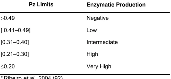
Some authors (92) proposed a classification for proteolytic activity, according to its
Table 1. Characterization of proteolytic activity (Pz), as a function of the ratio
between colony diameter (Dc) and colony diameter plus degradation zone diameter
(Dc + Dz)*.
Pz Limits Enzymatic Production
>0.49 Negative
[ 0.41–0.49] Low
[0.31–0.40] Intermediate
[0.21–0.30] High
≤0.20 Very High
*Ribeiro et al., 2004 (92).
2. Phospholipases
Phospholipases are hydrolytic enzymes capable of degrading phospholipids.
Phospholipase B is the dominant fraction, being responsible for 90% of the
extracellular activity of phospholipase produced by Candida spp. (97). The lipolytic
activity of phospholipase is concentrated at the yeast cell poles, facilitating tissue
adhesion and acting on lipidic constituents of the host’s cell membrane, as
demonstrated for phospholipase A and lipophospholipase (90).
Evaluation of the activity of phospholipase produced by C. albicans was standardized
by Price et al. (89) using egg yolk which is rich in phospholipids, mainly
phosphatidylcholine and phosphatidylethanolamine that serve as substrates for the
enzyme. Phospholipase activity is assessed by measuring the precipitation halo
represented by a calcium complex and fatty acids released due to the enzymatic
activity.
Phospholipase activity (Pz) is defined as the ratio between colony diameter and
colony diameter plus precipitation zone diameter, as follows:
Pz = Dc / Dc + Dz
Thus, Pz=1.00 indicates absence of phospholipase activity, whereas Pz=0.63, for
example, means that the Candida spp. isolate is releasing high levels of
phospholipase.
Candida spp. isolates from different sites and organic fluids of patients with
disseminated candidiasis were noticed to have the same phospholipase activity.
Such finding suggests that phospholipase production is not influenced by the
infection site and that the presence of C. albicans in different organs and/or organic
fluids of the same patient is indicative of systemic infection. Thus, phospholipase
activity can be used for characterizing strains (82, 122).
Samaranayake et al. (97) evaluated the influence of incubation period, pH and sugar
concentration in the culture medium on phospholipase activity. They demonstrated a
direct correlation between phospholipase activity and incubation period until the
fourth day, when enzymatic activity stabilized. Similarly, phospholipase activity was
higher at pH 4.4 than at pH 3.3, but not at pH 5.1 or 6.3, although there was a great
fungal growth at higher pH values. There was also a direct correlation between
phospholipase activity and galactose or sucrose concentration in the culture medium.
Contrastingly, increased glucose concentration (above 100mM) results in the
complete cessation of phospholipase activity.
It is important to mention that the works of Price et al. (89) and Samaranayake et al.
(97) demonstrated that: variation in the inoculum size does not interfere with
phospholipase activity; Pz value of each isolate is relatively constant; and
phospholipase-negative C. albicans isolates keep negative independently of the
culture conditions.
A study with C. albicans in mice revealed a direct correlation between pathogenicity
and phospholipase activity; 90% of C. albicans isolates showing high phospholipase
production were pathogenic to mice (52).
Clancy et al. (22) showed that Candida species other than C. albicans alsoproduced
extracellular phospholipases, however, at lower levels than those secreted by C.
albicans. Such difference can be attributed to variations in the isolates or differences
in the medium preparation or in the substrate employed for phospholipase detection.
Ghannoum (40), in an excellent review about the role of phospholipases on virulence
and pathogeny of several fungus genera including Candida, suggested that fungal
Research on phospholipase production by Candida spp. isolates is commonly carried
out using the method of Price et al. (89), with modifications suggested by the results
of Samaranayake et al. (97).
Candida albicans isolates are seeded at equidistant points on the culture medium
and the plates should be incubated at 37ºC for four days. A turbid precipitation zone
formed around the colony indicates the presence of the enzyme.
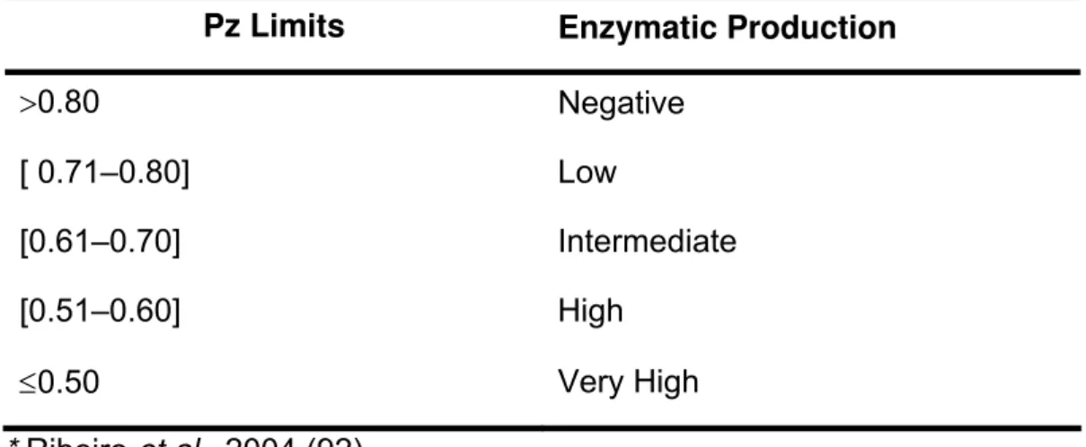
Some authors (92)proposed the classification of phospholipase activity, according to
its intensity, at the ranges presented in Table 2.
Table 2. Characterization of phospholipase activity (Pz), as a function of the ratio
between colony diameter (Dc) and colony diameter plus precipitation zone diameter
(Dc + Dz)*.
Pz Limits Enzymatic Production
>0.80 Negative
[ 0.71–0.80] Low
[0.61–0.70] Intermediate
[0.51–0.60] High
≤0.50 Very High
* Ribeiro et al., 2004 (92).
Pathogeny
The process by which Candida spp. colonizes and penetrates the epithelium of the
digestive tube can be analyzed in four stages: initial adhesion to the epithelium;
replication and colonization; formation of hyphae; and epithelial lesion and
penetration(14, 45). Genes INT1 and PLB1 have been related to colonization (6, 60).
Mechanical barriers, inflammatory cells and cellular immunity restrict Candida spp. to
non-sterile superficial sites. Besides, the resident bacterial microbiota generally limits
the number of fungal cells, blocks their adhesion to epithelial cells, competes with
them for nutrients, and prevents the fungus conversion into its most invasive form,
the filamentous form. When one or more of these defenses become deficient,
Mucosal surfaces have a well regulated defense system, which is still little known.
The mucous immune system has both a localized and a diffused portion. In the
former, foreigner antigens are taken to highly structured sites, where the immune
response begins. The diffused portion is composed of effector cells such as B and T
lymphocytes, differentiated plasmatic cells, macrophages and other
antigen-presenting cells, eosinophils, basophils, and mastocytes. Together, these two
portions induce mucosal response, antibody production, cellular immune response or
even tolerance, characterized by anergy.
A mucosal surface can include a huge area like the human gastrointestinal tract, with
more than 300m2, which requires a significant participation of lymphoid cells and
effector molecules related to cellular immunity (124).
The pathogenicity of Candida spp. depends on the composition of their cell wall as
the external surface is responsible for their interface with the host.
A study on C. albicans interaction with macrophages revealed a rapid phagocytosis
of the fungus which undergoes morphological transformation, forming elongated
germ tubs that penetrate the macrophage membrane, destroying it. This way, the
fungal cell goes back to its environment. Such study demonstrated the importance of
polymorphism, as mutants that did not form filaments showed to be avirulent (62).
Candida spp. antigens stimulate specific cellular and humoral immune response. The
interest in studying antibody response in Candida spp.-infected animals has been
renewed. The mannoprotein of 58 kDa found at C. albicans surface is highly
immunogenic; it is expressed by all C. albicans isolates and stimulates intense
antibody response during candidemias (16). The gene that codifies it is FBP1/PRH1.
However, the role of humoral immunity in the defense against fungal infections,
including those caused by Candida spp., is still highly controversial (44).
With regard to innate immunity, macrophages and neutrophils seem not to have a
relevant role.
The Th1 branch of cell-mediated immune response is considered fundamental for
such defense. Thus, when the number of CD4+ T lymphocytes is below a protective
level, like in AIDS patients, other defense mechanisms are needed to protect against
oral candidiasis.
Regulation of cytokines related to Th1 and Th2 branches plays an important role in
the host response to deep infections caused by Candida spp. Interferon-γ, tumor
increasing phagocytosis, superoxide production, and antifungal activity of
mononuclear cells and neutrophils. Contrastingly, Th2 branch-related cytokines, IL-4
and IL-10, present immunosuppressive effect by compromising the last phase of
monocytes response against C. albicans, i.e. microbicidal activity and hyphal lesions
are reduced, whereas phagocytosis is not affected or even increased by the action of IL-10 (121). Transforming growth factor-β (TGF-β), a Th2-type cytokine, is expressed
at high concentrations by hepatocytes and mononuclear cells of granulomatous
lesions in chronic disseminated candidiasis. Candida albicans also induces, in vitro, the expression of TGF-β by circulating blood monocytes.
Besides, human and animal epithelial cells present antifungal activity. In vitro
coculture of epithelial cells with Candida spp. reveals inhibition of fungal growth by
human epithelial cells within only nine hours (33, 79, 109, 110). The inhibitory effect
of oral epithelial cells on fungal growth is much higher than that of vaginal epithelial
cells. However, such epithelial cells cannot destroy fungal cells. Thus, epithelial cells
may represent an important mechanism of innate resistance against mucous
candidiasis as they inhibit fungal cell growth, which is attributed to a portion
constituted of carbohydrates (still not determined but existent) at the cells surface.
On the other hand, C. albicans wild-type strains incubated with a layer of CaCO2
epithelial cells in a TranswellR system reduced transepithelial resistance after 7.5
hours. Such reduction was specific for C. albicans and required the transcription of
factors CPH1 and EFG1 as well as proteinases SAP 4–6 (63).
Imam et al. (48)demonstrated that severe vaginal candidiasis can affect HIV-infected
patients with low or no degree of immunosuppression of CD4+ T lymphocytes or
CD4+ T/CD8+ T cell ratio. As immunosuppression increases, oral candidiasis can be
observed, and in more immunocompromised patients, esophageal candidiasis is
noticed. Thus, those authors suggested a hierarchy of mucosal infections caused by
Candida spp. in HIV-infected patients.
IDENTIFICATION OF Candida SPECIES
There are several available procedures to identify Candida spp., most of which
consist in the association of morphological and biochemical traits like the form and
size of blastoconidia, the production of chlamydoconidia, pseudohyphae and true
hyphae, and the capacity to assimilate carbohydrate and nitrogen and to ferment
In Sabouraud agar and blood agar, Candida spp. grow within 24h–48h at 37ºC in the
form of marble-white, humid, circular, convex colonies. The colonies composed of
blastoconidia can produce pseudohyphae. Chlamydospores and true hyphae can be
formed under special nutrient and oxygen conditions. True hyphae are formed from
yeast-like cells through development of the germ tube. Germ tubes are formed within
three hours at 37ºC, in the presence of albumin, only in C. albicans (56) and C.
dubliniensis cultures (112, 114). Such characteristic is used in routine laboratory for
identification.
About 95% of C. albicans isolates produce germ tube. In its absence, biochemical
tests must be carried out. The difference between germ tube and pseudohypha is
that the former does not present septa but parallel walls and has no constriction at
the mother cell neck (91).
Chlamydospore or chlamydoconidium is a resistance structure formed by thick cell
wall and condensed cytoplasm. It is generally produced when the yeast is under
unfavorable growth conditions. Microculture, i.e. culture in slides, is carried out to
verify the micromorphology of Candida spp.,the existence or not of filamentation, the
production of chlamydoconidia and blastoconidia, as well as the layout of such
elements at the mycelium. Candia albicans and C. dubliniensis produce round
chlamydoconidia at intercalary or terminal position to the pseudohypha (29).
Assimilation test, also called auxanogram, evaluates the capability of certain yeasts
to utilize several compounds as the only source of carbon or nitrogen in the presence
of oxygen. Each species has its own assimilation pattern. Such tests are highly
sensitive and useful for yeast identification and are conditioned to permeability
factors, enzymatic systems that catalyze carbon hydrate degradation, and the
reductase system which intervenes in nitrate reduction.
The capability of a yeast species to ferment certain sugars (which is determined by
the fermentation test or zymogram) depends on the presence of a transportation
system that allows sugar absorption under low O2 tension and on the existence of an
enzymatic system that acts on sugar glycolytic degradation with formation of ethanol
and carbon anhydride. The fermentation of a certain sugar contributes to distinguish
between species(53).
In the recent past, manual and automated boards were commercialized for yeast
identification by evaluating the assimilative capacity of biochemical and enzymatic
conventional methods auxanogram and zymogram (100, 104, 106). The systems API
20C AUX and ID32 BioMeriux (St. Louis, Mo), which contain dehydrated substrates
for assimilation tests, are examples of semi-automated methods. Positive and
negative results correspond to presence and absence of turbidity, respectively, in the
wells of each substrate. Reading of these reactions is carried out by comparison with
growth controls. Hypha or pseudohypha formation is also evaluated and, after
reading, the biochemical profile is converted into a numerical profile composed of a
seven-digit code. Interpretation is based on the analytical catalogue API 20 C AUX,
in which the found numerical profile is compared with a database of 44 taxa. Such
taxa can be a genus, a species, a biotype within a species or multiple species, i.e. a
group of different species that cannot be distinguished by the performed tests. The identification is interpreted as: excellent (%id≥99.0 and T≥0.75), very good (%id≥99.0 and T≥0.5), acceptable (%id>90.0 and T>0.25) or unacceptable (%id≥80.0 and T≥0), in which %id is the percentage of identification or an estimate of how closely the
profile corresponds to the taxon, relative to all other taxa in the database, and the
index T is an estimate of how closely the profile corresponds to the most typical set
of reactions for each taxon.
The Vitek system, bioMérieux Laboratories, is an automated process for the
identification of several microorganisms, among which are Candida yeasts. The Vitek
Yeast Biochemical Card (YBC) contains thirty tests, 26 substrates and four controls.
Carbohydrate assimilation, urea hydrolysis, resistance to cycloheximide, and nitrate
and nitrite reduction are evaluated (72).
It must be emphasized that the morphological and biochemical tests and the manual
and automated boards for yeast identification do not allow differentiation between C.
albicans and C. dubliniensis.
Recently, different chromogenic culture media capable of distinguishing C. albicans
from other yeasts of clinical interest have been commercialized. Such media are
based on the color alteration yielded by the colonies, which is measured by using
indicators of pH and fermentation of specific compounds or chromogenic substrates
for presumptive identification of C. albicans, C. tropicalis and C. krusei.
β-glucosaminidase is used as substrate and yeasts are differentiated according to the
and identification of the above-mentioned yeasts, providing presumptive results in
less time than conventional methods and confirming the isolates purity (34, 35, 85).
It must also be highlighted that chromogenic media do not allow a safe differentiation
between C. albicans and C. dubliniensis.
MOLECULAR BIOLOGY
The molecular methods used for the study of candidiasis are mostly designed to
verify the genetic similarity between isolates of the same species (107). Such
methods can also be used to confirm phenotypic identification, once each species
presents characteristic biochemical profile and morphological aspects.
Molecular typing methods based on different principles have been developed and
applicable for various Candida species. Among several techniques are those based
on pulsed field gel electrophoresis (PFGE) and polymerase chain reaction (PCR).
PFGE karyotyping and PFGE of fragments generated by cutting restriction
endonucleases were highly reproducible but expensive, laborious and
time-consuming. These techniques were already applied for several Candida species (21,
87, 105).
Among the PCR-based methods, randomly amplified polymorphic DNA (RAPD) is
the most used. It is easy and rapid but presents reproducibility problems inter and
intra laboratories (107). There are reports about its application for several Candida
species (86, 116). Recently, two new techniques arouse: amplified fragment length
polymorphism (AFLP) (3) and multilocus sequence typing (MLST) (12, 32, 115).
Ideally, more than one method or two or more random primers (in the case of RAPD)
should be used in order to verify which of them has the best discriminating power for
that species.
Because of the high degree of similarity between phenotypic characteristics of C.
albicans and C. dubliniensis, various molecular techniques have been successfully
proposed to identify C. dubliniensis (20, 65, 74).
COLONIZATION AND INFECTION BY Candida spp.
Candida spp. constitute the normal microbiota of the mouth of 25%–50% healthy
individuals; this condition is called colonization (81). Candida albicans is the most
prevalent species among colonizers; however, species other than C. albicans are
Several yeast species of Candida genus,as well as some other microorganisms, can
colonize mucosa, skin and digestive tract, transitorily or for long periods. However,
when they are present at different sites and when the culture repeatedly reveals the
same type of biological isolate, microbiota alteration is suggested. Excessive growth
of Candida spp. at those colonization sites may facilitate tissue invasion, mainly in
predisposed hosts (8).
About 70% of the healthy population has the gastrointestinal tract colonized by
Candida spp., which can precede fungemia due to alteration in the resident
microbiota and translocation of the pathogen through the gastrointestinal mucosa.
The vast majority of candidemias follow colonization by the same yeast species.
Genotyping methods for Candida spp. showed similarity between colonizing and
infecting strains, indicating a probable endogenous origin of most infections (24, 80).
Any factor causing microbiota disequilibrium or mucosa lesion can be considered an
agent that facilitates Candida spp. translocation to mesenteric capillaries. Factors
that increase intestinal colonization by Candida spp. (such as antibiotic use and
intestinal occlusion) or those that cause mucosa atrophy or lesion (such as prolonged
fasting, total parenteral nutrition, arterial hypotension and chemotherapy) can
reinforce the translocation phenomenon across the intestinal epithelium (1).
Rodero et al. (93) suggested that candidiasis begins with an endogenous commensal
agent which converts into a pathogen in an immunocompromised host. The most
frequent microorganism in these infections is again C. albicans, especially because it
is the predominant species in the gastrointestinal tract. Candidiasis presents varied
clinical manifestation. Cutaneous candidosis involves intertriginous areas of skin in
the hands, groins and armpits; mucocutaneous candidiasis affects tissues of the oral
and vaginal mucosa. The latter infects individuals with or without predisposing
factors; however, its chronic form is one of the most severe forms of the disease,
affecting patients with genetic or metabolic defects. Systemic candidiasis develops
after hematogenic dissemination in susceptible hosts with predisposing factors,
especially neutropenic patients. In such cases, tissue invasion may occur, leading to
pulmonary candidiasis, nephritis, suppurative phlebitis, arthritis, osteomyelitis,
endophtalmitis, balanitis, SNC lesions, myocarditis, pericarditis and endocarditis, in
which obstruction of cardiac valves and embolism may also occur due to the large
In the last years, the number of infections caused by species other than C. albicans
has increased. In 1963, there were five Candida species capable of causing disease
to human beings, C. albicans, C. parapsilosis, C. tropicalis, C. stellatoidea and C.
guilliermondii. Nowadays, about 20 species are known to cause infections in humans
(25, 31).
Candida albicans, C. parapsilosis, C. tropicalis, C. glabrata, C. krusei, C.
guilliermondii and C. lusitaniae are the main species of medical interest. However,
cases of superficial or invasive diseases related to emerging Candida species have
been reported including C. kefyr, C. rugosa, C. famata, C. utilis, C. lipolytica, C.
norvegensis, C. inconspicua , C. dubliniensis(25) and C. peliiculosa (Pichia anomala)
(66).
Candida albicans is the species most frequently isolated as colonizer or superficial
and invasive infection agent at different anatomical sites all over the world. It has a
well-known pathogenic potential and its main pathogenicity and virulence factors are:
capacity to adhere to different mucosae and epithelia; dimorphism, with production of
pseudohyphae helping tissue invasion; thermotolerance; and production of
exoenzymes like proteinases and phospholipases (31). This species is naturally
susceptible to all systemic-use drugs; however, cases of developed resistance to
azoles have been described, mainly in subjects that had been exposed to such
antifungals for a long time. Resistance to amphotericin B is rare (101).
Candida tropicalis can behave as an opportunistic agent when the host is
neutropenic, when there is bacterial flora suppression due to antimicrobial use or
when there is gastrointestinal mucosa injury. This species is responsible for
candidemias in neoplastic patients and is more frequent in leukemia than in solid
tumors (123).
Clinical isolates of C. tropicalis are susceptible to amphotericin B and, most of the
times, to triazoles. In Latin American countries, especially Brazil, it is frequent even in
non-cancer patients and constitutes the second or third most important cause of
candidemia (26, 37, 38).
Since the 1980s, Candida parapsilosis has been an important cause of fungemia,
being responsible for 7%–15% of candidemias in the United States (84) and Europe
(120). It proliferates in solutions containing glucose, produces biofilm and frequently
colonizes the skin. Fungemia by C. parapsilosis is associated with the utilization of
amphotericin B and triazoles (101). This species has been recognized as the second
main cause of invasive infection in Latin American countries (26, 37, 38).
The epidemiology of C. glabrata and C. krusei infections is different from that of other
species. These infections mostly begin after prophylactic use of antifungals (e.g.
fluconazole) in patients with hematological diseases and are not associated with
catheter use or previous antibiotic therapy. Candida krusei presents the highest
lethality rate, followed by C. glabrata (84). It has intrinsic resistance to fluconazole,
which may explain its increase in neutropenic patients exposed to this antifungal
(123). Candida glabrata can develop resistance with exposure to fluconazole (84).
Consequently, an increase in the colonization or infection rate by C. glabrata has
been observed in different groups of patients subjected to prolonged exposure to
fluconazole (84). Lower susceptibility to amphotericin B has also been recorded for
this species(120).
Candida dubliniensis, described in Ireland in 1995 by Sullivan et al. (114), presents
biochemical and morphological traits very similar to those of C. albicans, which
requires molecular methods to distinguish between them. This new species was
isolated from the oral cavity of AIDS/HIV-infected patients, among which 17%–35%
are colonized or infected with C. dubliniensis (113). The risk factors for the presence
of such species are not well established, but its virulence seems to be similar to that
of C. albicans.
Proteinase activity is known to be higher in Candida dubliniensis than in C. albicans.
The former also presents higher adherence to the oral mucosa but slower hypha
formation, suggesting less invasive capacity (113). This new Candida species had
probably been present in the community for a long time mistakenly identified as C.
albicans.
Cases of systemic diseases related to this new species are still rare, and most of
them are associated with oral mucosa infection. Such emerging species seems to be
less pathogenic than C. albicans but more capable ofdeveloping resistance to azoles
(55).
Candida lusitaniae is a yeast species less frequently isolated as a causative agent of
invasive diseases but has been reported as cause of candidemia in
immunocompromised patients. Clinical isolates of C. lusitaniae generally show
natural resistance to amphotericin B or rapidly develop it, but are susceptible to
Candida guilliermondii has been recognized as an emerging agent, although its
invasive infections are not frequent yet. There are reports about in vitro resistance of
clinical isolates to amphotericin B and reduced clinical response of patients treated
with such polyene (43).
HIV infection is associated with an increase in the rate of subjects colonized by
Candida spp., which can be observed even before the establishment of evident
immunosuppression. The frequency of isolation of Candida spp. from the oral cavity
of such patients increases with the cell immune compromising progress (88).
AIDS-CANDIDIASIS COINFECTION
AIDS is characterized by the host’s intensive and progressive immunodepression,
which is manifested by diverse symptoms. Oral alterations in AIDS patients include
more than 40 types of lesions, which very frequently constitute the main
manifestations of the disease (59, 71, 78, 108).
Oral candidiasis occurs in 90% AIDS patients, being the most prevalent opportunistic
infection and an important indicator of the disease progress and the
immunosuppression increase (71).
In a study about the oral manifestations observed in 100 AIDS or HIV-infected
patients in Natal, Rio Grande do Norte State, Brazil, oral candidiasis was the most
common manifestation (99 patients), presenting pseudomembranous (the most
frequent form), followed by erythematous, hyperplastic and angular cheilitis forms
(71), which confirms the findings of other authors (59).
HIV-infected women with low CD4+ T cell counts presented higher risk of oral C.
albicans colonization and infection than those with high CD4+ T cell counts (118).
Following the introduction of highly active antiretroviral therapy (HAART), there was a
reduction in occurrence of opportunistic infections, prevalence of oral manifestations
and oral candidiasis (18). Since then, the frequency of candidiasis has decreased,
even in the presence of viral resistance to the treatment or in the absence of
noticeable CD4+ T cells recovery (77).
Arribas et al. (2) suggested that the reduction in the frequency of oral candidiasis was
only related to immunological improvement after introduction of antiretroviral therapy
However, some HIV-positive patients with relatively high CD4+ T cell counts
developed oral candidiasis (49). On the other hand, Hoegh et al. (44) suggested that
inhibitors of HIV protease inhibited the prevalence of oral candidiasis in HIV-infected
patients by their direct action on the secretion of SAPs by Candida spp., which are
considered to belong to the same class of HIV aspartic proteases. Thus, the success
of antiretroviral therapy could be attributed to several factors such as inhibition of one
or more C. albicans virulence factors and recovery of the immunological state of
infected patients (2, 18, 41, 42, 46, 49, 77). SAPs by C. albicans was evaluated in 18
isolates from HIV-infected subjects with CD4+ cell counts inferior to 400 cells/mm3
and in other 18 isolates from HIV-seronegative subjects, which was the control group
(125). The authors observed that all C. albicans isolates from HIV-positive patients
secreted proteinase whereas only 56% of those from the control group secreted it.
Proteolytic activity was higher in isolates obtained from the HIV-infected group. They
also evaluated the inhibitory effect of polyene and imidazole antifungals, at levels
below the minimal inhibitory concentration – MIC (1/4 and 1/16), on proteinase
production in seven isolates from each group. There was a less intense inhibitory
effect on proteinase production by isolates from HIV-infected patients, compared with
that by isolates from the control group and, in general, there was a dose-dependent
effect (125).
Candida spp. COLONIZATION IN HIV-POSITIVE PATIENTS
In 1998, Schuman et al. (103) conducted a multicenter study in which they evaluated
oral mucosa colonization by Candida spp. in 518 HIV-infected women, who had not
developed AIDS, and in 207 at-risk HIV-seronegative women.
Colonization was more prevalent (p<0.001) among HIV-infected (60.4%) than among
seronegative women (47.5%). However, the prevalence of C. albicans colonization
did not differ between groups, being 87.3% in the HIV-infected and 85.8% in the
seronegative group. The prevalence of Candida species did not vary among
seropositive women, according to CD4+ T lymphocyte counts. However, CD4+ T
lymphocyte count was lower (p<0.004) in colonized (363 cells/mm3) than in
non-colonized seropositive women (405 cells/mm3).
Previous use of antifungals was associated with higher colonization by species other
Among seropositive women, multivariate logistic regression analysis revealed an
association of Candida spp. colonization with recent cigarette smoking and
intravenous drug use. More than one Candida species could be isolated from the
same sample in 11% cultures; this prevalence did not vary between groups.
Gottfredsson et al. (39) verified the intensity of oropharyngeal Candida spp.
colonization and correlated the findings with viral load, CD4+ T lymphocyte counts,
prophylaxis against pneumonia by P. jiroveci prior to fungal infection, and number of
administered antiretroviral drugs. The authors revealed that 70% out of 63 studied
patients were colonized, 56 out of 78 patients presented one single Candida species,
and 92% of these species were C. albicans. The only factor related to colonization,
assessed by multivariable regression analysis, was viral load.
However, Klein et al. (50) suggested that, besides the factors demonstrated by
Gottfredsson et al. (39), the direct action of HIV on Candida spp. as well as the direct
effect of PIs on Candida SAPs should also be taken into consideration. The direct
action of HIV on C. albicans was revealed by Gruber et al. (42), in 1997, while
showing that HIV glycoproteins increase the fungus virulence. Finally, the direct
effect of PIs on Candida SAPs (which favors mucosal invasion) was demonstrated by
Cassone et al. (18) and Blanco et al. (9). Several studies have proved that PIs are
not only directed against HIV proteases but also against the production of C. albicans
aspartyl proteinases, which belong to the same class of viral proteases (11, 17-19,
41, 42, 51, 75).
In 2002, Barchiesi et al. (4) studied Candida sp. colonization in 102 HIV-infected
patients, observing 67% prevalence and isolating C. albicans from 63 out of 68
colonized patients. The prevalence of colonization was evaluated as a function of
viral load, CD4+ T lymphocyte count, adopted antiretroviral regimen and previous
oropharyngeal candidiasis. The only factor that influenced colonization was prior
oropharyngeal candidiasis (p=0.009). The authors also demonstrated that 93% of the
isolates were susceptible to fluconazole and 7% were classified as dose-dependent
susceptible isolates.
Ribeiro et al.(92) studied the prevalence of Candida spp. in the oral mucosa of 332
women infected or colonized by this fungus; out of them, 127 were also infected with
HIV (group I), whereas the remaining 205 were seronegative for such virus (group II).
The authors observed that among HIV-infected women, 68% were colonized by
candidiasis. Among HIV-seronegative women, these values were 32% and 80%,
respectively. There was high predominance of C. albicans among the species
isolated from the oral mucosa in both groups: 79% in the HIV-negative and 78% in
the HIV-positive group. At a decreasing frequency rate, C. tropicalis and C.
parapsilosis were observed in group I, and C. tropicalis, C. parapsilosis and C.
guilliermondii in group II. Isolates from HIV-infected patients had higher enzymatic
activity than those from patients not infected with HIV. Patients under HAART
treatment including PI presented C. albicans isolates with lower enzymatic activity
which was equivalent to that observed in HIV-negative individuals.
Moris (76) assessed the prevalence of Candida spp. colonization in HIV-infected
subjects as a function of species, T CD4+ and T CD8+ lymphocyte counts, plasma
HIV viral load, use of viral PIs in antiretroviral treatment, and enzymatic activity of C.
albicans isolates. A prevalence-period study was carried out in 156 HIV-infected
subjects and 92 healthy subjects (control). The prevalence of colonization was higher
in HIV-infected (84.0%) than in healthy individuals (28.25%). Candida albicans was
more prevalent than non-albicans Candida both in HIV-infected (82.1% vs 17.9%)
and in healthy individuals (85.2% vs 14.8%, all C. parapsilosis). The prevalence of
Candida sp. colonization, according to CD4+ T lymphocyte count, independently of
the HIV infection stage, tended to be higher in patients with counts<200 cells/mm3
(0.05<p<0.10). Candida albicans isolates showing low proteinase activity were
prevalent, especially in HIV-infected non-AIDS individuals without the use of PIs. The
prevalence of colonization by Candida spp., mainly C. albicans, showed a direct
association with plasma HIV viral load. Candida albicans isolates presenting low
proteolytic activity were more prevalent in AIDS patients who had undetectable viral
load.
As it could be observed, few works have evaluated oral Candida spp. colonization in
HIV-infected patients, especially in Brazil.
Campisi et al. (15) studied oral Candida spp. colonization in 42 HIV-infected
individuals and 41 volunteers that were not at risk for AIDS by using the concentrated
oral rinse technique. Results revealed that the carriage rate was higher in
HIV-infected (61.9%) than in healthy individuals (29.3%) [p=0.003] and that the density
carriage was also higher in the HIV-positive group [p=0.0002].
Carriage rate and density were not associated with HIV-1 viral load, TCD4+ cell
Candida carriage was influenced by Plaque Index [p=0.009]. Among HIV-positive
patients, after adjustment for TCD4+ counts and viral loads, cigarette smoking
correlated with carriage rate [p=0.011] but not with density carriage, which was not
associated with number of cigarettes per day.
Species identification, performed only for isolates from the HIV-positive group,
showed predominance of C. albicans (73.1%) and only one C. glabrata (3.8%);
candidal combinations always included C. albicans with one or more non-C. albicans
species (seven carriers, 19.2%).
The authors suggested that smoking and bad oral hygiene status might act
synergistically with HIV-related predisposing factors, such as saliva pH, composition
and flow rate as well as mucosal immune dysfunction, to elicit increased Candida
colonization.
Costa et al. (27) collected swabs from 99 HIV-infected patients, 62 of them were
colonized by Candida spp. Candida albicans presented the highest frequency (50%)
whereas non-C. albicans species were represented by C. tropicalis (20.9%), C.
parapsilosis (19.3%), C. guilliermondii (4.8%), C. lusitaniae (1.6%), C. krusei (1.6%),
and C. kefyr (1.6%). Association between Candida species and use of HAART was
not observed.
Resistance to fluconazole was observed in 8.1% of Candida isolates, 8.2% of which
were obtained from 49 patients under HAART. Considering the break points for
resistance determined in NCCLS A27-A2 document for itraconazole (≥1.0μg/ml) and
fluconazole (≥64μg/ml), the values suggested for amphotericin B (>1.0μg/ml) and
voriconazole (≥1.0μg/ml) and the observed MIC90, susceptibility to voriconazole was
higher than that to fluconazole. All isolates were susceptible to amphotericin B.
Using the same material, Costa et al. (28) also demonstrated no correlation between
Candida spp. colonization and TCD4+ cell count or HIV-1 viral load.
Menezes et al. (69) studied the prevalence of Candida spp. in the oral cavity of 100
patients from Fortaleza (Ceará State, Brazil). Swabs were collected from patients
with or without lesions characteristic of oral candidiasis. Among Candida spp.
isolates from 80 patients, 65% were C. albicans, 27.5% C. tropicalis, 2.5% C.
glabrata, 2.5% C. krusei and 2.5% C. guilliermondii. Assessment of the enzymatic
activity of all 52 C. albicans isolates indicated intermediate proteolytic activity in
such findings correspond to colonization plus infection and were not compared with
those of a control group from the same region, the positivity rate seems to be high.
In 2006, Yang et al. (126) published the results of a prospective study which
evaluated the effects of HAART on HIV-infected patients. Increase in TCD4+ count
from 232.5 to 316.0 cells/mm3 [p=0.0003], decrease in the frequency of patients with
TCD4+ count lower than 200 cells/mm3 from 50.0% to 28.9% [p=0.0003], decrease in
the prevalence of oral candidiasis from 10.6% to 2.1% [p=0.0004] and a tendency
towards decrease in Candida spp. colonization from 57.8% to 46.5% [p=0.06] were
observed. There was also a decrease in the percentage of patients who were
hospitalized or received antifungal compounds and antibiotics – risk factors for
Candida spp. acquisition. These findings suggest that most patients under HAART
continue being colonized by Candida spp. but do not develop oral candidiasis.
Blignaut (10) studied colonization and infection by Candida spp. in the oral mucous
membrane of 87 children from orphanages and 330 not-hospitalized, AIDS or
HIV-infected adults in Gauteng (South Africa). Clinical material was collected, using
swabs, from the dorsal surface of the tongue. Prevalence of colonization or infection
was 41.7% among children, predominating non-C. albicans species, and 91.5%
among adults, clearly predominating C. albicans. The frequency of C. dubliniensis
isolation was high; however, it must be emphasized that such species was identified
based only on phenotypic methods. None of the children or adults was receiving
antiretroviral treatment at the time of the study.
Candida spp. colonization and infection were evaluated in Mexico by
Sánchez-Vargas et al. (98, 99) in 111 HIV-positive patients (51 adults and 60 children) and in
201 healthy controls (109 adults and 92 children). Swabs were taken from oral
lesions (when present), oral mucous membranes and dorsal surface of the tongue.
Yeasts were identified to species level by standard methods. Candida albicans
isolates were serotyped by an indirect immunofluorescent assay (IIA) with the IgM
monoclonal antibody B9E. Presumptive diagnosis of C. dubliniensis was based on
the growth of all C. albicans isolates at 45oC for 48h in Sabouraud dextrose agar, on
chamydoconidia production in Casein agar, and on an IIA with a polyclonal antiserum
against this species. Antifungal susceptibility tests were carried out using
standardized methods.
Yeast isolation was higher in adults both with and without HIV infection (74.5% and
respectively). Oral carriage in HIV-positive patients was not associated with TCD4+
cell count (623 cells/μl in colonized and 643 cells/μl in non-colonized patients), or
viral load (52,275 copies/ml in colonized and 55,173 copies/ml in non-colonized
patients). In addition, the antiretroviral regimen was not associated with the
colonization/infection status – 69.4% of patients undergoing HAART and 59.5% of
patients without this regimen. Oral candidiasis in HIV/AIDS patients was not
associated with TCD4+ cell count (775 cells/μl in patients with oral candidosis and
643 cells/μl in those without candidosis), or viral load (60,995 and 55,173 copies/ml
in patients with and without candidiasis, respectively). However, candidiasis was less
frequent in patients undergoing HAART (36.1%) than in patients without antiretroviral
therapy (45.9%) [p<0.05]. This finding was also observed in children with TCD4+ cell
count lower than 500 cells/μl, when compared with children not treated with
antiretrovirals.
Candida albicans was the most prevalent species in colonized and infected patients
as well as in healthy individuals; serotype A predominated in all four groups.
Non-albicans species, mainly C. glabrata and C. tropicalis, were isolated from 16.5%
colonized patients and from 38.5% candidiasis patients. Isolation of multiple species
was observed in only nine episodes of infection or colonization, seven of which
included C. albicans and non-C. albicans, and two showed association of two
non-albicans species.
According to resistance and intermediate susceptibility, the following results were
obtained, respectively: itraconazole (ITZ) – 10.7% and 28.9%; ketoconazole (KTZ) –
10.2% and 20.8%; miconazole (MCZ) – 2.7% and 17.6%; fluconazole (FCZ) – 3.2%
and 11.2%; amphotericin B (AMB) – 0.0 and 2.1%; 5-fluorcytosine (5-FC) – 0.0 and
0.0%. The most resistant isolates were C. glabrata, from HIV-negative individuals,
and C. albicans, from HIV-positive patients with pseudomembranous candidiasis.
Results of the evaluation of C. albicans isolates for resistance and intermediate
susceptibility were, respectively, 10.3% and 28.7% to KTZ; 8.1% and 21.3% to ITZ;
0.7% and 28.7% to MCZ; and 2.2% and 5.9% to FCZ. Identification of serotype B in 5
out of 14 ketoconazole-resistant C. albicans was significant.
Results of the analysis of factors involved in colonization are many times
contradictory, demonstrating how the pathogeny of candidiasis is little known, which
indicates that further studies are needed to better understand colonization and
REFERENCES
1 ALEXANDER JW., BOYCE ST., BABCOCK GF. The process of microbial
translocation. Ann. Surg., 1990, 212, 496-510.
2 ARRIBAS JR., HERNANDEZ-ALBUJAR S., GONZALES-GARCIA JJ., PEÑA JM.,
GONZÁLES A., CAÑEDO T., MADERO R., VAZQUEZ JJ., POWDERLY WG. Impact
of protease inhibitor therapy on HIV-related oropharyngeal candidiasis. AIDS, 2000,
14, 979-85.
3 BALL LM., BES MA., THEELEN B., BOEKHOUT T., EGELER RM., KUIJPER EJ.
Significance of amplified fragment length polymorphism in identification and
epidemiological examination of Candida species colonization in children undergoing
allogeneic stem cell transplantation. J. Clin. Microbiol., 2004, 42, 1673-9.
4 BARCHIESI F., MARACCI M., RADI B., ARZENI D., BALDISSARI I., GIACOMETTI
A., SCALISE G. Point prevalence, microbiology and fluconazole susceptibility
patterns of yeast isolates colonization the oral cavities of HIV-infected patients in the
era of highly active antiretroviral therapy. J. Antimicrobl. Chemother., 2002, 50,
999-1002.
5 BAUER TM., OFNER E., JUST H., FASCHNER FD. An epidemiological study
assessing the relative importance of airborne and direct contact transmission of
microorganisms in a medical intensive care unit. J. Hosp. Infect., 1990, 15, 301-9.
6 BENDEL CM., KINNEBERG KM., JECHOREK RP., ERLANDSEN SL., SAHAR
DE., WELLS CL. The Candida albicans INT1 gene facilitates cecal colonization in
endotoxin-treated mice. Shock, 2000, 13, 453-8.
7 BERKHOUT CM. De Schimmelgeschlachten Monília, Oidium, Oospora em Torula.
Utrecht: Universiteit Utrecht, 1923. 71p. [PhD Thesis].
8 BERNHARDT H., KNOKE M. Mycological aspects of gastrintestinal microflora.
Scand. J. Gastroenterol., 1997, 222, 102-6.
9 BLANCO MT., HURTADO C., PÉREZ-GIRALDO C., MORÁN FL.,
GONZALEZ-VELASCO C., GÓMEZ-GARCÍA AC. Effect of ritonavir and saquinavir on Candida
albicans growth rate and in vitro activity of aspartyl proteinases. Med. Mycol., 2003,
41, 167-70.
10 BLIGNAUT E. Oral candidiasis and oral yeast carriage among institutionalised
11 BORG-VON ZEPELIN M., MEYER I., THOMSSEN R., WURNER R., SANGLARD
D. HIV-protease inhibitors reduce cell adherence of Candida albicans strain by
inhibition of yeast secreted aspartic proteases. J. Invest. Dermatol., 1999, 113,
747-51.
12 BOUGNOUX ME., TAVANTI A., BOUHIER C., GROW AR., MAGNIER A.,
DAVIDSON AD., MAIDEN MCJ., D’ENFERT C., ODDS FC. Collaborative Consensus
for Optimization Multilocus Sequence Typing of C. albicans.J. Clin. Microbiol., 2003,
41, 5265-6.
13 CALDERONE RA. Candida and Candidosis. Washington: Ed. Calderoni, 2002.
14 CALDERONE RA., BRAUN P. Adherence and receptor relationship of Candida
albicans. Microbiol. Rev., 1991, 55, 1-20.
15 CAMPISI G., PIZZO G., MILICI ME., MANCUSO S., MARGIOTTA V. Candidal
carriage in the oral cavity of human immunodeficiency virus-infected subjects. Oral
Surg. Oral Med. Oral Path. Oral Radiol. Oral Endod., 2002, 93, 281-6.
16 CASADEVALL A. Antibody immunity and invasive fungal infections. Infect.
Immun., 1995, 63, 4211-8.
17 CASSONE A., CAUDA R. Response: HIV proteinase inhibitors: do they really
work against Candida in a clinical setting? Trends. Microbiol., 2002, 10, 1778.
18 CASSONE A., DE BERNARDIS F., TOROSANTUCCI A., TACCONELLI E.,
TUMBARELLO M., CAUDA R. In vitro and in vivo anticandidal activity of human
immunodeficiency virus protease inhibitors. J. Infect. Dis., 1999, 180, 448-53.
19 CASSONE A., TACCONELLI E., DE BERNARDIS F., TUMBARELLO M.,
TOROSANTUCCI A., CHIANI P., CAUDA R. Antiretroviral therapy with protease
inhibitors has an early immune reconstitution-independent beneficial effect on
Candida virulence and oral candidiasis in human immunodeficiency virus-infected
subjects. J. Infect. Dis., 2002, 185, 188-95.
20 CHAVASCO JK., PAULA CR., HIRATA MH., ALEVA NA., MELO CE., GAMBALI
W., RUIZ LS., FRANCO MC. Molecular identification of Candida dubliniensis isolated
from oral lesions of HIV-negative patients in São Paulo, Brazil. Rev. Inst. Med. Trop.
São Paulo, 2006, 48, 21-6.
21 CHEN KW., LO HJ., LIN YH., LI SY. Comparison of four typing methods to assess
genetic relatedness of Candida albicans clinical isolates in Taiwan. J. Med.
22 CLANCY CJ., GHANNOUM MA., NGUYEN MH. Implication of extracellular
phospholipase activity as a virulence factor in patients with candidemia caused by
non-C. albicans spp. In: Annu. Meet. Infect. Dis. Soc. Am., 36, Programs Abstr, 1998.
Abstract. p.317.
23 COHEN R., ROTH JF., DELGADO E., AHEARN DG., KALSER MH. Fungal flora
of the normal human small and large intestine. N. Engl. J. Med., 1968, 279, 340-4.
24 COLE GT., HALAWA AA., ANAISSE EJ. The role of the gastrointestinal tract in
hematogenous candidiasis: from the laboratory to the bedside. Clin. Infect. Dis.,
1996, 22, 73-88.
25 COLEMAN DC., RINALDI MG., HAYNES KA., REX JH., SEMMURBELL RC.,
ANAISSE E., LI A., SULLIVAN DJ. Importance of Candida species other than
Candida albicans as opportunistic pathogens. Med. Mycol., 1998, 36, 156-65.
26 COLOMBO AL., NUCCI M., SALOMÃO R., BRANCHINI ML., RICHTMANN R.,
DEROSSI A., WEY SB. High rate of non-albicans candidemia in Brazilian tertiary
care hospitals. Diag. Microbiol. Infect. Dis., 1999, 34, 281-6.
27 COSTA CR., COHEN AJ., FERNANDES OFL., MIRANDA KC., PASSOS XS.,
SOUZA AKH., SILVA RR. Asymptomatic oral carriage of Candida species in
HIV-infected patients in the highly active antiretroviral therapy era. Rev. Inst. Med. Trop.
São Paulo, 2006, 48, 257-61.
28 COSTA CR., LEMOS JA., PASSOS XS., ARAUJO CR., COHEN AJ., SOUZA
LKH., SILVA MRR. Species distribution and antifungal susceptibility profile of oral
Candida isolates from HIV-infected patients in the antiretroviral therapy era.
Mycopathologia, 2006, 162, 45-50.
29 DALMAU LM. Remarques sur la technique mycologique. Ann. Parasitol. Hum.
Comp., 1929, 7, 536-45.
30 DELGADO W., AGUIRRE JM. Las micosis orales em la era del sida. Rev.
Iberoam. Micol., 1997, 14, 14-22.
31 DIGNANI MC., SOLOMKIN JS., ANAISSE E. Candida. In: ANAISSE E.,
MCGINNIS MR., PFALLER MA. Eds. Medical Mycology. Philadelphia: Churchill
Livingstone, 2003: 95-239.
32 DODGSON AR., PUJOL C., DENNING DW., SOLL DR., FOX AJ. Multilocus
sequence typing of Candida glabrata reveals geographically enrich clades. J. Clin.
33 FIDEL JR PL. Host defense against Candida at the oral and vaginal mucosa. In:
INTERNATIONAL CONGRESS FOR HUMAN AND ANIMAL MYCOLOGY, 15, San
Antonio, 2003. Proceedings... San Antonio: ISHAM, 2003: 229.
34 FOTEDAR R., AL-HEDAITHY SSA. Identification of chlamydospore-negative
Candida albicans using CHROMagar Candida medium. Mycoses, 2002, 46, 96-103.
35 GARCÍA-MARTOS P., GARCÍA-AGUDO R., HERNÁNDES-MOLINA JM., MARÍN
P., TARELLO E., MIRA J. Identification de leveduras de interés clínico en el medio
de cultivo CHROMagar Candida. Rev. Iberoam. Micol., 1998, 15,131-5.
36 GHANNOUM MA., ABU-ELTEEN KH. Pathogenicity determinants in Candida: a
review. J. Mycol. Med., 1990, 33, 265-82.
37 GODOY P., TIRABOSCHI IN., SEVERO LC., BUSTAMANTE B., CALVO B.,
ALMEIDA LP., MATTA DA., COLOMBO AL. Species distribution and antifungal
susceptibility profile of Candida spp. bloodstream isolates from Latin American
hospitals. Mem. Inst. Oswaldo Cruz, 2003, 98, 401-5.
38 GOLDANI LZ., MARIO PS. Candida tropicalis fungemia in a tertiary care hospital.
J. Infect., 2003, 46, 150-60.
39 GOTTFREDSSON M., COX GM., INDRIDASON OS., ALMEIDA GMD., HEALD
AE., PERFECT JR. Association of plasma levels of human immunodeficiency virus
type 1 RNA and oropharyngeal Candida colonization. J. Infect. Dis., 1999, 180,
534-7.
40 GHANNOUM MA. Potential role of phospholipases in virulence and fungal
pathogenesis. Clin. Microbiol. Rev., 2000, 13, 122-43.
41 GRUBER A., BERLIT J., SPETH C., LASS-FLORL C., KOFFER G., NAGL M.,
BORG-VON ZEPELIN M., DIERICH MP., WURZNER R. Dissimilar attenuation of
Candida albicans virulence properties by human immunodeficiency virus type 1
protease inhibitors. Immunobiology, 1999, 201, 133-44.
42 GRUBER A., LAKASSER-VOGL E., ZEPELIN MB., DIERICH., WÜZNER R.
Human immunodeficiency virus type 1 gp 160 and gp 41 binding to Candida albicans
selectively enhances candidal virulence in vitro. J. Infect. Dis., 1997, 177, 1057-63.
43 HAZEN KC. New and emerging yeast pathogens. Clin. Microbiol. Rev., 1995, 8,
462-78.
44 HOEGL L., THOMA-GREBER E., ROCKEN M., KORTING HC. HIV protease
inhibitors influence the prevalence of oral candidiasis in HIV-infected patients: a
45 HOSTETTER M. Molecular mechanisms underlying candidemia. In:
INTERNATIONAL CONGRESS FOR HUMAN AND ANIMAL MYCOLOGY, 15, San
Antonio, 2003. Proceedings... San Antonio: ISHAM, 2003: 127.
46 HUBE B. Possible role of secreted proteinases in Candida albicans infections.
Rev. Iberoam. Micol., 1998, 15, 65-8.
47 IBRAHIM AS., MIRBORD F., FILLER SG., BANNO Y., COLE GT., KITAJIMA Y.,
EDWARDS JR JE., NOZAWA Y., GHANNOUM MA. Evidence implicating
phospholipases as a virulence factor of Candida albicans. Infect. Immun., 1995, 63,
1993-8.
48 IMAM N., CARPENTER CCJ., MAYER KH., FISHER A., STEIN M., DANFORTH
SB. Hierarchical pattern of mucosal Candida infections in HIV-seropositive women.
Am. J. Med., 1990, 89, 42-6.
49 KAHN JO., WALKER BD. Acute human immunodeficiency virus type1 infection.
N. Engl. J. Med., 1998, 339, 33-9.
50 KLEIN RS., ARNSTEN JH., SOBEL JD. Oropharyngeal Candida colonization and
human immunodeficiency virus type 1 infection [letter]. J. Infect. Dis., 2000,181, 812.
51 KORTING HC., SCHALLER M., EDER G., HAMM G., BOHMER U., HUBE B.
Effects of the human immunodeficiency virus (HIV) protease inhibitors saquinavir and
indinavir on in vitro activities of secreted aspartyl proteases of Candida albicans
isolates from HIV-infected patients. Antimicrob. Agents Chemother., 1999, 43,
2038-42.
52 KOTHOVADE RJ., PANTHAKI MH.. Evaluation of phospholipase activity of
Candida albicans and its correlation with pathogenicity in mice. J. Med. Microbiol.,
1998, 47, 99-102.
53 KREGER–VAN RIJ NJW. The yeasts: a taxonomic study. In: KREGER–VAN RIJ
NJW. Ed. The yeasts. 3.ed. Amsterdam: Elsevier Science Publishers, 1984.
54 KRETSCHAMAR M., HUBE B., BERTSCH T., SANGLARD D., MERKER R.,
SHRODER M., HOF H., NICHTERLEIN T. Germ tubes and proteinase contribute to
virulence of Candida albicans in murine peritonitis. Infect. Immun., 1999, 67,
6637-42.
55 KROMERY V., BARNES AJ. Non-albicans Candida spp. causing fungemia:
pathogenicity and antifungal resistance. J. Hosp. Infect., 2002, 50, 243-60.
56 KURTZMAN CPK., FELL JW. The yeasts: a taxonomic study. Amsterdam:
57 LACAZ CS., PORTO E., MARTINS JCE., HEINS-VACCARI EM., MELO NT.
Micologia Médica. 8.ed. São Paulo: Sarvier, 2002: 123-73.
58 LACAZ CS., SALEBIAN A., MENDES MJS., TAKAHASHI N., NAGÃO MT.
Ecologia das leveduras do gênero Candida. In: LACAZ CS., PETTINATI AH.,
SALEBIAN A., PADILHA-GONÇALVES A., SIQUEIRA MA., SALVATORE CA.,
PORTO E., HEINS EM., FONSECA JB., MARTINS JEC., PORTUGAL MASC.,
RODRIGUES MC., MENDES MJS., BRACCIALLLI ML., NAGÃO MT., GRINBERG
M., TAKAHASHI N. Candidiases. São Paulo: EPU, EDUSP, 1980, 47-54.
59 LASKARIS G., HADJIVASSILIOU M., STRATIGOS J. Oral signs and symptoms in
160 HIV-infected patients. J. Oral Pathol. Med., 1992, 21, 120-3.
60 LEIDICH SD., IBRAHIM AS., FU Y., KOUL A., JESSUP C., VITULLO J., FONZI
W., MIRBOD F., NAKASHIMA S., NOZAWA., CHANNOUM MA. Cloning and
disruption of caPLB1, a phospholipase B gene involved in the pathogenicity of
Candidaalbicans. J. Biol. Chem., 1998, 273, 26078-86.
61 LELARGE P., MARIOT J. Systemic candidiasis [Review]. Ann. Fr. Anesth.
Reanim., 1992, 11, 558-75.
62 LO HJ., KOHLER JR., DIDOMENICO B., LOEBENBERG D., CACCIAPUOTI A.,
FINK GR. Non-filamentous C. albicans mutants are avirulent. Cell., 1997, 90, 939-49.
63 LORENZ M. A genomic view of phagocyte-Candida interactions. In:
INTERNATIONAL CONGRESS FOR HUMAN AND ANIMAL MYCOLOGY, 15, San
Antonio, 2003. Proceedings... San Antonio: ISHAM, 2003, 85.
64 MACDONALD F., ODDS FC. Virulence for mice of a proteinase-secreting strain of
Candida albicans and proteinase-deficient mutant. J. Gen. Microbiol., 1983, 129,
431-8.
65 MANNARELLI BM., KURTZMAN CP. Rapid identification of Candidaalbicans and
other human pathogenic yeasts by using short oligonucleotides in a PCR. J. Clin.
Microbiol., 1998, 36, 1634-41.
66 MATTA VLR., MELHEM MSC., COLOMBO AL., MORETTI ML., RODERO L.,
ALMEIDA GMD., MARTINS MA., COSTA SF., SOUZA DIAS MBG., NUCCI M.,
LEVIN ASS. Antifungal drug susceptibility profile of Pichia anomala isolates from
patients presenting with nosocomial fungemia. Antimicrob. Agents Chemother., 2007,
67 MCGHEE JR., MERSTECKY J. In defense of mucosal surfaces. Development of
novel vaccines for IgA responses protective at the portals of entry of microbial
pathogens [Review]. Infect. Dis. Clin. North Am., 1990, 4, 315, 41.
68 MENDES-GIANNINI MJS., MELHEM MSC. Infecções fúngicas. In: FERREIRA
AWF., ÁVILA SLM. Eds. Diagnóstico laboratorial das principais doenças infecciosas
e auto-imunes. Rio de Janeiro: Guanabara Koogan, 1996: 219-75.
69 MENEZES EA., MONTEIRO MNR., PARENTE TMA., CUNHA FA., AUGUSTO
KL., FREIRE CCF. Frequency and enzymatic activity of Candida albicans isolated
from the oral cavity of HIV-positive patients at Fortaleza, Ceará. J. Bras. Patol. Med.
Lab., 2006, 42, 253-6.
70 MERZ WG. Candida lusitaneae: frequency of recovery, colonization, infection and
amphotericin B resistance. J. Clin. Microbiol., 1984, 20, 1194-5.
71 MESQUITA RA., AGUIAR MCF., TARQUINO SBC. Candidiase oral com a
infecção HIV. Rev. Cons. Reg. Odontol. Minas Gerais, 1998, 4, 27-31.
72 MEURMAN O., KOSKENSALO A., RANTAKOKKO-JALAVA K. Evaluation of
Vitek 2 for identification of yeasts in the clinical laboratory. Clin. Microbiol. Infect.,
2006, 12, 591-3.
73 MEYER SA., PAYNE RW., YARROW D. Candida Berkhout. In: KURTSMAN CP.,
FELL JW. Eds. The yeasts: a taxonomic study. 4.ed. Amsterdam: Elsevier Science
Publishers, 1998: 454-573.
74 MEYER W., MASZEWSKA K., SORRELL TC. PCR fingerprinting: a convenient
molecular tool to distinguish between Candida dubliniensis and Candida albicans.
Med. Mycol., 2001, 39, 185-93.
75 MONOD M., BORG-VON ZEPELIN M., TELENTI A., SANGLARD D. The
inhibition of Candida albicans-secreted aspartic proteases by three different HIV
protease inhibitors. Dermatology, 1999, 198, 412-4.
76 MORIS DV. Isolamento e identificação de Candida sp. da cavidade oral de
indivíduos com infecção pelo vírus da imunodeficiência humana. Botucatu:
Universidade Estadual Paulista, Faculdade de Medicina, 2006. 94p. [Master’s
Thesis].
77 MUNRO CA., HUBE B. Anti-fungal therapy at the HAART of viral therapy. Trends.
Microbiol., 2002, 10,173-7.
78 NAZARIN N., EPSTEIN JB. Classification of oral lesions in HIV infection. J. Clin.