Cheila Martins Brito
Licenciada em Biologia
Clinical implications of
PIK3CA
mutations in gliomas
molecular subgroups
Dissertação para obtenção do Grau de Mestre em
Genética Molecular e Biomedicina
Orientadora: Doutora Marta Sofia Pojo Sousa, Instituto Português de
Oncologia
Co-Orientadora: Doutora Maria Lúcia Primo Nobre de Oliveira Roque,
Instituto Português de Oncologia
Júri:
Presidente: Prof. Doutora Paula Maria Theriaga Mendes Bernardes Gonçalves Arguente: Doutor Bruno Filipe Marques Costa
Vogal:Doutora Marta Sofia Pojo Sousa
Cheila Martins Brito
Licenciada em Biologia
Clinical implications of
PIK3CA
mutations in gliomas
molecular subgroups
Dissertação para obtenção do Grau de Mestre em
Genética Molecular e Biomedicina
Orientadora: Doutora Marta Sofia Pojo Sousa, Instituto Português de
Oncologia
Co-Orientadora:Doutora Maria Lúcia Primo Nobre de Oliveira Roque,
Instituto Português de Oncologia
Clinical implications of
PIK3CA
mutations in gliomas molecular subgroups
Copyright Cheila Martins Brito, FCT/UNL,UNL
Agradecimentos
Ao terminar este trabalho gostaria de agradecer a todas as pessoas que, de uma forma ou de outra contribuíram para a sua realização.
Quero agradecer principalmente às minhas orientadoras Doutora Lúcia Roque e Doutora Marta Pojo, pela excelente oportunidade de realizar este trabalho que me proporcionou uma aprendizagem muito enriquecida e me ajudou a crescer enquanto profissional. Um agradecimento especial por todo o apoio, simpatia, disponibilidade e incentivos demonstrados, imprescindíveis à realização deste trabalho. O meu muito obrigado por me terem acompanhado nesta jornada e por estimularem o meu interesse por esta área tão fascinante.
À Dra. Ana Azevedo, por todo o seu empenho e apoio demonstrado, que foi imprescindível para a concretização deste projeto e também pela sua amabilidade, simpatia e vontade de ajudar ao longo desta etapa.
À Doutora Carmo Martins, à Doutora Ana Rita Marques e ao Doutor Vasco por toda a disponibilidade constante, simpatia, boa disposição e vontade de ajudar, foram contributos essenciais ao longo deste ano.
Aos restantes membros da UIPM, que de alguma forma contribuíram para a realização deste trabalho, em especial à Sofia Fragoso, Patrícia Machado e à Sidónia Santos pelo seu apoio e palavras amáveis de incentivo.
À Doutora Susana Esteves, pela disponibilidade, simpatia e apoio no tratamento estatístico dos resultados obtidos neste trabalho.
À Filipa por todas as palavras de conforto e incentivo ao longo desta fase, por todos os seus conselhos e boa disposição.
Resumo
Os gliomas são os tumores malignos mais comuns e letais do sistema nervoso central. Em 2016, a classificação da organização mundial de saúde (OMS) incluiu as mutações no gene IDH e a codeleção 1p/19q como critérios de diagnóstico para os gliomas. Contudo, novos biomarcadores de diagnóstico, prognostico e resposta à terapia são necessários. Assim, as mutações no gene PIK3CA foram recentemente descritas como constitutivas, tornando-se um potencial alvo terapêutico.
O objetivo deste trabalho foi clarificar a relevância clínica das mutações no gene PIK3CA de acordo com a nova classificação da OMS, assim como o impacto de vários biomarcadores no diagnóstico, prognóstico e resposta à terapia em 437 amostras de gliomas.
A análise multivariada demonstrou que os grupos moleculares de gliomas têm maior valor de prognóstico que os histológicos (P<0.001). As deleções no gene PTEN constituem fatores de pior prognóstico em astrocitomas IDH wildtype, e de melhor prognóstico em GBM IDH wildtype. Contrariamente, a amplificação no gene EGFR e as mutações no gene TERT não tiveram impacto na sobrevivência dos doentes. Foi verificado que a amplificação no gene EGFR tem valor preditivo de resposta à radioterapia (P=0.007).
As mutações no gene PIK3CA foram mais frequentes em oligodendrogliomas (10%). H1047R e E542K foram as mutações mais comuns nos restantes subgrupos moleculares. Foram identificadas 3 variantes patogénicas não descritas no exão 20 (c.3112T>C, c.2988T>C, c.3040C>T) e uma polimórfica (c.3210A>G). Foi identificado, pela primeira vez, o polimorfismo rs45455192 (16%-24%) nos diferentes subgrupos moleculares, contudo sem valor de prognóstico. A análise das recidivas de gliomas mostrou que as mutações neste gene constituem eventos precoces mantidos durante a progressão tumoral.
Este estudo mostrou que a classificação molecular constitui um método preciso de previsão do outcome clínico e que as mutações no gene PIK3CA são pouco frequentes em gliomas, contudo parecem ser importantes na progressão tumoral.
Abstract
Gliomas are the most common and lethal malignant tumors of central nervous system. In 2016, World Health Organization (WHO) classification included IDH mutations and 1p/19q codeletion as a diagnostic criteria to define gliomas. However new biomarkers of diagnosis, prognosis and response to therapy are needed. In this context, PIK3CA mutations have been described as constitutive mutations seeming to be a good therapeutic target.
Our objective was to clarify the clinical importance of PIK3CA mutations according to the 2016 WHO classification, as well as the impact of several biomarkers on diagnosis, prognosis and response to therapy in 437 glioma samples.
According to the multivariate analysis performed, gliomas molecular subgroups have higher prognostic value than histological subgroups (P<0.001). PTEN deletions were considered prognostic factors of poor outcomes in astrocytomas IDH wildtype, while in GBM IDH wildtype were associated with better prognosis. On opposite, EGFR amplification and TERT mutations had no impact in the overall survival of patients. We verified that EGFR amplification had a predictive value of response to radiotherapy (P=0.007).
PIK3CA mutations were most common in IDH mutant + 1p/19q codeletion (oligodendrogliomas) (10%). H1047R and E542K were the most frequent mutations identified in the remaining gliomas molecular subgroups. Importantly, we found 3 unreported pathogenic variants in exon 20 of PIK3CA (c.3112T>C, c.2988T>C, c.3040C>T) and one polymorphic variant (c.3210A>G). For the first time, it was identified the rs45455192 polymorphism (16% - 24%) in the different gliomas molecular subgroups, although this polymorphism did not showed prognostic value. The recurrences analysis demonstrated that PIK3CA mutations constitute early events maintained during tumor progression.
Overall, this study showed molecular classification is a more accurate method to predict clinical outcome and despite PIK3CA mutations being present at low frequency in gliomas, they seem to be important for tumor progression.
Table of contents
Agradecimentos ... V Resumo ... VII Abstract... IX Table of contents ... XI List of figures ... XIII List of tables ...XV List of abbreviations, symbols and conventions ...XVII
1. Introduction ... 1
1. Principles of carcinogenesis ... 1
2. The tumors of Central Nervous System ... 2
3. Gliomas ... 3
4. Molecular alterations of adult gliomas ... 5
4.1. Isocitrate Dehydrogenase (IDH) ... 5
4.2. 1p/19q Codeletion ... 5
4.3. Alpha-Thalassemia/ Mental Retardation Syndrome X-linked (ATRX) ... 6
4.4. Tumor Protein (TP53) ... 6
4.5. Telomerase Reverse Transcriptase (TERT) ... 7
4.6. Epidermal Growth Factor Receptor (EGFR) ... 8
4.7 Phosphatase and Tensin homolog (PTEN) ... 9
4.8. O-6-methylguanine-DNA methyltransferase (MGMT)... 9
5. The evolution of WHO Classification for adult gliomas based on molecular characterization ... 10
6. Importance of new biomarkers for gliomas classification ... 13
6.1. PIK3CA (phosphatidylinositol-4,5-bisphosphate 3-kinase catalytic subunit alpha) ... 13
6.2. PI3K/Akt signaling pathway ... 14
6.3. Why PIK3CA could be a good therapeutic target? ... 16
2. Objectives ... 18
3. Material and Methods ... 19
1. Study population ... 19
2. DNA extraction ... 20
3. Polymerase Chain Reaction (PCR) ... 21
3.1. Enzymatic digestion /purification ... 22
4. Sequencing ... 22
4.1. Analysis of the functional impact of the variants identified... 22
5. Statistical Analysis ... 23
4. Results ... 25
2. Survival analysis based on the 2007 and 2016 WHO classifications ... 27
3. Study of the importance of EGFR amplification, PTEN deletion, TERT mutations and MGMT methylation for gliomas diagnosis and prognosis ... 30
3.1. The percentage of TERT mutations, PTEN deletions, EGFR amplification, MGMT methylation in each glioma molecular subgroup ... 30
3.2. Prognostic impact of EGFR amplification, PTEN deletion, TERT mutations and MGMT methylation ... 31
3.2.1. EGFR amplification ... 32
3.2.2 - TERT mutations... 32
3.2.3. PTEN deletion... 33
3.2.4. MGMT Methylation ... 34
3.3. The effect of EGFR amplification, PTEN deletion and MGMT methylation in response to therapy ... 35
3.3.1. EGFR amplification ... 35
3.3.2. PTEN deletion... 36
3.3.3. MGMT methylation ... 37
4. PIK3CA mutational analysis ... 39
4.1. PIK3CA mutations already reported... 40
5. PIK3CA mutations and other biomarkers: TERT mutations, PTEN deletion, EGFR amplification and MGMT methylation ... 42
6. Impact of PIK3CA mutations in patient’s prognosis ... 45
7. Rs45455192 single nucleotide polymorphism (SNP) ... 45
8. PIK3CA mutational analysis in recurrent gliomas ... 47
5. Discussion ... 51
1. Impact of the 2016 WHO classification in gliomas characterization ... 51
2. PIK3CA mutational analysis ... 55
6. Conclusions ... 61
7. Future Perspectives ... 63
List of figures
Figure 1.1. Schematic representation of the 10 hallmarks of cancer and their respective targeting therapies. ... 1 Figure 1.2. Schematic representation of the glial cell types interacting with neurons ... 4 Figure 1.3. Schematic representation of the 2016 WHO classification for brain tumors with the specific criteria for the molecular definition of each subgroup of gliomas ... 12 Figure 1.4. Overview of PI3K signaling pathway and their downstream effects. ... 15 Figure 3.1. Standard program used for PCR amplification of several genes in brain tumors………….21 Figure 4.1. Kaplan-Meier Curves of overall survival for the subgroups of gliomas established according to the 2007 WHO classification………..27 Figure 4.2. Kaplan-Meier Curves of overall survival for the glioma molecular subgroups established according to the 2016 WHO classification………28 Figure 4.3. Frequency of EGFR amplification, PTEN deletion, TERT mutations, 1p/19q codeletion and
MGMT methylated in glioma molecular subgroups……….30
Figure 4.4. Kaplan – Meier curves of overall survival to determine the EGFR amplification effect in patients with GBM IDH wildtype and Astrocytomas IDH wildtype……….32 Figure 4.5. Kaplan – Meier survival analysis to determine TERT mutations prognostic impact in patients
with GBM IDH wildtype………33
Figure 4.6. Kaplan – Meier survival analysis to determine PTEN prognostic impact in patients with GBM IDH wildtype, astrocytomas IDH wildtype and astrocytomas IDH mutated……….33 Figure 4.7. Kaplan – Meier survival analysis to determine MGMT methylation prognostic impact in
patients with GBM IDH wildtype……….34
Figure 4.8. Kaplan- Meier survival analysis categorized according to the type of adjuvant therapy administered in patients with GBM IDH wildtype………..35 Figure 4.9. Kaplan - Meier survival estimates of overall survival according to the EGFR status and random assignment to chemoradiotherapy or radiotherapy………...36 Figure 4.10. Kaplan - Meier survival estimates of overall survival according to the PTEN status and random assignement to radiotherapy………....36 Figure 4.11. Kaplan - Meier survival estimates of overall survival according to the MGMT promoter methylation status and random assignement to chemoradiotherapy or radiotherapy………37 Figure 4.12. Kaplan – Meier curves of overall survival to determine the prognostic effect of Rs45455192
………46
List of tables
Table 3.1. Glioma samples. ... 19
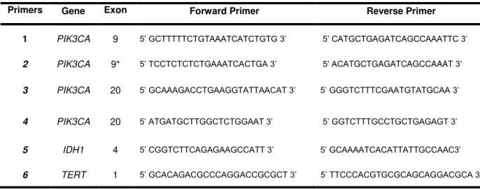
Table 3.2. Sequence of the PIK3CA, IDH1 and TERT promoter primers used for PCR reactions ... 21
Table 3.3. PCR conditions used for each pair of primers targeting PIK3CA, IDH1 and TERT ... 22
Table 4.1. Effect of the 2016 WHO classification in the subdivision of gliomas samples………...25 Table 4.2. Prevalence of gliomas molecular subgroups characterized according to the 2016 WHO classification………..25
Table 4.3. Patients characterization based on the molecular subgroup established…….………26
Table 4.4. Medians for the survival time of each histological subgroup.………..27
Table 4.5. Medians for the survival time of each glioma molecular subgroup………...28
Table 4.6. Univariate and Multivariate Cox regression for the 2007 and 2016 WHO classifications…29 Table 4.7. Ratios used to calculate the percentages of EGFR amplification, PTEN deletion, TERT mutations, 1p/19q codeletion and MGMT methylated………31
Table 4.8. Univariate and multivariate Cox Regression analysis of overall survival………...38
Table 4.9. Percentage of PIK3CA mutations identified in the cohort in study……….39
Table 4.10. Description of the PIK3CA mutations already reported identified in our cohort……….40
Table 4.11. Unreported variants identified in PIK3CA and estimation of their impact through in silico analysis………..41
Table 4.12. Clinical and molecular data from samples with PIK3CA mutated GBM in our cohort…….42
Table 4.13. Clinical and molecular data from samples with PIK3CA mutated oligodendrogliomas……43
Table 4.14. Clinical and molecular data from samples with PIK3CA mutated astrocytomas ………...44
Table 4.15. Frequency of Rs45455192 in glioma molecular subgroups………45
List of abbreviations, symbols and conventions
A - Adenine
ALT - Alternative Lengthening of Telomeres amp - amplification
ATRX - Alpha-Thalassemia Syndrome X-linked bp - Base pairs
CIC - Capicua transcriptional repressor C - Cytosine
CNS - Central Nervous System
COSMIC - Catalogue of Somatic Mutations in Cancer
CRISPR- CAS9 - Clustered Regularly Interspaced Short Palindromic Repeats associated protein 9 nuclease
CRT - Chemoradiotherapy
DAXX- Death-associated protein 6 del - deletion
DNA - Deoxyribonucleic acid
dNTP's- Deoxynucleotide triphosphates ddNTP's-Dideoxynucleotide triphosphates EDTA - Ethylene Diamine Tetraacetic Acid EGF - Epidermal Growth Factor
EGFR - Epidermal Growth Factor Receptor
ERK - Extracellular signal- Regulated Kinase F- Female
FTB - Flow-through buffer
FUBP1- Far upstream element binding protein- 1 G - Guanine
GBM - Glioblastoma
HGMB - Human Gene Mutation Database
IDH - Isocitrate Desidrogenase
IPOLFG - Instituto Português de Oncologia de Lisboa Francisco Gentil JNK - Jun K-terminal Kinase
M- Male
MAPK - Mitogen Activated Protein Kinase
Mdm2 - murine double minute 2
MGMT - O-6-methylguanine-DNA methyltransferase
MgCl2 - Magnesium Chloride min - minutes
n - number of samples
NA - Not available NaAc - Sodium Acetate NaCl - Sodium Chloride
NADP - Nicotinamide Adenine Dinucleotide Phosphate
NADPH - Reduced Nicotinamide Adenine Dinucleotide Phosphate NOS - Not Otherwise Specified
p- short arm
P - P-value
p110- catalytic subunit p85 - regulatory subunit
PCR - Polymerase chain reaction
PCV - Procarbazine, lomustine, and vincristine PDK - Phosphoinositide – dependent kinases PH - Pleckstrin Homology
PI3K - Phosphatidylinositol 3-kinase
PIK3CA - Phosphatidylinositol-4,5-bisphosphate 3-kinase catalytic subunit alpha PIP3 - Phosphatidylinositol 3,4,5 – triphosphate
PIP2 - Phosphatidylinositol 4,5- bisphosphate
PTEN - Phosphatase and Tensin homolog deleted on chromosome 10 q- long arm
QT - Chemotherapy
RAS -Rat Sarcoma Virus Homolog RT - radiotherapy
SDS - Sodium Dodecyl Sulfate sec - seconds
SIFT-Sorting Intolerant From Tolerant SNP - Single Nucleotide Polymorphism
STAT - Signal Transducer and Activator of Transcription proteins Std. - Standard
T- Thymidine
Ta - annealing temperature TBE - Tris/Borate/EDTA TE - Tris - EDTA buffer
TET - Ten-Eleven Translocation
TERT- Telomerase Reverse Transcriptase Promoter
TP53 - Tumor protein TMZ- Temozolomide
3'UTR - Untranslated region VEP - Variant Effect Predictor WHO - World Health Organization wt - wildtype
1. Introduction
1. Principles of carcinogenesis
Cancer is one of the major public health problems worldwide, being the second leading cause of death affecting countries with different income levels (World Health Organization (WHO), 2018). Recently, it was predicted the total number of new cancer cases diagnosed will increase around 70% over the next two decades paralleled by an increasing number of deaths (Montagnana and Lippi, 2017).
Carcinogenesis is described as a multistep process resulting from the influence of genetic and environmental factors, which drive to the progressive transition of a normal cell to a neoplastic state, due to the acquisition of several hallmarks (Hanahan and Weinberg, 2000). The hallmarks of cancer correspond to eight distinctive and complementary capabilities, purposed in 2000 and then complemented in 2011 by Hanahan and Weinberg (Hanahan and Weinberg, 2000; Hanahan and Weinberg, 2011).
Figure 1.1 – Schematic representation of the 10 hallmarks of cancer and their respective targeting therapies. The scheme
highlights the drugs which could interfere with each of the capabilities needed for tumor growth and progression. These therapeutic approaches correspond to strategies purposed by many investigators in an attempt to block the cancer progression, based on their transversal features. Adapted from Hanahan, D., (2011) Cell, 144(5), 646-674.
Firstly in 2000, it was described six hallmarks of cancer to understand the features shared between the different types of neoplasms that explain the transition of normal cells into a neoplastic state (Hanahan and Weinberg, 2000). Sustaining the proliferative signaling, evading growth suppressors, resistance to cell death , enabling replicative immortality, capability to induce angiogenesis and activating invasion and metastasis were the six hallmarks of cancer defined (Figure 1.1) (Hanahan and Weinberg, 2000). These were the physiological changes noticed during the development of the malignant tumors, which play important roles in the malignant growth of cancer cells.
and tumor promoting inflammation (Hanahan and Weinberg, 2000). The breakdown in one or several genes belonging to the repair machinery and the increased sensitivity to mutagenic agents are the main reasons for the genome instability (Salk et al., 2010). Lately, it was also reported tumor associated inflammatory response stimulates tumorigenesis and tumor progression, because inflammation release bioactive molecules that facilitate invasion and metastasis (Colotta et al., 2009).
Then in 2011, Hanahan and Weinberg, introduced two new emerging hallmarks which are, capability to reprogram energy of metabolism and evade immune destruction (Figure 1.1), to contextualize the hallmarks according to the conceptual progress and recent advances on the last decade (Hanahan and Weinberg, 2011). Otto Warburg verified for the first time, that even in the presence of oxygen, cancer cells can reprogram their glucose metabolism and consequently the way how to produce energy, through glycolysis (Warburg, 1956a; Warburg, 1956b). In addition, the immune destruction is another barrier that tumor cells need to overcome, because the immune system is the primary entity responsible for the detection and elimination of cancer cells, preventing tumor formation (Hanahan and Weinberg, 2011).
Currently, these hallmarks continue to be an important source to understand the cancer biology, although this knowledge has been expanded and updated over the years.
Recently, the concept of tumorigenesis has been described as tissue and cell type specific, suggesting that this process is influenced by a context (Schneider et al., 2017). Many studies have been investigated common therapeutic targets between the different types of tumors, but according to this statement those kinds of treatments are increasingly useless (Seshacharyulu et al., 2012; Zhang, 2012; Dillon and Miller, 2014). Even at the intratumoral level there is a high heterogeneity of cells, it is believed that cancer cells act as communities and that this cooperative behavior of subclones can influence disease progression (Tabassum and Polyak, 2015). The big challenge is to understand how these communities interact with each other, and how these dynamics change according to the tumor microenvironment. It is necessary to study these events and all the mechanisms by which cancer evolves to develop efficient therapies.
2. The tumors of Central Nervous System
The concept of brain tumors refers to a mixed group of neoplasms originating from intracranial tissues and meninges with degrees of malignancy ranging from benign to malignant (McKinney, 2004). Brain cancer constitutes a complex and heterogeneous group of tumors responsible for 3% of cancer cases worldwide (Miranda-Filho et al., 2016). Over time, the incidence of these tumors has increased and differs according to gender, age, race, ethnicity, and geography (Miranda-Filho et al., 2016; Ostrom et al., 2015). These tumors are rare when compared with other types of cancers, although they constitute an important source of morbidity and mortality (Miranda-Filho et al., 2016; De Robles et al., 2014). Recent studies have reported that primary brain malignancies represent only 1.4% of all cancers in adults, this incidence increases when benign and metastatic brain tumors are considered, which are more common (Vargo, 2017).
common solid tumors, representing more than 20% of all pediatric cases (Peris-Bonet et al., 2006). The annual incidence of pediatric brain tumors is estimated to be approximately 2.9 and 5.05/100,000 children in Europe and in the United States (US), respectively (Ostrom et al., 2015). Thus, it is possible to understand that pediatric and adult central nervous system tumors are two distinct realities, differing significantly not only in the epidemiology but also in the type of malignancies (Bondy et al., 2008; Santos et al., 2016; Lannering et al., 2009).
Several types of CNS malignancies are described such as meningiomas, gliomas, medulloblastomas, choroid plexus tumors, germ cell tumors, neuronal and mixed neuronal - glial tumors, melanocytic tumors, lymphomas, between others (Louis et al., 2016). Considering the set of benign tumors, meningiomas correspond to the most frequent type of brain tumor in the adult population (Wiemels et al., 2010). On the other hand, the incidence of medulloblastomas, ependymomas and pilocytic astrocytoma decreases with age, since these neoplasms are more prevalent in children than in adults (Santos et al., 2016; Lannering et al., 2009; Bauchet et al., 2008; Alexiou et al., 2011).
In addition, epidemiological studies in many parts of the world have reported that brain tumors occur more frequently in men than in women, exhibiting a male: female incidence ratio that range from 1.5:1 to 3:1 (Sun et al., 2015; Sun e Rubin, 2012; Sun et al., 2014). Regarding risk factors, the exposure to moderate - high dose ionizing radiation is the single unequivocal environmental risk factor established for brain tumors (Thompson et al., 1994; Braganza et al., 2012). Besides this, another types of pathologies could elevate the tendency to develop brain tumors (Reilly, 2009).
3. Gliomas
Gliomas are among the most common malignant primary tumors of the CNS in adults, representing 81% of all malignant brain tumors (Ostrom et al., 2014; Ferris et al., 2017). This group of tumors comprises a great clinical, histological and genetic heterogeneity.
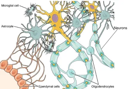
The term glioma defines any tumor that arises from the transformation of glial cells, mainly astrocytes, oligodendrocytes and ependymal cells or its precursors (Canoll and Goldman, 2008). Astrocytes are star shaped cells which represent the most abundant fraction of glial cells in the adult brain (Figure 1.2). These cells are involved in a wide range of functions: formation of the blood – brain barrier, detoxification of toxic compounds, modulation of neuronal damages, physical and metabolic support to neurons, buffering of neurotransmitter levels, maintenance of CNS homeostasis and participation in the formation of synapses (Purves et al., 2012). Astrocytomas derived from astrocytes, which constitute the supportive tissue of neurons.
Oligodendrocytes are myelin producing cells that insulate neurons, leading to the formation of myelin sheaths, allowing a rapid saltatory conduction of the electric impulse in neurons (Figure 1.2) (Purves et al., 2012). Oligodendrogliomas arrive from oligodendrocytes, being the third most common type of glioma, representing approximately 4.2% of all primary brain tumors (Mørk et al., 1985).
2-3 cases per 100000 in Europe and North Europe (Jemal et al., 2010; Rock et al., 2012
)
. GBM constitute the most common type of malignant brain tumors, accounting for more than half of all newly diagnosed gliomas (Reifenberger et al., 2016). Despite the efforts to reach efficient therapies against GBM, it continues to be the most aggressive and lethal brain tumor associated with a dismal prognosis. Around 95% of these tumors are located in cerebral hemispheres, while few percent emerge from cerebellum and brainstem (Nakada et al., 2011). The patients with GBM usually have a median overall survival of 12 to 15 months from diagnosis (Stupp et al., 2005; Koshy et al., 2011).The other group of glial cells is responsible for coating the brain ventricles and the central canal of the spinal cord. Ependymal cells can also facilitate the cerebrospinal fluid movement (Purves et al., 2012). Microglial cells are mainly derived from hematopoietic precursor cells, being categorized as a type of macrophage, capable of removing cellular debris from sites of injury, controlling pathogen invasion and tissue damage by inducing an inflammatory response (Purves et al., 2012). These cells seem to contribute to glioma growth and invasion, having tumor-supporting roles which result from the effect of glioma derived molecules (Li and Graeber, 2012). Thus, these complex and heterogeneous tumors can arrive from different types of brain cells, when they are exposed to damages. After being exposed to damages, the glial cells have the capacity to proliferate and propagate the lesion (Purves et al., 2012). This is the reason why gliomas are the most common malignant type of brain tumors.
Figure 1.2 – Schematic representation of the glial cell types interacting with neurons. It is represented the myelin sheathes formed by oligodendrocytes and the metabolic and physic support function of astrocytes to neurons. Ependymal cells have essentially a structural function, forming cellular compartments, as evidenced in the figure. Adapted from https://courses.lumenlearning.com/wm-biology2/chapter/glial-cells/.
4. Molecular alterations of adult gliomas
Over the last decades, it has been found molecular alterations frequently present in gliomas, some of them are referred as playing a central role as biomarkers for diagnosis and the others as an important complement to the diagnosis and prognosis (van den bent et al. 2017). Even if these genes do not confer additional information about patient’s clinical features, they contribute to the understanding of the tumor development. In this section are mentioned the main molecular alterations found in gliomas.
4.1. Isocitrate Dehydrogenase (IDH)
The isocitrate dehydrogenase (IDH) is a nicotinamide adenine dinucleotide phosphate (NADP) dependent enzyme that catalyzes the oxidative decarboxylation of isocitrate to form α-ketoglutarate in the tricarboxylic acid cycle producing reduced nicotinamide adenine dinucleotide phosphate (NADPH) (Keys and McAlister-Henn, 1990). IDH1 is located within the cytoplasm and peroxisomes and IDH2 is located in the mitochondria. In addition, IDH mutations lead to the production of altered isocitrate dehydrogenases forms, which convert α-ketoglutarate into 2-hydroxyglutarate (Dang et al., 2009). Under these conditions the oncometabolite 2-hydroxyglutarate is produced and the levels of α -ketoglutarate decrease.
Furthermore, 2-hydroxyglutarate is responsible by the inhibition of α-ketoglutarate dependent dioxygenases such as ten-eleven translocation (TET) family, 5-methylcytosine hydroxylases and the Jumonji- C-domain-containing histone-lysine demethylases (Xu et al., 2011). This metabolic alteration lead to a global methylated state of CpG islands, which consequently induce the methylation of MGMT gene promoter and a better response to chemotherapy. Another possible hypothesis is that 2-hydroxyglutarate inhibits the α-ketoglutarate- dependent allkB homolog DNA repair enzymes, inducing the accumulation of DNA damages, which sensitize cells to the alkylating agents (Wang et al., 2015).
In gliomas, it has been identified two types of IDH mutations (in the IDH1 and IDH2 genes) and all of them are missense mutations involving a single amino acid change at arginine 132 (R132) of IDH1 or the analogous residue in IDH2 (R172) (Balss et al., 2008). The amino acidic alteration occurs in the active site of the enzyme, impairing the isocitrate binding. The IDH2 mutations are relatively rare compared to the IDH1 R132H mutations that represent 90% of all IDH mutations (Balss et al., 2008). In summary, IDH mutations are associated with a favorable prognosis of gliomas and it is speculated that they could select patients who would benefit from chemotherapy.
4.2. 1p/19q Codeletion
association between 1p/19q codeletion and the genesis of oligodendrogliomas (Louis et al., 2016). Besides this, it has been noticed that 1p/19q codeled tumors are associated with a better response to chemotherapy and consequently to patient’s better prognosis, however the mechanism involved in this increased sensitivity is unclear (Griffin et al., 2006).
Furthermore, other molecular alterations have been investigated in order to understand their association with oligodendrogliomas and 1p/19q loss. For example, capicua transcriptional repressor (CIC) located at the long arm of chromosome 19, has been described as frequently mutated in oligodendrogliomas (69% of the cases analyzed) (Yip et al., 2011). In other studies it is also mentioned that all CIC mutations occur in tumors 1p/19q codeleted, and IDH mutated (Sahm et al., 2012).
Far upstream element binding protein- 1 (FUBP1) located at the short arm of chromosome 1, is another gene that sometimes appear mutated in oligodendrogliomas (Yip et al., 2011). However, the CIC mutations are much more common than FUBP1 mutations in oligodendrogliomas, and usually FUBP1 mutations only appear in tumors CIC mutated (Sahm et al., 2012). However, the correlation of FUBP1 mutations and 1p/19q codeletion is unclear.
4.3. Alpha-Thalassemia/ Mental Retardation Syndrome X-linked (ATRX)
The ATRX gene was identified for the first time in patients with x-linked mental retardation syndrome characterized by psychomotor difficulties and facial dimorphism (Gibbons et al., 1995). It is known that ATRX encode a histone chaperone protein involved in chromatin remodeling and transcription through the formation of a complex with DAXX (death-associated protein 6) responsible by the inclusion of H3.3 histone proteins into the telomeric regions (Goldberg et al., 2010). The disruption of this complex lead to the alternative lengthening of telomeres (ALT) and genomic instability (Lovejoy et al., 2012).
In gliomas, ATRX alterations are present in around 70% of IDH mutated tumors from astrocytic lineage, although ATRX loss and 1p/19q codeletion seem to be mutually exclusive (Kannan et al., 2012). This fact suggests that ATRX analysis may help to differentiate the astrocytic from oligodendroglial lineages. Furthermore, it has been described an association between ATRX alterations, TP53 mutations and ALT phenotype in astrocytic tumors (Liu et al., 2012). Patients with astrocytomas containing ATRX loss and IDH mutated have prolonged overall survival, seeming that ATRX loss defines a subgroup of astrocytic tumors with favorable prognosis (Wiestler et al., 2013). In GBM, ATRX loss is identified in a reduced percentage of tumors, and it seems that its role is not specific (Nandakumar et al., 2017).
Therefore, ATRX loss is an important alteration to validate the diagnosis of astrocytomas, and it may be useful in doubtful cases to distinguish the different gliomas subgroups (Louis et al., 2016).
4.4. Tumor Protein (TP53)
human cancers and its inactivation lead to tumor cells invasion, proliferation and survival (Muller and Vousden, 2013).
Moreover, these mutations are present in 70% of astrocytomas, more specifically in 95% of IDH mutated tumors without 1p/19q codeletion (Takami et al., 2014). However these alterations are also found in other gliomas subgroups even in a low percentage. Currently, this gene is not analyzed for routine diagnosis of gliomas for two reasons: it is correlated with distinct gliomas entities, so by itself cannot discriminate the glioma sample and because this is a long gene, becoming difficult its analysis recurring to the conventional molecular techniques (van den bent et al., 2017). TP53 mutations as well as ATRX loss are two molecular alterations, analyzed only in particular situations when the diagnosis of gliomas need to be confirmed, assuring the correct validation and differentiation of the tumor type.
4.5. Telomerase Reverse Transcriptase (TERT)
Telomerase Reverse Transcriptase (TERT) Promoteris a gene involved in telomerase activation, being responsible for the generation of its catalytic subunit (Bryan and Cech, 1999). Telomerase activity is important to maintain telomeres length, since this large multicomponent reverse transcriptase is able to recognize, bind and elongate telomeres (Bryan and Cech, 1999). However, telomerase expression is reduced in the most normal tissues (Kim et al., 1994), except for cells that contain high rates of self-renewal such as intestinal epithelium and hematopoietic stem cells (Chiu et al., 1996).
Nevertheless, it is described that tumor cells can proliferate indefinitely, maintaining the length of their telomeres, due to increased levels of telomerase activity or by ALT (Kim et al, 1994; Bell et al., 2016; Amorim et al., 2016). In addition, TERT promoter mutations were associated with increased TERT expression and consequently elevated levels of telomerase activity due to recruiting transcriptional factors that usually did not bind to TERT promoter (Bell et al., 2016 Amorim et al., 2016).
Around 80-90% of gliomas have TERT promoter mutations, suggesting that this is the main mechanism of telomerase activation (Bollam et al., 2018). Over 85% of GBM IDH wildtype and 77% of GBM IDH mutated have TERT promoter mutations (Lee et al., 2017). In addition, oligodendrogliomas also have an elevated percentage of TERT promoter mutations in approximately 97% of cases (Lee et al., 2017). The incidence of TERT promoter mutations in the astrocytoma group is less common, 20% in anaplastic astrocytomas IDH wildtype and 4.4% in anaplastic astrocytomas IDH mutated (Lee et al., 2017). All these percentages are distinct between the different studies, because of that it is needed a large cohort of well classified and characterized samples according to the 2016 WHO classification to establish the standard incidences. In gliomas as in many types of cancer, the most common mutations in TERT are C228T and C250T map -124 and -146 bp upstream of TERT ATG site (Huang et al., 2013).
survival of IDH mutated gliomas (Vuong et al., 2017). Therefore, Eckel - Passow reported that grade II and III gliomas patients with IDH and TERT promoter wildtype have worse overall survival compared to patients with IDH and TERT promoter mutated or IDH mutated alone, but showed greater overall survival compared with patients only with TERT promoter mutation (Eckel-Passow et al., 2015). Based on these results, TERT promoter mutations could be used as a biomarker in association with IDH analysis to predict patient’s prognosis. In the future, it is crucial new studies to ensure the viability of these observations.
4.6. Epidermal Growth Factor Receptor (EGFR)
Epidermal Growth Factor Receptor (EGFR) also designated as HER1 or ERBB1, is a transmembrane glycoprotein belonging to the HER superfamily of receptor tyrosine kinases (Downward et al., 1984). The EGFR activation occurs through the binding of epidermal growth factor (EGF) ligands and growth factors to its extracellular domain. This connection between the ligand and the extracellular domain leads to the dimerization of EGFR and consequently to a conformational change that activates the intracellular domain of the receptor. Once the intracellular domain is phosphorylated, the receptor activates PI3K/Akt (protein kinase B), ERK and JAK/STAT signaling pathways to ensure cell survival (Downward et al., 1984; Yarden and Pines, 2012). These pathways regulate a wide range of cellular processes such as proliferation, apoptosis, angiogenesis, metabolism, protein synthesis, autophagy, cell migration and differentiation (Hobbs et al., 2012). EGFR amplification is a structural alteration that determines the deregulation of all these cellular processes, inducing the constitutive activation of downstream signaling pathways that enhance tumor growth, migration, angiogenesis and metastatic spread. Therefore, EGFR constitutes a proto-oncogene, whose structural alterations are present in different types of cancers (Hobbs et al., 2012). EGFR amplification is a relatively common alteration identified in approximately 40%-50% of all the GBM cases (Libermann et al., 1985; Wong et al., 1987; Decker, 1990), although this amplification is predominant in GBM IDH wildtype (Sturm et al., 2012).
There is no unanimity regarding the prognostic value of EGFR amplifications in GBM, some studies have documented that EGFR amplifications in GBM do not have prognostic value (Chen et al., 2015; Quan et al., 2005) others have reported that these alterations are associated with unfavorable outcomes (Shinojima et al., 2003). Thus, the independent prognostic role of EGFR was not clearly proved.
inhibitors (Taylor et al., 2012). In the future it will be crucial to study new alternatives to inhibit the compensatory mechanisms developed in GBM, improving the efficiency of these therapies.
4.7 Phosphatase and Tensin homolog (PTEN)
Phosphatase and Tensin homolog (PTEN) deletions are frequent in gliomas, particularly in 30%-40% of GBM cases (McLendon et al., 2008; Verhaak et al., 2010). The inactivation of this gene could result from genomic alterations such as the entire or partial chromosomal loss, specific allelic loss and inactivating mutations (Srividya et al., 2010). These alterations are not restrict to CNS malignancies but also to other types of tumors such as breast, kidney and lung cancers (Simpson and Parsons, 2001).
PTEN is an important tumor suppressor gene localized in chromosome 10, which is involved in the phosphoinositol – 3 - Kinase (PI3K) pathway, the most mutated signaling pathway during gliomas development (Maehama e Dixon, 1998). This gene encode a phosphatase that converts phosphatidylinositol 3, 4, 5 – triphosphate (PIP3) into phosphatidylinositol 4,5- bisphosphate (PIP2), having the opposite function of PI3K. PTEN functions as a regulator of PIP3 levels in cells, to prevent the excessive activation of Akt, and consequently the deregulation of all the intracellular mechanisms necessary to cell survival and homeostasis (Yang et al., 2017).
The impact of PTEN deletion in patient’s prognosis is not well established, the results obtained seem to be controversial (Srividya et al., 2010; Carico et al., 2012). PTEN deletion was one of the first cytogenetic alterations introduced in gliomas diagnosis due to its high frequency and importance as an identifying feature (McLendon et al., 2008). PTEN deletion by itself is present in a higher percentage than all the other PTEN mutations in gliomas (Srividya et al., 2010). The data about these alterations are controversial, different authors are not sure about the clinical relevance of PTEN alterations or if they should be routinely assessed (van den Bent et al., 2017).
4.8. O-6-methylguanine-DNA methyltransferase (MGMT)
In addition to the biomarkers used to establish the diagnosis and characterization of gliomas, there are biomarkers that allow the prediction of the most appropriate therapies to be applied after surgery (Reifenberger et al., 2016). O-6-methylguanine-DNA methyltransferase (MGMT) promoter methylation is a predictive biomarker of benefit from alkylating-agents chemotherapy mainly in patients with IDH wildtype gliomas, particularly in elderly patients (aged ≥70 years) (Laperriere et al., 2013; Wick et al., 2012; Reifenberger et al., 2016).
of CpG islands of its promoter (Watts et al., 1997). Therefore, MGMT promoter methylation leads to decreased levels of MGMT, maintaining the alkyl groups in DNA, which is directly associated with a greater responsiveness of tumors to alkylating agents (Esteller et al., 2000; Watts et al., 1997 ).
Additionally, high levels of MGMT promoter methylation are significantly correlated with prolonged overall survival in patients with GBM treated with neoadjuvant temozolomide (Hegi et al., 2004; Chinot et al., 2007) and increased progression free survival in patients with gliomas treated upfront with temozolomide (Friedman et al., 1998). Around 40% of GBM IDH wildtype have hypermethylation of MGMT promoter, suggesting that these patients have a better response to temozolomide (Wick et al., 2014). In GBM IDH mutated, MGMT promoter methylation is present in the majority of cases, predicting a favorable prognosis, but this predictive biomarker cannot differentiate which therapy, radiotherapy or temozolomide chemotherapy, is better to be administered in patients with gliomas (Wick et al., 2013). In gliomas grade II, it was also noticed that MGMT methylated tumors respond better to temozolomide treatment applied neoadjuvant, being a good predictor of favorable progression free survival (Everhard et al., 2006). Overall, many studies have shown high levels of MGMT methylation in patients with gliomas predict prolonged survival time compared to patients with MGMT unmethylated (Li et al., 2017). Thus, the molecular analysis to determine the percent of MGMT promoter methylation is an important indicator in the determination of patients who would benefit from temozolomide chemotherapy. This predictive biomarker helps to estimate the possible behavior of patients when exposed to therapy and how the course of disease will be.
5. The evolution of WHO Classification for adult gliomas based on molecular
characterization
The international classification of human tumors published by World Health Organization (WHO) since 1956, intended to establish a classification and grade system for brain tumors that could be accepted and used worldwide (Louis et al., 2007). In the 2007 WHO classification a histological grading system was implemented as a form of predicting the behavior of each neoplasm. The histopathological grading established a hierarchic organization of brain tumors according to the malignancy level visualized by the pathologist (Kleihues et al., 1993). The malignancy scale goes from grade I to IV, whereas grade I is associated with low levels of anaplasia and better prognosis and grade IV is correlated with the highest levels of anaplasia and worst prognosis (Louis et al., 2007).
Oligoastrocytomas were an entity described in 1993 WHO classification as “tumors showing a conspicuous mixture of 2 distinct neoplastic cell types resembling the tumor cells in oligodendroglioma and diffuse astrocytoma” (Kleihues et al., 1993). This group of tumors, also designated as mixed gliomas, are brain tumors originate from both oligodendrocytes and astrocytes. The oligoastrocytoma classification has been severely contested because there are no immunohistochemical markers and molecular genetic alterations to distinguish this group of tumors from diffuse astrocytomas or oligodendrogliomas (Kleihues et al., 2002). It is not possible to establish the accurate diagnosis of oligoastrocytomas without confusing them with other glioma entities.
similarities with their origin cells (Louis et al., 2007). For many years, this methodology has been used as the gold standard to analyze the cells morphology and their degree of differentiation (van den Bent et al., 2017).
However, the long-term application of 2007 WHO classification demonstrated some weaknesses and doubts in the differentiation of glioma entities. Thus, it seems that the histopathological classification was no longer meeting the current clinical needs due to different reasons (van den Bent et al., 2017). Firstly, this classification was based on the interobserver variability, mainly in grade II and III gliomas differentiation (van den Bent, 2010). The lack of restrictive criteria, establishing the differentiation between the grades of gliomas constitute a great concern. Additionally, the oligoastrocytoma concept has been severely contested owing the necessity to validate the differences between this entity and astrocytomas and oligodendrogliomas (Sahm et al., 2014).
Based on the limitations demonstrated by the 2007 classification, several studies have highlighted the importance of introducing molecular alterations in the diagnosis and classification of brain tumors (Foote et al., 2015). During the last two decades, most studies, have defended that molecular alterations associated with histological data confers a more accurate classification and a better prediction of clinical outcome compared to the histological classification alone. This association between histological and molecular features is considered because brain tumors have a complex origin and may appear similar in terms of histology but have different underlying molecular profiles, which is only seen through molecular genetics (Louis et al., 2016).
All this insight lead to the 2016 WHO classification for brain tumors that finally includes molecular alterations in the diagnosis of gliomas, reinforcing the importance of molecular biomarkers in gliomas classification (Louis et al., 2016). This new classification breaks the century-old principle of diagnosis based entirely on microscopy. Presently, the new classification includes IDH mutations and 1p/19q codeletion as central biomarkers to define glioma subgroups (Louis et al., 2016).
In this perspective, based on the 2016 WHO classification astrocytomas were subdivided into IDH-wildtype and IDH-mutated. The great majority (70-80%) of astrocytomas correspond to the IDH mutated category (Balss et al, 2008; Hartmann et al., 2009). It has been described that the IDH mutated subgroup seems to be correlated with a better prognosis in both astrocytoma grades compared to IDH wildtype subgroup (Louis et al., 2016).
Oligodendrogliomas are now characterized by the presence of a codeletion in the 1p and 19q chromosomal arms combined with a mutation in the IDH gene family (Louis et al., 2016).
It is important to highlight that in the 2016 WHO classification the concept of mixed oligoastrocytoma disappear, remaining only oligoastrocytomas (NOS) cases in which the molecular analysis was not concluded or was inconclusive, becoming impossible to include these gliomas in the appropriate subgroup (Louis et al., 2016).
In addition to the biomarkers referenced above, other molecular alterations such as ATRX and TP53 mutations were considered in the 2016 WHO classification of gliomas (Louis et al., 2016). The molecular analysis of these two biomarkers can be done to confirm the astrocytoma diagnosis, however this analysis is not mandatory and by itself is not enough to determine the final diagnosis of an astrocytoma. Thus, the new classification included IDH mutations and 1p/19q codeletion as part of the standard diagnosis, which means that gliomas diagnosis depends on the analysis of these alterations, being the two most important biomarkers of this classification (Louis et al., 2016). The remaining molecular alterations referenced (ATRX and TP53) are complementary and may be useful for define and characterize the gliomas groups, particularly in doubtful cases (Figure 1.3).
Many other studies have been developed to investigate biomarkers which could confer some additional information about the prognosis of patients and ultimately their response to therapy. The introduction of a biomarker in a universal classification requires many studies to assure the viability and veracity of its impact. Because of this, many genes frequently mutated in gliomas such as telomerase reverse transcriptase promoter (TERT), epidermal growth factor receptor (EGFR) and phosphatase and tensin homologue (PTEN) have been studied (van den Bent et al., 2017). These genes which are not currently present in the new classification may have some importance to the routine diagnostics, to understand the overall picture, even if they are not essential for any diagnosis. This brings the problem of deciding, which genes should be routinely analyzed, which genes are optional but give additional information and which genes do not have clinical significance(van den Bent et al., 2017).
Figure 1.3 - Schematic representation of 2016 WHO classification for brain tumors, with the specific criteria to the molecular definition of each subgroup of gliomas. IDH mutations, 1p/19q codeletion, ATRX and TP53 mutations are the main biomarkers featured in this new classification. Adapted from Louis, D. et.al (2016). Acta Neuropathologica, 131(6),803-820
Astrocytoma Oligoastrocytoma Oligodendroglioma Glioblastoma
IDH mutant IDH wildtype
1p/19q codeletion ATRX loss*
TP53 mutation
IDH mutant IDH wildtype
Glioblastoma, IDH mutant
Glioblastoma, IDH wildtype
Diffuse astrocytoma, IDH mutant
Oligodendroglioma, IDH mutant and 1p/19q codeleted
After exclusion of other entities: Diffuse astrocytoma, IDH wildtype Oligodendroglioma NOS
Genetic test not done or inconclusive
Diffuse astrocytoma, NOS Oligodendroglioma, NOS Oligoastrocytoma, NOS Glioblastoma NOS * Characteristic but
not required for diagnosis
Histology
IDH
6. Importance of new biomarkers for gliomas classification
Additional biomarkers will be needed to better understand the prognostic role of these alterations in gliomas and for stratify these very complex and heterogeneous tumor entities. In this perspective, it is important to discover new possible pathways and targets that could be helpful to determine better choices of treatments (Louis et al., 2016).
In this context, it seems that are missing biomarkers that for example could clearly distinguish the astrocytomas group from the GBM group and molecular alterations that may help to characterize the GBM IDH mutated subgroup and GBM IDH wildtype subgroup, in an attempt to create subdivisions in these lethal group of tumors. GBM is the most common type of malignant brain tumors associated with fatal outcomes, so characterizing these tumors in subgroups that can impart additional information about patients is a big step.
In addition to IDH mutations, it would be interesting to identify molecular alterations that could characterize and divide astrocytomas, in order to create subgroups that would provide some additional information about their aggressiveness. The need of new biomarkers is evidenced in the 2016 WHO classification for brain tumors, since some categories are defined as NOS (not otherwise specified), because the molecular markers available are not enough to achieve the most correct glioma diagnosis (Louis et al., 2016).
In sum, it is crucial to study additional biomarkers that may help to clarify and define the different subtypes of gliomas, understanding the impact of these molecular alterations in patient’s diagnosis, prognosis and response to therapy.
6.1.
PIK3CA
(
phosphatidylinositol-4,5-bisphosphate 3-kinase catalytic subunit alpha)
In this context, it was already known the importance of PIK3CA in the aggressiveness of different types of tumors. PIK3CA mutations are frequently identified in patients with breast cancer 25%-40% (Samuels and Velculescu, 2004), endometrial 36% (Oda et al., 2005) and colon cancer 32% (Samuels et al., 2004). Nevertheless in gliomas, the percentage of PIK3CA mutations is not well established, which means that there is no concordance between the different studies.
2004). In addition, Broderick and co-authors were the only group to define the percentage of these oncogenic mutations in anaplastic astrocytomas, which was around 3% (1/31) (Broderick et al., 2004).
PIK3CA is a gene with 21 exons located on the long arm of chromosome 3 (3q26) which encodes the p110α catalytic subunit of class IA PI3K lipid kinases (Volinia et al., 1994). PI3K are important enzymes involved in the PI3K/Akt signaling pathway, which regulate many cellular activities such as proliferation, angiogenesis, growth, motility and survival (Katso et al., 2001). These enzymes
are organized in three main classes (I, II and III) based on its structure and substrate affinity (MacDougall, et al., 1995). The class I is subdivided into class IA and Class IB, the first includes p110α, p110β and p110δ catalytic subunits and the second contains p110γ catalytic subunit (Kang et al., 2006). The class IA of PI3K enzymes is the most studied, being the main focus due to its impact on tumorigenesis. Moreover, this class is characterized by the formation of a heterodimeric complex between a catalytic subunit (p110) and a regulatory subunit (p85) (Fry et al., 1992). The PIK3CA gene contains a regulatory subunit binding domain (p85), which allows the integration of signals from the receptor to the catalytic subunit (Samuels, 2004). Furthermore, this gene also contains a RAS binding domain important to perform the ERK signaling pathway activation, helical and catalytic domains (Samuels, 2004).
According to Samuel et al., the most PIK3CA mutations occurred in two small clusters in the helical (exon 9) and kinase (exon 20) domains. (Samuels et al., 2004). Exon 9 and 20 of PIK3CA are defined in distinct types of cancer as the main observed hotspots of PIK3CA mutations (Broderick et al., 2004; Samuels et al., 2004). Many articles referred E542K and E545K as the two most frequent mutations found in exon 9 of PIK3CA and H1047R and H1047Y as the main PIK3CA mutations found in exon 20 (Broderick et al., 2004; Samuels et al., 2004; Gallia et al., 2006).
Previously, it was documented that PIK3CA mutations led to the constitutive activation of class IA PI3K kinases. However, the mechanisms by which mutations in exon 9 and exon 20 of PIK3CA induce the PI3K gain of function are distinct. PIK3CA mutations in exon 9 induce the gain of function in a Ras-GTP binding dependent manner, while exon 20 mutations are dependent from the conformational change induced by the regulatory subunit (Zhao and Vogt, 2008). Moreover, the mutations in exon 20 interfere with the kinase domain of PIK3CA, inducing the independence of PI3K from the upstream signaling. On the other hand, the helical domain seems to be the local of interaction between the regulatory and the catalytic subunit, suggesting that mutations in exon 9 could block the connection between the both subunits, inhibiting the effect of the regulatory subunit (Miled et al., 2007).
Overall, the exons 9 and 20 of PIK3CA have important roles in the functionality of p110α, explaining why the mutations in these spots are the most reported.
6.2. PI3K/Akt signaling pathway
important role in the regulation of PI3K/Akt upstream and downstream signaling (Maehama and Dixon, 1998). When the PI3K/Akt signaling pathway is regulated, some PIP3 is accumulated near the plasmatic membrane of cell, leading to the recruitment of proteins, such as Akt, which contains a lipid binding domain, Pleckstrin Homology (PH) (Lai et al., 2015). The Akt protein is recruited by PIP3, but only becomes active after being phosphorylated by phosphoinositide – dependent kinases (PDK) (Vara et al., 2004). After activation, Akt phosphorylates effector proteins, such as mammalian target of rapamycin (mTOR) and murine double minute 2 (Mdm2), regulating innumerable cell activities including proliferation, apoptosis, autophagy, angiogenesis, protein synthesis and metabolism (Vara et al., 2004). In figure 1.4, it is represented all the PI3K/AKT signaling pathways and its main players.
Therefore, to prevent the development of tumors it is important to maintain the regulation of PI3K/Akt signaling pathway. The deregulation of this pathway is mainly associated with PTEN, PIK3CA and tyrosine kinases receptors genetic alterations (Lai et al., 2015). The tyrosine kinase receptors alterations, such as EGFR amplification, induce the constitutive activation of the downstream signaling pathway. PTEN inactivation, due to mutations or deletions, stimulates an excessive accumulation of PIP3 in the intracellular medium due to the loss of its ability to regulate PIP3 levels (Lai et al., 2015). PIK3CA mutations lead to conformational changes in the p110α catalytic subunit, becoming its associated PI3K independent on upstream signaling, making them constitutively active (Lai et al., 2015). In these three situations, there is an excessive accumulation of PIP3 which culminates in the over activation of Akt, causing an excessive increase in proliferation and angiogenesis and a decrease in apoptosis.
Figure 1.4 - Overview of PI3K/Akt signaling pathway and their downstream effects. The blue boxes indicate the three main genes (EGFR, PIK3CA and PTEN) involved in the deregulation of this pathway. The regulation of this enzymatic cascade is crucial to prevent the development of tumors. Adapted from Kai, L., et al. (2015) Journal Of Clinical Pathology, 68(4), 253-257.
Angiogenesis
p110α p85 PI3K (class IA)
PIK3CA
PIP2 PIP3
PTEN
PIP3
PDK
PIP3
AKT
Inhibits Promotes
Proliferation Metabolism & Protein Synthesis
6.3. Why
PIK3CA
could be a good therapeutic target?
The PI3K/Akt signaling pathway is one of the most mutated pathways during the development of gliomas (Mao et al., 2012). Therefore, all the efforts should be done to efficiently target possible molecules involved in this important pathway. Several clinical trials have been performed to generate drugs targeting EGFR, when this receptor is mutated. Although as EGFR is located upstream of this pathway, its inhibition has not been successful due to compensatory mechanisms developed by cancer cells (Westphal et al., 2017). In this point of view, maybe PIK3CA could be a potential target, since it is located more downstream compared to EGFR.
Recently, oncogenic mutations of PIK3CA have been referred as possible initial events in GBM, which means that these mutations could be an important player in GBM initiation (Lee et al., 2017). Additionally, PIK3CA mutations were referred as associated with a role in tumor multiplicity and in the heterogeneous patterns of drug-response (Lee et al., 2017). Furthermore, these mutations were defined as clonal mutations shared by all sectors of the tumor (Lee et al., 2017). Altogether these new data, seem to highlight the role of PIK3CA in GBM. Nevertheless, it is not clear if these mutations remain during GBM progression or if they are passenger mutations with an important role in the initiation of the tumor. Neither, its concrete role in response to therapy. In addition, the impact of these alterations in the other groups of gliomas is unknown, as well as in gliomas recurrences.
In a previous pioneer study, Verhaak and co-authors reported that PIK3CA mutations were found mainly in the GBM proneural subtype, which is associated with IDH mutations, and which have no survival advantage from aggressive treatment protocols (Verhaak et al., 2010) .Besides this, in 2015 The Cancer Genome Atlas Research Network, also showed that this gene is mutated in 20% of low grade gliomas with an IDH-mutation and 1p/19q codeletion (Brat., et al., 2015).
Recently, it has been developed several inhibitors targeting PI3K/Akt signaling pathway members. The problem is that the majority of inhibitors target PI3K kinases (Lai et al., 2015). As mentioned previously, PI3K are enzymes included in three classes and all of them play vital roles in cellular activities, which means that the general inhibition of these enzymes could cause adverse effects on healthy cells. In this perspective, inhibitors targeting specifically the p110α catalytic subunit could maximizes their therapeutic potential in patients with PIK3CA mutations without causing the side effects of interfering with other PI3K classes (Lai et al., 2015). For example, alpelisib (BYL719) is a specific inhibitor to PIK3CA protein, which was tested in breast cancer and currently is in phase II clinical trials (Massacesi et al., 2016). According to this statements, PIK3CA mutations may be a positive predictor biomarker for the clinical use of PIK3CA-selective inhibitors (Fritsch et al., 2014).
Until now, the difficulties in achieving a concordant PIK3CA mutational frequency are related to sample size, which introduces a great variability between the different studies (Samuels, 2004; Broderick et al., 2004; Gallia et al., 2006; Hartmann et al., 2005; Wen et al., 2012; Lee et al., 2017). As we know, gliomas are relatively rare tumors, becoming difficult to obtain a cohort with a considerable number of samples. In addition, despite the number of samples, the major concern was to acquire gliomas samples well characterized and classified, which was not easy using the 2007 WHO rules (van den Bent et al., 2017; van den Bent, 2010; Sahm et al., 2014; Louis et al., 2007). Thus, the proper classification used also potentiate this variation in the frequencies of PIK3CA mutations.
Since the new classification has been stablished, several questions were raised to finally understand the possible role of this gene as a biomarker. Presently, it is not clear, what is the role of PIK3CA mutations in the prognosis of diffuse gliomas with codeletion and /or IDH mutations, neither its role in GBM IDH wildtype versus GBM IDH mutated. It would be interesting, analyze to which subgroup of GBM these mutations are most associated. Furthermore, it is unknown if PIK3CA mutations have prognostic value in the different gliomas subtypes or if these alterations could be associated with a preferential response to therapy.
It is increasingly believed that the behavior of a tumor is explained not only by a single molecular alteration in a gene but by the interaction between different alterations in distinct genes (Selleck et al., 2017; Kraus, 2018). In this perspective, it is unknown the possible correlation between the mutations in PIK3CA and alterations in other genes. It would be useful, understand if there is any correlation between the onset of these mutations and PTEN deletion, EGFR amplification, TERT mutations, 1p/19q codeletion and MGMT methylation. Additionally, PIK3CA mutations are possibly early events in the development of tumors (Lee et al., 2017), although it is not clear the role of these mutations in gliomas clonal expansion. Importantly, the question that remains to answer, is will these mutations remain during tumor progression and relapses or are they passenger mutations.
2. Objectives
Considering the role of PIK3CA mutations in several types of cancer as well as their relevance as a putative druggable target, here, we propose to clarify the clinical impact of PIK3CA mutations in the molecular subgroups of gliomas. In this work, we intend to analyze the importance of PIK3CA mutations in gliomas using a cohort of 437 gliomas referred at the Instituto Português de Oncologia Francisco Gentil de Lisboa, from 2011 to 2016, the most were molecularly characterized for PTEN loss, EGFR amplification, IDH mutation and TERT promoter mutations, 1p/19q codelection, and MGMT methylation.
The specific aims are:
i) to reorganize the gliomas samples according to the new 2016 WHO classification;
ii) to analyse the impact of the 2016 WHO classification in the reclassification of gliomas cohort;
iii) to evaluate the impact of many biomarkers, such as IDH, TERT, PTEN, EGFR, MGMT and 1p/19q codeletion in diagnosis, prognosis and response to therapy on the Portuguese cohort of gliomas;
iv) to analyse the impact of PIK3CA mutations on the molecular stratification according with WHO 2016 classification;
v) to evaluate the impact of PIK3CA mutations on the prognosis, diagnosis, aggressiveness and response to therapy in molecular subgroups of gliomas;
vi) to investigate the potential correlation between PIK3CA mutations and other biomarkers such as: TERT mutations, PTEN loss, EGFR amplification, and MGMT methylation;
3. Material and Methods
1. Study population
Firstly, we elaborated a database of adult glioma samples, diagnosed from 2011 to 2016, referred to the Cytogenetics Lab of Instituto Português de Oncologia de Lisboa Francisco Gentil (IPOLFG). Initially, we had 486 primary glioma samples, although in this work we included only 419 primary tumors, 15 first recurrences and 3 second recurrences samples. These 67 primary samples were excluded for two reasons: lack of material to perform the molecular analysis and/or because they were histologically or molecularly doubtful cases with an uncertain diagnosis. For example cases with 1p or 19q codeletion or IDH wildtype with 1p/19q codeletion.
Additionally, the glioma cohort was reclassified and organized according the 2016 WHO classification, considering the IDH mutations and 1p/19q codeletion as central biomarkers. Furthermore, this characterization also included the genetic analysis of several markers which are frequently altered in gliomas such as: TERT mutations, MGMT methylation, PTEN deletion and EGFR amplification. The majority of this genetic analysis was previously performed by the Cytogenetic group.
The database also included clinical information from patients: age, sex, treatments administered, recurrences, overall survival (OS), and follow-up. Tumor samples were received as fresh tissue or paraffin-preserved tissue for DNA extraction.
Table 3.1. Glioma samples. This table represents all the gliomas samples studied.
n- Number of samples per group; +10 – samples already included in the astrocytoma group
Variable No
Types of gliomas GBM (n) Oligodendrogliomas (n) Astrocytomas (n) Recurrences (n)
Number of samples