PCR-Based Simple Subgrouping Is Validated
for Classification of Gliomas and Defines
Negative Prognostic Copy Number
Aberrations in
IDH
Mutant Gliomas
Shunsuke Nakae1, Hikaru Sasaki2, Saeko Hayashi2, Natsuki Hattori1, Masanobu Kumon1, Yuya Nishiyama1, Kazuhide Adachi1, Shinya Nagahisa1, Takuro Hayashi1, Joji Inamasu1, Masato Abe3, Mitsuhiro Hasegawa1, Yuichi Hirose1*
1Department of Neurosurgery, Fujita Health University, Toyoake, Aichi, Japan,2Department of
Neurosurgery, Keio University, Tokyo, Japan,3Department of Pathology, Fujita Health University, Toyoake, Aichi, Japan
Abstract
Genetic subgrouping of gliomas has been emphasized recently, particularly after the find-ing ofisocitrate dehydrogenase 1(IDH1) mutations. In a previous study, we investigated whole-chromosome copy number aberrations (CNAs) of gliomas and have described genetic subgrouping based on CNAs andIDH1mutations. Subsequently, we classified gli-omas using simple polymerase chain reaction (PCR)-based methods to improve the avail-ability of genetic subgrouping. We selectedIDH1/2andTP53as markers and analyzed 237 adult supratentorial gliomas using Sanger sequencing. Using these markers, we clas-sified gliomas into three subgroups that were strongly associated with patient prognoses. These includedIDHmutant gliomas withoutTP53mutations,IDHmutant gliomas with TP53mutations, andIDHwild-type gliomas.IDHmutant gliomas withoutTP53mutations, which mostly corresponded to gliomas carrying 1p19q co-deletions, showed lower recur-rence rates than the other 2 groups. In the other high-recurrecur-rence groups, the median pro-gression-free survival (PFS) and overall survival (OS) of patients withIDHmutant gliomas withTP53mutations were significantly longer than those of patients withIDHwild-type glio-mas. Notably, mostIDHmutant gliomas withTP53mutations had at least one of the CNAs +7q, +8q,−9p, and−11p. Moreover,IDHmutant gliomas with at least one of these CNAs had a significantly worse prognosis than did otherIDHmutant gliomas. PCR-based muta-tion analyses ofIDHandTP53were sufficient for simple genetic diagnosis of glioma that were strongly associated with prognosis of patients and enabled us to detect negative CNAs inIDHmutant gliomas.
OPEN ACCESS
Citation:Nakae S, Sasaki H, Hayashi S, Hattori N, Kumon M, Nishiyama Y, et al. (2015) PCR-Based Simple Subgrouping Is Validated for Classification of Gliomas and Defines Negative Prognostic Copy Number Aberrations inIDHMutant Gliomas. PLoS ONE 10(11): e0142750. doi:10.1371/journal. pone.0142750
Editor:Marta M. Alonso, University Hospital of Navarra, SPAIN
Received:September 4, 2015
Accepted:October 26, 2015
Published:November 11, 2015
Copyright:© 2015 Nakae et al. This is an open access article distributed under the terms of the
Creative Commons Attribution License, which permits unrestricted use, distribution, and reproduction in any medium, provided the original author and source are credited.
Data Availability Statement:All relevant data are within the paper and its Supporting Information files.
Funding:The authors received no specific funding for this work.
Introduction
Gliomas are currently classified according to their histological appearance, and the associated malignancy is defined by the World Health Organization (WHO) grading system. In cases of high grade gliomas, patients tend to show high recurrence rates and a worse prognosis. However, in some cases, the clinical course does not reflect the histological classification, warranting the use of genetic diagnoses and subgroups. We previously reported that adult supratentorial gliomas could be classified into genetic subgroups on the basis of their copy number aberrations (CNAs) using comparative genomic hybridization (CGH) and suggested that gliomas with +7q and 1p/ 19q co-deletions may have a better prognosis than those with−9p,−10q, and +7 CNAs [1].
The clinical significance ofisocitrate dehydrogenase 1(IDH1) point mutation in gliomas was first reported in 2008, the overall survival (OS) of glioblastoma patients withIDH1 -mutated glioblastoma was demonstrated to be significantly longer than that of patients with wild-typeIDH1glioblastoma [2]. Various subsequent studies confirmed the prognostic impor-tance ofIDH1mutations [3–6]. Therefore, we combined a CGH analysis with theIDH1 muta-tion status to propose the genetic subgrouping of gliomas [5]. The data demonstrated that
IDH1mutant gliomas with−1p/19q and +7q CNAs are associated with a better prognosis than that associated withIDH1wild-type gliomas.
Although these genetic subgroups were clinically informative, copy number-independent and simplified methods are desirable for genetic classification in clinical use. Therefore, in the present study we aimed to identify simpler and more widely available methods by which glio-mas could be diagnosed at many clinical institutions. We focused on Sanger sequencing to address this problem and selectedIDH1/2andTP53as markers for polymerase chain reaction (PCR) analyses.IDH2mutations were first detected in gliomas by Yan et al. [3]; similar to
IDH1mutations,IDH2mutation were associated with a better prognosis, although these muta-tions occurred at considerable lower frequency. Moreover,TP53mutations are often detected in astrocytic tumors [7] and it has been shown that these are mutually exclusive with 1p/19q co-deleted gliomas [8]. Therefore, we hypothesized that mostIDHmutant gliomas without
TP53mutations carry 1p/19q co-deletions.
Given the increase in CNAs with tumor regrowth or progression to high grade gliomas, according to our CGH analyses, the identification of common and specific CNAs for each genetic subgroup should facilitate an oncological understanding of gliomas. Although we pre-viously reported that 1p/19q co-deletions and +7q are frequently detected inIDHmutant glio-mas [5] according to our CGH data, only 68% ofIDHmutant gliomas harbored 1p/19q co-deletions and/or +7q. Therefore, in this study, we also aimed to identify common CNAs in
IDHmutant gliomas, particularly those harboringTP53mutation.
In the present study, we analyzedIDH1/2andTP53mutations in adult supratentorial glio-mas via direct sequencing and characterized these malignancies using PCR-based genetic sub-grouping, achieving greater prognostic accuracy than that achieved with pathological
classifications. In addition, we confirmed that mostIDHmutant gliomas withTP53mutations contained at least one of the CNAs +7q, +8q,−9p, and−11p. BecauseIDHmutant gliomas withTP53mutations showed high recurrence rates, we suggest that these CNAs are negative prognostic factors for patients withIDHmutant gliomas.
Materials and Methods
Patients, samples, and DNA preparation
histological diagnoses included (anaplastic) astrocytomas, (anaplastic) oligodendrogliomas, (anaplastic) oligoastrocytomas, and glioblastomas. Some patients underwent2 surgeries dur-ing their clinical course, thereby contributdur-ing to multiple glioma samples. The samples were evaluated by neuropathologists and were classified according to the WHO criteria. Tumor samples were available as frozen tissues and/or as formalin-fixed paraffin-embedded (FFPE) samples. DNA was extracted from freshly frozen tissue using DNeasy blood and tissue kits (QIAGEN) and from FFPE samples with DNA FFPE tissue kits (QIAGEN) or REPLI-g kits (QIAGEN). DNA quality was assessed via absorptiometric analyses. This study was approved by the Ethics Committee of the Fujita Health University (Approval number: 11–106). Written informed consent was obtained from each patient.
CGH
The CGH analysis was conducted as described by Hirose et al. [1]. Tumor tissues were removed from FFPE samples according to pathological appearance or MIB-1 density and tumor DNA was amplified via degenerate oligonucleotide-primed PCR (DOP-PCR). DNA from peripheral blood lymphocytes was obtained from healthy donors and was used as a con-trol. DNA from these samples was labeled with biotin–deoxyuridine triphosphate (Roche) after amplification. Subsequently, labeled DNA from tumors and normal tissues was hybrid-ized to normal metaphase spreads. After unhybridhybrid-ized probes were washed away, the spreads were counterstained with 4,6-diamino-2-phenylindole and the fluorescence intensity ratios for each chromosome were assessed using CytoVision software (Applied Imaging).
As described previously, total chromosomal gains and partial gains, such as +7 and +7q, were interpreted as different CNAs [5]; +7 was interpreted as a typical copy number change for
IDHwild-type gliomas and +7q was often detected inIDHmutant gliomas. Because gliomas with and withoutIDHmutations are thought to be evolved through different lineages [5], we assumed that the total and partial chromosomal gains would reflect different processes. How-ever, we considered total loss and partial losses (such as−10 and−10q) to be identical CNAs, although they were frequently detected inIDHwild-type gliomas that did not show differences in prognosis or histology. Accordingly, we categorized−10 and−10q as−10q.
Mutation analysis
Sanger sequencing was used to detectIDH1/2andTP53mutations in the samples. We analyzed the sequence of codon 132 forIDH1and codon 172 forIDH2. In previous studies, mostTP53
missense mutation hotspots were found in exons 5–8 [9–11], and missense mutations in the DNA-binding domains affected the prognosis of patients with breast carcinoma [12]; therefore, we investigated exons 5–8 inTP53mutation analyses. The primers used in our study were selected according to previous studies [10,13–15], and sequence analyses were conducted using ABI 3100 apparatus (Applied Biosystems).
Statistical analysis
recurrence since the median PFS ofIDHmutant gliomas ranged from 42 to 51 months according to our patient data and a previous study [16] and excluded cases if follow-up months were less than 36 months. We defined subtotal resection (STR) as a tumor resection volume of>90%.
Results
Comparison of the PCR-based genetic classification and histological
classification
The histological diagnosis of the 237 adult supratentorial gliomas evaluated in this study included astrocytomas, oligodendrogliomas, oligoastrocytomas, and glioblastomas;IDH1/2
andTP53mutation statuses were determined via direct sequencing (S1 Fig). Among the 113
IDHmutant gliomas, 42 harboredTP53mutations and 42 did not. The remaining 29 samples were from biopsies or were very old; thus those samples provided DNA of insufficient quantity or quality for analyses. BecauseTP53mutations did not affect the prognosis ofIDHwild-type gliomas according to our study, we classified gliomas asIDHmutant gliomas withoutTP53
mutations,IDHmutant gliomas withTP53mutations, andIDHwild-type gliomas. Fig 1shows the prognoses of patients grouped according to histology or genetics.
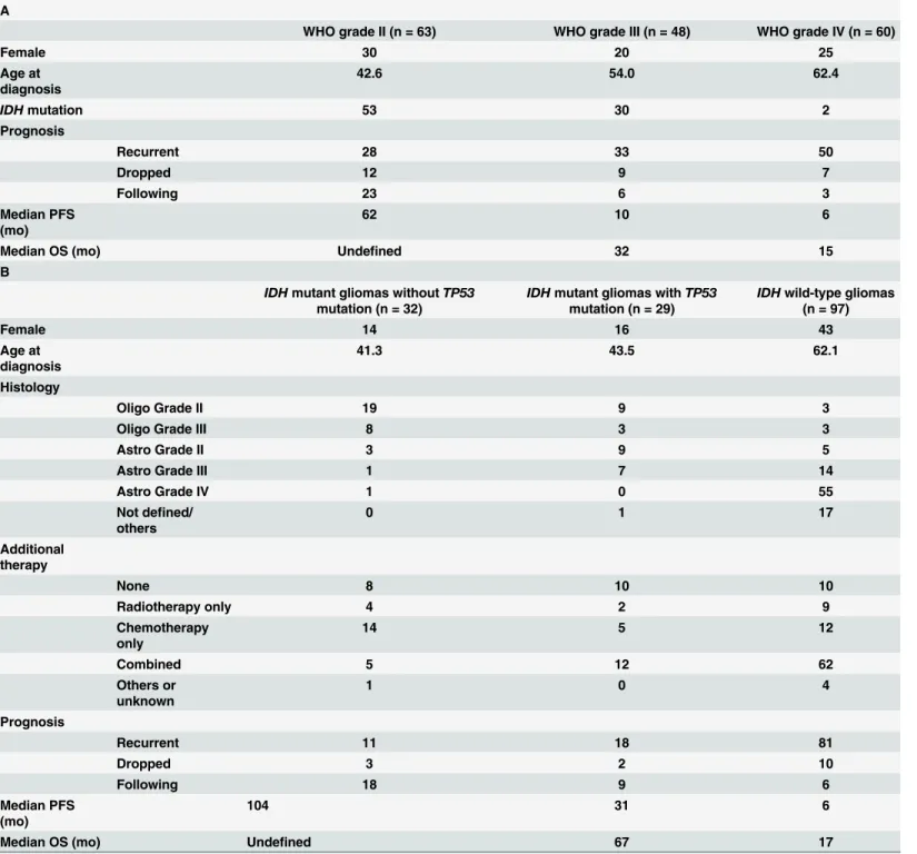
Table present patients background information, including gender, age at diagnosis, recurrence rates, median PFS, and median OS, for each histological and genetic subgroup, respectively. As this was a retrospective study, the study cases had not undergone adjuvant therapy according to strict regimen. Patients withIDHwild-type gliomas were significantly older than those withIDH
mutant gliomas (p<0.05). Those harboringIDHmutant gliomas withoutTP53mutations had a
lower recurrence rate relative to the other two subgroups (p<0.05). AlthoughIDHmutant
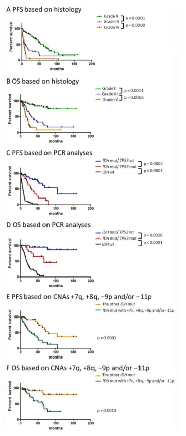
glio-mas withTP53mutations andIDHwild-type gliomas were both associated with high recurrence rates, the median PFS in the latter group was significantly shorter than that in the former group (Table 1BandFig 1C; hazard ratio = 0.229; 95% confidence interval [CI]: 0.142–0.368;
p<0.0001). In these two genetic subgroups, the median OS of patients withIDHwild-type
glio-mas was also significantly shorter than that of patients withIDHandTP53mutant gliomas (Table 1BandFig 1D; hazard ratio = 0.270; 95% CI: 0.155–0.460; p<0.0001). On the other hand,
the median OSs for grade III and IV gliomas were relatively similar (10 and 6 months, respec-tively) although the difference between grade III and IV gliomas were statistically significant. These results suggests that PCR-based genetic classification provides more precise clinical infor-mation, which includes recurrence rates and PFS, than that provided by histological classification.
Results of CGH analysis in
IDH
mutant gliomas
In a previous study, we reported a high frequency of +7 and−10q CNAs among patients with
IDH1wild-type gliomas [5]. In this study, we analyzed whole-chromosome gains and losses and identified the CNAs frequently observed inIDHmutant gliomas with and withoutTP53
mutations (Fig 2). Notably,−1p was uniquely observed inIDHmutant gliomas withoutTP53
mutations and was always accompanied by−19q. Moreover, the CNAs−4q, +7,−14q, and −19q were mainly detected inIDHmutant gliomas withoutTP53mutations. However, +7q and−9p were more frequently found inIDHmutant gliomas withTP53mutations than in otherIDHmutant gliomas; +8q,−11p, and +12p were almost exclusively detected inIDH
mutant gliomas withTP53mutations.
Fig 1. Kaplan–Meier curves of progression-free survival (PFS) according to subgroups.A comparison
of PFS and overall survival (OS) according to (A and B, respectively) pathological (n = 171) and (C and D, respectively) genetic classification (n = 158). Kaplan–Meier curves comparing PFS (E) and OS (F) associated withIDHmutant gliomas harboring CNAs +7q, +8q,−9p, and/or−11p with the PFS and OS of
otherIDHmutant gliomas (n = 73). Only patients who underwent an initial surgical intervention were included in these analyses. Abbreviations: mut; mutation, wt; wild-type.
Table 1. Background of patients who underwent initial surgical intervention.
A
WHO grade II (n = 63) WHO grade III (n = 48) WHO grade IV (n = 60)
Female 30 20 25
Age at diagnosis
42.6 54.0 62.4
IDHmutation 53 30 2
Prognosis
Recurrent 28 33 50
Dropped 12 9 7
Following 23 6 3
Median PFS (mo)
62 10 6
Median OS (mo) Undefined 32 15
B
IDHmutant gliomas withoutTP53 mutation (n = 32)
IDHmutant gliomas withTP53 mutation (n = 29)
IDHwild-type gliomas (n = 97)
Female 14 16 43
Age at diagnosis
41.3 43.5 62.1
Histology
Oligo Grade II 19 9 3
Oligo Grade III 8 3 3
Astro Grade II 3 9 5
Astro Grade III 1 7 14
Astro Grade IV 1 0 55
Not defined/ others
0 1 17
Additional therapy
None 8 10 10
Radiotherapy only 4 2 9
Chemotherapy only
14 5 12
Combined 5 12 62
Others or unknown
1 0 4
Prognosis
Recurrent 11 18 81
Dropped 3 2 10
Following 18 9 6
Median PFS (mo)
104 31 6
Median OS (mo) Undefined 67 17
A comparison of patient backgrounds according to histological classification (A) and genetic classification (B). In this table, oligoastrocytomas were classified as oligodendroglial tumor.
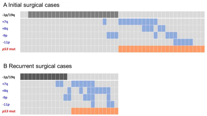
mutant gliomas withTP53mutations had at least one of the CNAs +7q, +8q,−9p, and−11p (Fig 3), and those CNAs overlapped with−1p/19q in tumors lackedTP53mutations.IDH
mutant gliomas with and without +7q, +8q,−9p, and−11p are summarized inTable 2. BecauseTP53mutations inIDHmutant gliomas were indicative of a poor prognosis and as +7q, +8q,−9p, and−11p were frequently observed inIDHmutant gliomas withTP53 muta-tions, we hypothesized that these CNAs were associated with a poor prognosis in patients with
IDHmutant gliomas. Accordingly, patients withIDHmutant gliomas who harbored at least one of the abovementioned CNAs had a significantly worse prognosis than did patients with
IDHmutant gliomas without these CNAs (p<0.0001;Fig 1E and 1F). The median PFS was 31
months for patients withIDHmutant gliomas harboring +7q, +8q,−9p, and/or−11p com-pared with 78 months for all otherIDHmutant gliomas (hazard ratio = 0.254; 95% CI: 0.128– 0.506; p<0.0001). The median OS for patients withIDHmutant gliomas who harbored these
CNAs was 65 months, whereas the median OS for all others could not be defined (hazard ratio = 0.255; 95% CI: 0.111–0.586; p<0.0001). In addition, a multivariate logistic regression
analysis revealed that the 3-year recurrence rate was higher for patients with gliomas who Fig 2. CNAs frequently detected inIDHmutant gliomas.A comparison of CNAs found inIDHmutant gliomas (A) with wild-typeTP53and (B) mutant
harbored these CNAs than for patients with other types of gliomas (S1 Table). Therefore, +7q, +8q,−9p, and−11p should be considered negative prognostic factors inIDHmutant gliomas.
The CNAs +8q,
−
9p,
−
11p, and +12p are candidate markers for tumor
progression in
IDH
mutant gliomas
In the present copy number analyses, gliomas with +7q were mainly detected in cases involving first surgeries. However, tumors harboring +8q,−9p,−11p, and +12p were frequently found after subsequent surgeries (Fig 3); +7q, +8q,−9p,−11p, and +12p emerged between the initial surgery and recurrent surgical interventions in 0, 2, 4, 3, and 4 cases ofIDHmutant gliomas withTP53mutations, respectively. Moreover, +7q was frequently detected in grade II gliomas, whereas +8q,−9p,−11p, and +12p were observed in high grade cases. These observations sug-gest that +7q is an early event, whereas +8q,−9p,−11p, and +12p may reflect tumor progres-sion inIDHmutant gliomas withTP53mutations. Among patients withIDHmutant gliomas withTP53mutations, the median PFS was 31 months for gliomas harboring any one of +8q, −9p,−11p, or +12p compared with 47 months for patients harboring all other CNAs (n = 24; p = 0.067). On the other hands, the average MIB-1 indexes was 25.4% among cases harboring +8q,−9p,−11p, and/or +12p and 8.04% in gliomas without these CNAs (n = 31; p = 0.011). These results suggest that malignancy-related genes are present in these regions.
Fig 3. The correlation betweenTP53mutations and the CNAs−1p/19q, +7q, +8q,−9p, and−11p inIDHmutant gliomas.A comparison between (A) primary (n = 47) and (B) recurrent disease (n = 24). The CNA−1p/19q represents a favorable prognostic marker; the CNAs +7q, +8q,−9p, and−11p
represents unfavorable prognostic markers.
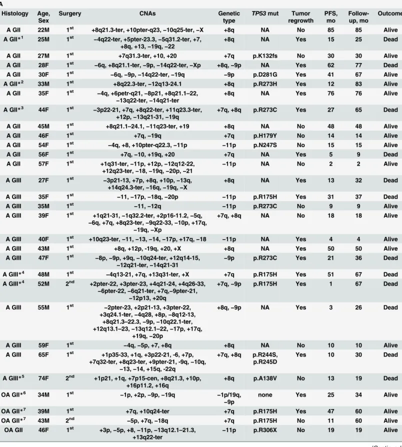
Table 2. A list ofIDHmutant glioma patients with (A) and without (B) +7q, +8q,−9p, and/or−11p according to comparative genomic hybridization
(CGH) analysis as well as their prognosis andTP53mutation status.
A
Histology Age, Sex
Surgery CNAs Genetic
type
TP53mut Tumor regrowth PFS, mo Follow-up, mo Outcome
A GII 22M 1st +8q21.3-ter, +10pter-q23,−10q25-ter,−X +8q NA No 85 85 Alive
A GII*1 25M 1st
−4q22-ter, +5pter-23.3,−5q31.2-ter, +7,
+8q, +13,−19q,−22
+8q NA Yes 15 25 Dead
A GII 27M 1st +7q31.3-ter, +10, +20 +7q p.K132fs No 30 30 Alive
A GII 28F 1st
−6q, +8q21.1-ter,−9p,−14q22-ter,−Xp +8q,−9p NA Yes 62 77 Dead
A GII 30F 1st −6q,−9p,−14q22-ter,−19q −9p p.D281G Yes 41 67 Alive
A GII*2 33M 1st +8q22.3-ter,−12q13-24.1 +8q p.R273H Yes 12 83 Alive
A GII 35F 1st
−4q, +6petr-q21,−8p21, +8q21.1–22,
−13q22-ter,−14q21-ter
+8q NA Yes 76 76 Alive
A GII*3 44F 1st
−3p22-21, +7q, +8q22-ter, +11q23.3-ter,
+12p,−13q21-31,−19q
+7q, +8q p.R273C Yes 27 65 Dead
A GII 45M 1st +8q21.1
–24.1,−11q23-ter, +19 +8q NA No 48 48 Alive
A GII 46F 1st +7q,
−19q +7q p.H179Y No 14 14 Alive
A GII 54F 1st −4q, +8, +10pter-q22.3,−11p −11p p.N247S No 15 15 Alive
A GII 56F 1st +7q,−10, +19q, +20 +7q NA Yes 5 9 Dead
A GII 57F 1st +1q31-ter,
−11p, +12p,−12q12-22,
+12q23-ter,−18,−19q,−20p,−21
−11p NA No 2 2 Alive
A GIII 27F 1st
−3p21-13, +7p, +8q, +10p,−13q,
+14q24.3-ter,−16q,−19q,−X
+8q NA Yes 13 32 Dead
A GIII 35F 1st
−11,−17p,−18q,−20p −11p p.R175H Yes 31 37 Dead
A GIII 35M 1st
−11,−12q −11p p.R273C No 9 9 Alive
A GIII 39F 1st +1q21-31,−1q32.2-ter, +2p16-11.2,−5q, −6q, +7q, +8q23-ter,−9q22-33,−10p, +17q,
−19q,−Xp
+7q, +8q NA No 18 18 Alive
A GIII 40F 1st +10q23-ter,
−11,−13,−14,−17p, +17q,−18 −11p NA Yes 4 4 Alive
A GIII 43M 1st +8q, +12p, -19q, +20, +X +8q NA Yes 50 50 Alive
A GIII 47F 1st −8p,−9p, +9q,−10q24-ter, +12q14-15, −12q21-ter,−14q21-31
−9p p.R273C Yes 21 36 Dead
A GIII*4 48M 1st −4q13-21, +7q, +13q31-ter, +X +7q p.R175H Yes 51 67 Dead
A GIII*4 52M 2nd +2pter-22, +3pter-23, +4q21-24, +4q26-33,
−6pter-22,−6q21-ter, +7q,−9pter-21, −12p13, +20q
+7q,−9p p.R175H Yes 1 67 Dead
A GIII 55M 1st −2pter-23, +2p21-13, +3pter-22,
+3q24.1-ter,−4q28, +8p,−8q12-13,
+8q21.3–22.3,−9p,−10q22.1-ter,
+12q13.1–23,−13q12.1–22,−17p, +17q,
+19q,−20p
+8q,−9p NA Yes 3 26 Dead
A GIII 59F 1st
−4q,−5p, +7, +8q +8q NA No 10 10 Alive
A GIII 65F 1st +1p35-33, +1q, +3p22-21, -6, +7p,
+7q32-ter, +8q23-ter, +9pter-21, -9q,−10q, −13,−14, +15q, -22q
+7q, +8q p.R244S, p.R245D
Yes 10 30 Dead
A GIII*5 74F 2nd +1p21, +1q, +7p15-cen, +8q21.3, +10p,
+16p11.2, +16q
+8q p.A138V No 13 19 Dead
OA GII*6 34M 1st
−1p, +2p,−9p,−19q −1p/19q, −9p
none Yes 25 34 Alive
OA GII*7 39M 1st +7q, +10q24-ter +7q p.R175H Yes 47 60 Alive
OA GII*7 43M 2nd
−5p, +7q,−18q +7q p.R175H No 11 60 Alive
OA GII 46F 1st +3p,−5p, +8,−11p,−13q12.1–21.3, +13q22-ter
−11p p.R306X No 19 19 Alive
Table 2. (Continued)
OA GI*8 61F 1st +7q31-ter,−X +7q p.Y163C Yes 20 74 Alive
OA GII*8 63F 2nd +7q31.1-ter, +12q22-ter,−Xp +7q p.Y163C No 7 74 Alive
OA GIII*9 30M 2nd
−1p,−4, +7q21.3-ter, +8, +11,−14q22-23, −18,−19q
−1p/19q,
+7q
none Yes 16 111 Alive
OA GIII*2 34M 2nd +4p,
−4q,−5qcen-13,−5q21-ter, +8q13-ter, −9pter-21.3,−11p, +12p,−12q22-23
+8q,−9p,
−11p
p.R273H Yes 48 83 Alive
OA GIII 36F 1st
−1q41-ter,−6q, +7q31-ter,−9p,−14q22-ter, −Xq21-ter
+7q,−9p p.G245S No 36 36 Alive
OA GIII 40F 1st
−1p, +1q, +3,−9, +12q14,−15q, +17, +18, −19q, +20
−1p/19q, −9p
none Yes 105 115 Alive
OA GIII*10 44F 3rd +2p,
−3p21.3–11.2, +7, +8q23-ter, +10pter-12.3,−19q13.2-ter
+8q p.Y220C Yes 2 80 Alive
OA GIII*3 47F 2nd
−4q28-ter, +7q, +8q23-ter, +12p,−Xq +7q, +8q p.R273C Yes 29 65 Dead
OA GIII*5 74F 1st +1p21, +1q, +2p16-ter, +2q,
−3p21, +7p21,
+7qcen-21, +8, +10p, +10qcen-24, +11, +12p, -12q14-23, +16q
+7q p.A138V Yes 6 19 Dead
O GII 57F 1st
−1p,−9pter-21,−18,−19q, +21 −1p/19q, −9p
none No 51 51 Alive
O GII 59M 1st +7q, +8q21.1-ter, +10p,
−13q14-32,−16q, −19q
+7q, +8q p.R248W No 36 36 Alive
O GII 71M 1st
−6pter-16, +8q21.1–21.3 +8q NA No 40 40 Alive
O GIII*11 37F 2nd
−1p, +1q,−2, +6, +7, +8,−9, +11,−16, +17, −18, +19p,−19q, +21,−22
−1p/19q, −9p
none No 8 26 Alive
O GIII 41M 5th
−1p, +7q, +8, +18p,−18q,−19q, +22q −1p/19q,
+7q
none Yes 5 76 Dead
O GIII 62M 1st
−1p, +7q31.1-ter,−19q −1p/19q,
+7q
none No 0 0 Dead
O GIII*12 64M 2nd
−1p, +2, +7,−9p, +9q,−15q,−19q −1p/19q, −9p
none Yes 27 134 Alive
O GIII 72M 1st +1q,
−2q37,−4p, +4q,−6pter-21.3, −6q16-ter,−8,−9pter-23,−11p,−14q,−17p,
+17q22-ter
−9p,−11p p.R248W Yes 4 15 Dead
GBM*1 26M 2nd
−3q11.2–24, +3q24.1-ter, +4p,−4q,
+5pter-5q23.3,−5q31.2-ter,−6pter-22.1, +6p22.2–
18.3,−6q21-26, +7, +8q,−9p, +13,−14q,
+16q, +17q,−18,−19q, +21,−22
+8q,−9p none Yes 1 25 Dead
GBM*13 28F 2nd −3pter-3q24,−5p, +7,−11p,−11q22-23.1, −13q,−19q,−22,−X
−11p p.Y220C,
p.R248W
No 24 102 Alive
GBM*14 28M 3rd −4q28-ter, +5pter-q23.3, +7q, +8q,−9p, −9q,−11pter-15.1,−11q23.1-ter, +12p,
−13q21.1–22, +13q31-ter
+7q, +8q,
−9p
p.Y236D Yes 4 67 Dead
GBM 62M 1st +2,
−6p,−7p, +7q, +8q22-ter,−9p, +9q, −11,−13q,−14q, +15q, +18q21
+7q, +8q,
−9p,−11p
NA Yes 3 4 Dead
ND*14 26M 1st +7q, +8q22.1-ter, +11q23.3-ter, +12p, +19 +7q, +8q NA Yes 27 67 Dead
ND*14 30M 4th +7q, +8q,−9p,−X +7q, +8q,
−9p
p.Y236D Yes 4 67 Dead
ND*2 38M 3rd
−5q31.1-ter, +8q22.3-ter, +10p,−10q, +12p +8q p.R273H No 20 83 Alive
B
A GII*13 22F 1st
−11q22-23.1 another p.Y220C,
p.R248W
Yes 78 102 Alive
A GII*15 22F 1st none none p.H193Y Yes 72 85 Alive
A GII 37M 1st +2q24-33,
−3p22-q25,−4q28-ter, +7 another NA No 7 7 Alive
A GII 38F 1st
−1p,−19q −1p/19q none No 14 14 Alive
Table 2. (Continued)
A GII 41M 1st −4 another p.H179R No 9 9 Alive
A GIII 55M 1st −1p, +7,−10p,−19q −1p/19q NA No 12 12 Alive
A GIII 60M 1st
−1p,−4,−9q22-ter,−14q21.3-ter,−19q −1p/19q NA No 118 118 Alive
OA GII*9 24M 1st
−1p,−19q −1p/19q none Yes 69 111 Alive
OA GII 31F 1st
−12q,−13q14.3–22 another none No 5 5 Alive
OA GII 34F 1st −1p,−19q −1p/19q none No 55 55 Alive
OA GII 34M 1st
−1p,−14q,−19q −1p/19q none No 161 161 Alive
OA GII*16 35F 1st
−1p,−19q −1p/19q none Yes 52 102 Alive
OA GII 37M 1st
−1p,−14,−19q −1p/19q none Yes 35 35 Alive
OA GII*10 40F 2nd +7 another p.Y220C Yes 22 80 Alive
OA GII 41M 1st
−4q26-ter,−5q21-ter, +7,−11q,−12q another none No 79 79 Alive
OA GII 41F 1st
−1p,−19q −1p/19q none No 43 43 Alive
OA GII 44F 1st
−1p,−14q,−19q −1p/19q none No 81 81 Alive
OA GII 45F 1st −1p, +7,−19q −1p/19q none No 5 5 Alive
OA GII 48M 1st
−1p,−19q −1p/19q none No 39 39 Alive
OA GIII*15 28F 2nd
−13q22, +18p, +19,−X another p.H193Y No 4 85 Alive
OA GIII*17 29M 1st
−1p,−19q −1p/19q none Yes 49 68 Alive
OA GIII*9 32M 3rd −1p, +3,−4, +5, +7, +9q, +10p,−10q,−13q, −15q11.2–22.3, +15q22.2-ter,−19q
−1p/19q none Yes 11 111 Alive
OA GIII*17 34M 2nd −1p,−19q −1p/19q none No 9 68 Alive
OA GIII 35M 1st
−1p,−4,−13,−18,−19q −1p/19q none No 40 40 Alive
OA GIII 36M 1st
−1p,−4, +11,−19q −1p/19q none No 116 116 Alive
OA GIII 37F 1st
−1p,−14q13-24,−19q −1p/19q none No 151 151 Alive
OA GIII 43M 1st −1p, +1q12-32.1,−1q32.2-ter, +11, +17,
+19p,−19q
−1p/19q none Yes 44 64 Dead
OA GIII 44M 1st −1p,−19q −1p/19q none No 11 11 Alive
OA GIII 74M 1st +1q, +3, +4q12-24,
−5q13.1–14, +9p,−14q, −18p
another NA No 3 3 Alive
O GII 33M 1st
−1p,−14q22-24.3,−19q −1p/19q NA No 15 15 Alive
O GII*18 34M 1st
−1p,−14,−19q −1p/19q none Yes 63 69 Alive
O GII 52F 1st
−1p,−4, +7, +11q,−15q21-ter,−18q,−19q −1p/19q NA No 12 12 Alive
O GII*12 53M 1st −1p, +7,−15q,−19q, +22q −1p/19q none Yes 104 134 Alive
O GIII*9 34M 4th
−1p,−4,−10q,−13q,−14q,−15qcen-21,
+15q24-ter,−19q
−1p/19q none Yes 9 111 Alive
O GIII*11 36F 1st
−1p,−19q −1p/19q none Yes 17 26 Alive
O GIII*6 36M 2nd
−1p,−17p,−18q,−19q −1p/19q none No 7 34 Alive
O GIII*18 39M 2nd
−1p,−14q,−19q, +21q −1p/19q none No 6 69 Alive
O GIII*16 40F 2nd −1p, +11,−14,−19q −1p/19q none No 48 102 Alive
O GIII*19 57M 2nd
−1p,−14q13-24,−19q −1p/19q none Yes 12 85 Alive
O GIII*19 58M 3rd
−1p, +7,−14q21-24.3,−15q15-22.1,−19q −1p/19q none Yes 6 85 Alive
O GIII 68F 1st
−1p, +2,−3p, +3q,−4, +5, +7, +8, +9q, 13q,
+14q, +17,−19q
−1p/19q none No 21 21 Alive
The genetic type indicates detected the CNAs which are regarded as a favorable CNA (–1p/19q) or unfavorable CNAs (+7q, +8q,−9p, and/or−11p). A
repeated number denoted by an asterisk indicates ta single patient who underwent multiple surgeries. Abbreviations: NA, not available.
Discussion
From this study, we report two major findings. First, we have shown that copy number-inde-pendent genetic subgroups determined usingIDH1/2andTP53as markers for Sanger sequenc-ing could sufficiently substitute for genetic classification with 1p/19 co-deletions. Second, via a whole-chromosome CNA analysis ofIDHmutant gliomas withTP53mutations, we have clari-fied the CNAs that contribute to poor prognosis in patients withIDHmutant gliomas.
Previous studies have confirmed that specific genetic features includingIDHmutation and 1p/19p co-deletions are excellent prognostic markers for gliomas [17,18]. In the present study, we aimed to identify copy number-independent methods that would allow a widespread clini-cal application of genetic classification of gliomas. Several previous studies reported various prognostic genes identified via mutation analyses, andATRXandTERTpromoters have recently been recognized as prognostic markers of gliomas [6,19,20]. In the present study, we selectedTP53as a prognostic marker because gliomas with 1p/19q co-deletions andTP53
mutations were previously shown to be mutually exclusive [8]; accordingly, we hypothesized thatIDHmutant gliomas with wild-typeTP53would predominantly harbor 1p/19q co-dele-tions. Our results support the previous finding that 1p/19q co-deletions andTP53mutation are mutually exclusive. The survival curves for patients with gliomas carrying 1p/19q co-dele-tions were almost identical to those of patients with wild-typeTP53, suggesting that wild-type
TP53is sufficiently indicative of 1p/19q co-deletions. In addition to the convenience of PCR-basedTP53mutation analysis, we are now investigating the relevance of prognosis, CNA, and the mutatedTP53exon for demonstrating the advantage of subgrouping according toTP53
mutation versus subgrouping according to 1p/19q co-deletions.IDHandTP53mutant gliomas that carry +7q also tend to carry mutations inTP53exon 5, suggesting that an exon 5 mutation is associated with a better prognosis inIDHandTP53mutant gliomas comparing with other types ofIDHandTP53mutant gliomas. However, the sample size is extremely low, and we would need to increase the number of analyzed samples to support this conclusion.
As shown inFig 1E and 1F,IDHmutant gliomas harboring any one of the CNAs +7q, +8q, −9p, or−11p were associated with a significantly worse survival when compared with other
IDHmutant gliomas, indicating that these CNAs are negative prognostic factors forIDH
mutant gliomas. Several studies previously reported that specific CNAs were candidate nega-tive prognostic markers in gliomas. Our previous studies suggested that gliomas carrying +7q were more likely to be associated with a shorter PFS than were gliomas carrying−1p/19q;−9p was found to be a negative prognostic factor in grade II and III gliomas [1]. Moreover, Kitange et al. and Trost et al. indicated that +8q was associated with short survival durations in patients with oligodendrogliomas [21,22], and recent studies have reported that−10q,−11p, and−19q were negative prognostic factors for low grade gliomas [16,23]. Via a whole-chromosome anal-ysis of CNAs forIDHmutant gliomas withTP53mutations, we clarified that +7q, +8q,−9p, and−11p are unfavorable prognostic factors forIDHmutant gliomas. In addition, because +12p was unique toIDHmutant gliomas withTP53mutations, we suspected that this CNA will be associated with poor survival in patients withIDHmutant gliomas. Accordingly, this CNA tended to emerge in cases involving recurrent surgical interventions or high grade glio-mas (Figs2and3). However, correlations between gliomas and +12p remain elusive. The chro-mosomal regions 7q, 8q, 9p, 11p, and 12p contain various oncogenes or tumor suppressor genes, includingMET(7q31),MYC(8q24.21),CDKN2A(9p21),CDKN1C(11p15.5), and
suppressor gene [24]. Recent studies have correlated the absence ofTP53with chromosome segregation errors and chromosomal instability [25,26], suggesting thatTP53mutations occur during the early phase of tumorigenesis inIDHmutant gliomas and cause chromosomal insta-bility and gene dysregulation in specific regions such as 7q, 8q, 9p, 11p, or 12p. Further studies of these chromosomal changes may facilitate interpretations of tumor growth processes inIDH
mutant gliomas withTP53mutations.
Our results confirmed that mostIDHmutant gliomas withTP53mutations involve at least one of the CNAs +7q, +8q,−9p, and−11p, and that mostIDHmutant gliomas with wild-type
TP53carry 1p/19q co-deletions. On the other hands, +7 and−10q are frequently detected in
IDHwild-type gliomas [5]. These results suggest that gliomas can be separated into different lineage depending onIDHmutation, andIDHmutant gliomas are further separated into two distinct linages according to theTP53mutation developing the specific CNAs in each lineage. As mentioned above,TP53mutation did not affect the prognosis of patients withIDH wild-type gliomas. In a comparison of prognosis betweenIDHwild-type gliomas and primary glio-blastomas, the median PFS (6 and 6 months, respectively) and median OS (17 and 15 months, respectively) were almost identical, suggesting that histological diagnosis can sufficiently pre-dict prognosis in cases of primary glioblastomas.
Given the high recurrence rate amongIDHmutant gliomas withTP53mutations, efforts are required to prevent progression to high grade gliomas or secondary glioblastomas, which are difficult to control with multidisciplinary treatments. To this end, studies are in progress now using OncoScan arrays (Affymetrix) for this type of glioma to identify specific regions with common losses, gains, or high copy number gains, and consequent changes in gene expression. In addition, some patients with 1p/19q co-deleted gliomas developed recurrence within a few years and these gliomas lackedTP53mutations, suggesting the presence of other genes that contribute to a poor prognoses in patients withIDHmutant gliomas.
In this study, we showed that PCR-based mutation analyses usingIDH1/2andTP53as markers could rapidly and simply classify glioma with prognostic relevance. Although patho-logical diagnoses facilitate evaluations of malignancy at the time of surgery, genetic classifica-tions provide better prognostic predicclassifica-tions, particularly in cases of WHO grade II and III gliomas. Specifically,IDHmutant gliomas carrying at least one of the CNAs +7q, +8q,−9p, or −11p were associated with a shorter survival and were predominantly associated withTP53
mutations. In conclusion, both pathological and genetic classifications are essential for glioma diagnosis and the present observations could be used to facilitate genetic classification.
Supporting Information
S1 Fig. Summary of the histology (A) andIDHandTP53mutation statuses (B) of
partici-pating patients. (TIF)
S1 Table. Multivariate analysis of 3-year recurrence amongIDHmutant gliomas (n = 53).
(DOCX)
Acknowledgments
Author Contributions
Conceived and designed the experiments: SN YH. Performed the experiments: SN MK NH HS SH. Analyzed the data: SN JI. Contributed reagents/materials/analysis tools: YN KA SN TH MH. Wrote the paper: SN JI YH. Contributed histological diagnoses of gliomas: MA.
References
1. Hirose Y, Sasaki H, Miwa T, Ohba S, Ikeda E, Abe M, et al. Whole genome analysis from microdis-sected tissue revealed adult supratentorial grade II-III gliomas are divided into clinically relevant sub-groups by genetic profile. Neurosurgery. 2011; 69: 376–90. doi:10.1227/NEU.0b013e318212bcd8 PMID:21358357
2. Parsons DW, Jones S, Zhang X, Lin JC, Leary RJ, Angenendt P, et al. An integrated genomic analysis of human glioblastoma multiforme. Science. 2008; 321: 1807–12. doi:10.1126/science.1164382 PMID:18772396
3. Yan H, Parsons DW, Jin G, McLendon R, Rasheed BA, Yuan W, et al. IDH1 and IDH2 mutations in glio-mas. N Engl J Med. 2009; 360: 765–73. doi:10.1056/NEJMoa0808710PMID:19228619
4. Hartmann C, Hentschel B, Simon M, Westphal M, Schackert G, Tonn JC, et al. Long-term survival in primary glioblastoma with versus without isocitrate dehydrogenase mutations. Clin Cancer Res. 2013; 19: 5146–57. doi:10.1158/1078-0432.CCR-13-0017PMID:23918605
5. Hirose Y, Sasaki H, Abe M, Hattori N, Adachi K, Nishiyama Y, et al. Subgrouping of gliomas on the basis of genetic profiles. Brain Tumor Pathol. 2013; 30: 203–8. doi:10.1007/s10014-013-0148-y PMID:23604523
6. Killela PJ, Pirozzi CJ, Healy P, Reitman ZJ, Lipp E, Rasheed BA, et al. Mutation inIDH1,IDH2, and in theTERTpromoter define clinically distinct subgroups of adult malignant gliomas. Oncotarget. 2014; 5: 1515–25. PMID:24722048
7. Huse JT, Aldape KD. The evolving role of molecular markers in the diagnosis and management of dif-fuse glioma. Clin Cancer Res. 2014; 20: 5601–11. doi:10.1158/1078-0432.CCR-14-0831PMID: 25398843
8. Ueki K, Nishikawa R, Nakazato Y, Hirose T, Hirato J, Funada N, et al. Correlation of histology and molecular genetic analysis of 1p, 19q, 10q,TP53,EGFR,CDK4, andCDKN2Ain 91 astrocytic and oli-godendroglial tumors. Clin Cancer Res. 2002; 8: 196–201. PMID:11801559
9. Walker DR, Bond JP, Tarone RE, Harris CC, Makalowski W, Boguski MS, et al. Evolutionary conserva-tion and somatic mutaconserva-tion hotspot maps ofp53: correlation with p53 protein structural and functional features. Oncogene. 1999; 19: 211–8.
10. Holstege H, Joosse SA, van Oostrom CT, Nederlof PM, de Vries A, Jonkers J. High incidence of pro-tein-truncatingTP53mutations in BRCA1-related breast cancer. Cancer Res. 2009; 69: 3625–33. doi: 10.1158/0008-5472.CAN-08-3426PMID:19336573
11. Leroy B, Anderson M, Soussi T. TP53 mutations in human cancer: Database reassessment and pros-pects for the next decade. Hum Mutat. 2014; 35: 672–88. doi:10.1002/humu.22552PMID:24665023
12. Vegran F, Rebucci M, Chevrier S, Cadouot M, Boidot R, Lizard-Nacol S. Only missense mutations affecting the DNA binding domain of P53 influence outcomes in patients with breast carcinoma. PLOS one. 2013; 8: 1–8.
13. Watanebe T, Nobusawa S, Kleihues P, Ohgaki H. IDH1 mutations are early events in the development of astrocytomas and oligodendrogliomas. Am. J. Pathol. 2009; 174: 1149–53. doi:10.2353/ajpath. 2009.080958PMID:19246647
14. Koga Y, Yasunaga M, Moriya Y, Akasu T, Fujita S, Yamamoto S, et al. Detection of the DNA point muta-tion of colorectal cancer cells isolated from feces stored under different condimuta-tions. Jpn J Clin Oncol. 2009; 39: 62–9. doi:10.1093/jjco/hyn129PMID:19042945
15. Solis OE, Mehta RI, Lai A, Mehta RI, Farchoukh LO, Green RM, et al. Rosette-forming glioneuronal tumor: a pineal region case with IDH1 and IDH2 mutation analyses and literature review of 43 cases. J Neuro Oncol. 2011; 102: 477–84.
16. Alentorn A, van Thuijl HF, Marie Y, Alshehhi H, Carpentier C, Boisselier B, et al. Clinical value of chro-mosome arms 19q and 11p losses in low grade gliomas. Neuro Oncol. 2014; 16: 400–8. doi:10.1093/ neuonc/not227PMID:24335697
18. McLendon RE, Herndon JE 2nd, West B, Reardon D, Wiltshire R, Rasheed BK, et al. Survival analysis of presumptive prognostic markers among oligodendrogliomas. Cancer. 2005; 104: 1693–9. PMID: 16116609
19. Jiao Y, Killela PJ, Reitman ZJ, Rasheed AB, Heaphy CM, de Wilde RF, et al. FrequentATRX,CIC,
FUBP1andIDH1mutations refine the classification of malignant gliomas. Oncotarget. 2012; 3: 709– 22. PMID:22869205
20. Yip S, Butterfield YS, Morozova O, Chittaranjan S, Blough MD, An J, et al. Concurrent CIC mutations, IDH mutations, and 1p/19q loss distinguish oligodendrogliomas from other cancers. J pathol. 2012; 226: 7–16. doi:10.1002/path.2995PMID:22072542
21. Kitange G, Misra A, Law M, Passe S, Kollmeyer TM, Maurer M, et al. Chromosomal imbalances detected by array comparative genomic hybridization in human oligodendrogliomas and mixed oligoas-trocytomas. Genes Chromosomes Cancer. 2005; 42: 68–77. PMID:15472895
22. Trost D, Ehrler M, Fimmers R, Felsberg J, Sabel MC, Kirsch L, et al. Identification of genomic aberra-tions associated with shorter overall survival in patients with oligodendroglial tumors. Int. J. Cancer. 2007; 120: 2368–76. PMID:17285580
23. van Thuijl HF, Scheinin I, Sie D, Alentorn A, van Essen HF, Cordes M, et al. Spatial and temporal evolu-tion of distal 10q deleevolu-tion, a prognostically unfavorable event in diffuse low-grade gliomas. Genome Biol. 2014; 15: 471. doi:10.1186/s13059-014-0471-6PMID:25245118
24. Shatz M, Menendez D, Resnick MA. The humanTLRinnate immune gene family is differentially influ-enced by DNA stress andp53status in cancer cells. Cancer Res. 2012; 72: 3948–57. doi:10.1158/ 0008-5472.CAN-11-4134PMID:22673234
25. de Carcer G, Malumbres M. A centrosomal route for cancer genome instability. Nat. Cell Biol. 2014; 16: 504–6. doi:10.1038/ncb2978PMID:24875738