Original Article
ISSN 0100-736X (Print) ISSN 1678-5150 (Online)
Pequenos Animais/Small Animals Diseases
RESUMO.-
[
Concentrações séricas de cortisol e tiroxina em
cadelas com carcinoma mamário em estádio inicial.
] Em
vista do fato de neoplasias serem consideradas doenças crônicas
que por meio de citocinas podem interferir na homeostase
hormonal, hipotetizou-se que o carcinoma mamário, mesmo
nos seus estádios iniciais, fosse capaz de alterar o equilíbrio
dos eixos hipofisário-tireóideo e
hipotalâmico-hipofisário-adrenal. Para tal, foram avaliadas as concentrações
séricas de cortisol basal, tiroxina (T4), tiroxina livre (fT4)
e tireotrofina (TSH) de 20 fêmeas caninas, inteiras, com
diagnóstico histopatológico de carcinoma mamário grau 1 e
estadiamento clínico I segundo a classificação da Organização
Mundial da Saúde - OMS (T1N0M0). Os animais controle
constituíram-se por 10 fêmeas caninas inteiras, em perfeitas
condições de higidez, as quais foram pareadas, por idade,
com aquelas portadoras de carcinoma mamário. Não foram
encontradas diferenças significativas nas concentrações
de cortisol basal, TSH, T4 e fT4 das cadelas portadoras de
carcinoma mamário em estádio inicial quando comparadas às
controles sugerindo que, mesmo considerados malignos, ainda
não apresentam a capacidade de alterar as concentrações dos
ABSTRACT.-
Salomão R.L., Crivellenti L.Z., Simões A.P.R., Brito M.B.S., Silva P.E.S., Costa P.B.,
Tinucci-Costa M., Santana A.E. & Borin-Crivellenti S. 2018.
Evaluation of serum concentrations
of cortisol and thyroxine in bitches with early-stage mammary carcinoma. Pesquisa
Veterinária Brasileira 38(10):1949-1954
. Faculdade de Medicina Veterinária, Universidade Federal
de Uberlândia, Avenida Pará 1720, bloco 2T, Umuarama, Uberlândia, MG 38400-902, Brazil.
E-mail:
sofiabcrivellenti@ufu.br
In view of the fact that cancer is considered a chronic disease that can interfere with
hormonal homeostasis by means of cytokines, we hypothesized that, even at early stages,
mammary carcinoma is able to alter the balance of the hypothalamic-pituitary-thyroid and
hypothalamic-pituitary-adrenal axes. To test this hypothesis, the serum concentrations of
basal cortisol, thyroxine (T4), free thyroxine (fT4), and thyroid-stimulating hormone (TSH)
were evaluated in 20 unspayed bitches that had a histopathological diagnosis of grade 1
mammary carcinoma at clinical stage I according to the World Health Organization (WHO)
classification (T1N0M0). The control animals comprised 10 unspayed bitches in perfect health
conditions that were matched with those with mammary carcinoma by age. No significant
differences regarding the concentrations of basal cortisol, TSH, t4, and fT4 were found between
the bitches carrying early stage mammary carcinoma when compared to the control group.
This suggests that, even if malignant, early-stage mammary carcinomas do not exhibit the
ability to alter the concentrations of hormones produced by the
hypothalamic-pituitary-adrenal or hypothalamic-pituitary-thyroid axes.
INDEX TERMS: Serum concentration, cortisol, thyroxine, bitches, mammary carcinoma, dogs, hormone quantification, mammary cancer, endocrine tests, clinics.
PVB-5264 AS
Evaluation of serum concentrations of cortisol and
thyroxine in bitches with early-stage mammary
carcinoma
1Ricardo L. Salomão
2, Leandro Z. Crivellenti
2, Ana Paula R. Simões
3,
Marina B.S. Brito
3, Paloma E.S. Silva
3, Paula B. Costa
2,4, Mirela Tinucci-Costa
3,
Aureo E. Santana
3e Sofia Borin-Crivellenti
2,4*
1 Received on July 7, 2017.
Accepted for publication on March 3, 2018.
2 Universidade de Franca (Unifran), Avenida Dr. Armando de Sáles Oliveira 201, Parque Universitário, Franca, SP 14404-600.
3 Faculdade de Ciências Agrárias e Veterinária (FCAV), Universidade Estadual Paulista (Unesp), Campus Jaboticabal, Via de Acesso Prof. Paulo Donato Castelane s/n, Vila Industrial, Jaboticabal, SP 14884-900, Brazil.
4 Faculdade de Medicina Veterinária, Universidade Federal de Uberlândia, Avenida Pará 1720, bloco 2T, Umuarama, Uberlândia, MG 38400-902, Brazil. *Corresponding author: sofiabcrivellenti@ufu.br
[Concentrações séricas de cortisol e tiroxina em cadelas
cinoma mamário em estádio inicial].
Salomão R.L., Crivellenti LZ., Simões A.P.R., Brito M.B.S.,
Silva P.E.S., Costa P.B., Santana A.E. & Borin-Crivellenti S.
hormônios produzidos pelos eixos
hipotalâmico-hipofisário-adrenal e tireóideo.
TERMOS DE INDEXAÇÃO: Concentração sérica, cortisol, tiroxina, cadelas, carcinoma mamário, caninos, determinações hormonais, neoplasia mamária, testes endócrinos, clínica.
INTRODUCTION
Breast cancer is considered one of the worst health issues
affecting women in the western world (Lacombe et al. 2013).
Likewise, it is a neoplastic disease that frequently affects adult
female dogs (De Nardi et al. 2009), which makes this species
an excellent model for understanding the physiopathology,
diagnostics, treatment, and prognostics of mammary carcinoma
in humans; the most frequent forms of this disease in both
humans and dogs are epithelial (carcinoma), and mixtures of
epithelial and connective tissue (carcinosarcomas or mixed
malignant tumors) (Ferreira et al. 2003, Lana et al. 2007).
Studies indicate that tumor size, involvement of lymph
nodes, and histological grade are among the factors associated
with a negative prognosis in cases of mammary neoplasia
(Novosad 2003). Several authors agree that early detection
and early therapeutic intervention are essential for preventing
spreading of this cancer to nearby or distant sites in the body
(Novosad 2003, Lana et al. 2007, De Nardi et al. 2009).
Even though bitches exhibit high mortality rates (Schneider et al.
1969) and reduced average survival time after diagnosis,
mammary cancers at early stages are frequently overlooked
by clinicians and tutors owing to the scarcity of research data
on the early stages of this disease (Sorenmo et al. 2003).
There is evidence that other factors are involved in
the progression of mammary cancer, even in early stages.
The influence of hormones on the onset, maintenance, and
progression of mammary tumors has been intensely studied,
especially in the last two or three decades (Rutteman & Misdorp
1993, Peña et al. 2003, Queiroga et al. 2005, Hardee et al.
2006, Illera et al. 2006, Obr & Edwards 2012, Brisken 2013,
Perry et al. 2013). Mammary cancer is a chronic disease that
elicits an inflammatory response; hormones and cytokines
potentially related to the occurrence of this disease include
cortisol, thyroxine, and proinflammatory cytokines (Baumann
& Gauldie 1994).
From the endocrinological standpoint, and similar to what
happens in other chronic diseases, changes in the balance
of these hormones are speculated to mislead interpretation
of hormonal tests, which could result in false positives for
important endocrine diseases such as hypothyroidism and
hypercotisolism (Buckingham et al. 1992, Carvalho & Jericó
2004), and, ultimately, lead to inadequate procedures and
treatments.
Therefore, since mammary carcinoma is a malignant
neoplasm, we hypothesized that, even at its early stages, this
disease is able to change the hormone production balance of the
pituitary-adrenal (HPA) and the
hypothalamic-pituitary-thyroid (HPT) axes.
MATERIALS AND METHODS
This experiment has been approved by the Ethics Committee on Animal Use (CEUA) of São Paulo State University (UNESP), Jaboticabal campus, SP, Brazil (protocol no. 010289/14).
Experimental groups. This prospective study included 20 unspayed bitches with a histological diagnosis of mammary carcinoma at clinical stage I according to the TNM classification of the World Health Organization. These bitches were treated at the “Governador Laudo Natel” Veterinary Hospital of FCAV/Unesp, Jaboticabal campus or at the Veterinary Hospital of University of Franca (Unifran), and were subjected to mastectomy and ovariohysterectomy (OH). The control group (CG) was composed of 10 healthy unspayed bitches that were selected and matched by age with their counterparts in the mammary carcinoma group (MCG). All the patients were evaluated by means of clinical and laboratory exams before mastectomy and/or OH. Animals were excluded from the CG whenever they exhibited any type of tumor, signs of metastasis in nearby or distant lymph nodes, and/or endocrinopathy within six months after the initial hormonal evaluation. Additional exclusion criteria in study were previous history of treatment with exogenous corticosteroid (or any other type or hormone replacement therapy), concomitant disease that involved thyroid or adrenal glands, pregnancy, presence of any tumors other than mammary carcinoma, and evidence of pulmonary, abdominal, and/or adjacent lymph node (axillary and/or inguinal) metastasis as assessed by radiographic, ultrasonographic or histopathological exams.
Sample collection. Before mastectomy, peripheral blood samples were collected by venipuncture of the jugular vein. A fraction of the blood was transferred to vacuum tubes containing K2-EDTA for hematological analyses; another fraction of the blood was transferred to vacuum tubes containing no anticoagulant, which were then centrifuged for 15 minutes at 800xg in a refrigerated centrifuge to provide serum. Serum samples were frozen at -80°C for subsequent biochemical analysis, including quantification of hormones and cytokines.
Laboratory analyses. All the hematological and biochemical laboratory analyses on samples of all the patients were performed at the “Professor Dr. Joaquim Martins Ferreira Neto” Veterinary Clinical Pathology Laboratory at “Governador Laudo Natel” Veterinary Hospital at UNESP, Jaboticabal campus, SP, Brazil, and at the Clinical Pathology Laboratory of Franca (UNIFRAN), Franca/SP, Brazil. Complete blood count was performed using automated cell counters (ABC Vet - HORIBA ABX - e pocH-100 Iv-Diff - MS Diagnóstica). Serum samples were processed using commercially available kits (Reagentes - Labtest Diagnóstica) following the manufacturer’s instructions in order to determine the concentrations of creatinine, urea, total protein, albumin, alanine aminotransferase, and alkaline phosphatase.
reference values were provided by the manufacturer or by the reference laboratory.
Statistical analysis. The data were submitted to Kolmogorov-Smirnov normality test. Comparisons between the two experimental groups were made using the Student’s t-test for variables with parametric distribution, and the Mann-Whitney test for non-parametric variables (band neutrophils, eosinophils, monocytes, creatinine, ALT, UPC, and TSH). P values less than 0.05 were considered significant. The variables are presented as mean ± standard error of the means (parametric distribution), or median with minimum and maximum interval (non-parametric variables).
RESULTS
Age and weight were used as criteria for selection of the
animals for the control group (CG), which were matched
with the animals of the mammary carcinoma group (MCG).
The average ages were 9.18±1.8 years in the MCG, and
7.80±2.97 years in the CG (P=0.1290). The average weights in
the MCG and in the CG were 10.90±6.97 kg and 10.54±4.56 kg,
respectively (P=0.8834).
In the MCG, 40% of the bitches exhibited nodules only in
the left mammary chain (n=8), 25% (n=5) of the bitches had
nodules only in the right mammary chain, and 35% (n=7)
had nodules in both chains. Most of the bitches exhibited a
single mammary nodule (40%, n=8), while 30% (n=6) had
6 nodules, 15% (n=3) had 2 nodules, and 5% had 4, 6, and
10 nodules each (n=1). The average diameter of the largest
mammary nodule of each bitch was 1.62±1.09cm. Only one
bitch had a nodule in the thoracic cranial mammary gland,
and two bitches had nodules in the thoracic and caudal
breasts. The remaining animals (n=17) had nodules in the
central, abdominal and/or inguinal breasts. Histological grade
evaluation categorized 19 tumors as grade 1, and 1 tumor
as grade 2 (Elston & Ellis 1991). The most frequent type of
tumor according to histological classification of mammary
tumors described by Misdorp et al. (1999) was tubular
carcinoma (35%, n=7), followed by complex carcinoma
(25%, n=5), mixed carcinoma (15%, n=3), complex tubular
carcinoma (10%, n=2), solid tubular carcinoma (5%, n=1),
papillary carcinoma (5%, n=1), and complex tubulopapillary
carcinoma (5%, n=1).
The results of the hematological, biochemical and urinary
analyses of the animals of the MCG and the CG are shown in
Tables 1 and 2.
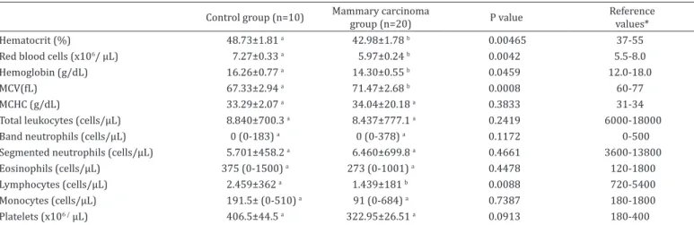
Complete blood count (CBC) results revealed that, albeit
within normal values for dogs, the variables hematocrit,
red blood cells, and hemoglobin were significantly reduced
in the MCG when compared to the CG (P=0.046, P=0.0042,
and P=0,046, respectively). The mean corpuscular volume
(MCV) was slightly higher in the MCG when compared to
the CG (P=0.0008). With the exception of lymphocytes, the
remaining variables of the leukogram did not differ significantly
between the experimental groups. Even though absolute
lymphocyte counts were within normal values for dogs, they
were significantly reduced in the MCG when compared to the
CG (P=0.0088).
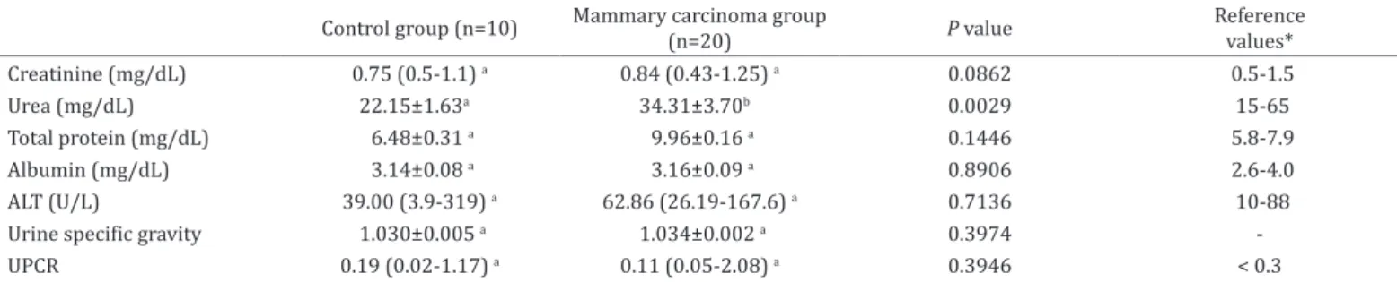
No qualitative or quantitative alterations were detected
in the urine samples from both experimental groups. With
exception of urea, the serum biochemical variables exhibited no
difference between the experimental groups (Table 2). Albeit
within the normal range for dogs, urea concentration in the
serum was significantly higher in the MCG when compared
to the CG (P=0.0294).
The hormonal evaluations revealed no alterations regarding
concentrations of cortisol (basal) or hormones that represent
the HPT axis, namely TSH, total T4, and free T4 by equilibrium
dialysis, in any of the experimental groups; all these values
were within the normal range for dogs according to the
reference values provided by the laboratory where these
analyses were performed (Table 3).
DISCUSSION
The ability of chemical mediators such as cytokines to effect
alterations on the productions and secretion of adrenal and
thyroid hormones has been known for decades (Drucker &
Shandling 1985, Baumann & Gauldie 1994). Since mammary
Table 1. Means and standard error of the means or medians (minimum-maximum) obtained for the erythrogram and leukogram of bitches in the control and mammary carcinoma groups
Control group (n=10) Mammary carcinoma group (n=20) P value Reference
values*
Hematocrit (%) 48.73±1.81 a 42.98±1.78 b 0.00465 37-55
Red blood cells (x106/ µL) 7.27±0.33 a 5.97±0.24 b 0.0042 5.5-8.0
Hemoglobin (g/dL) 16.26±0.77 a 14.30±0.55 b 0.0459 12.0-18.0
MCV(fL) 67.33±2.94 a 71.47±2.68 b 0.0008 60-77
MCHC (g/dL) 33.29±2.07 a 34.04±20.18 a 0.3833 31-34
Total leukocytes (cells/µL) 8.840±700.3 a 8.437±777.1 a 0.2419 6000-18000
Band neutrophils (cells/µL) 0 (0-183) a 0 (0-378) a 0.1172 0-500
Segmented neutrophils (cells/µL) 5.701±458.2 a 6.460±699.8 a 0.4661 3600-13800
Eosinophils (cells/µL) 375 (0-1500) a 273 (0-1001) a 0.4478 120-1800
Lymphocytes (cells/µL) 2.459±362 a 1.439±181 b 0.0088 720-5400
Monocytes (cells/µL) 191.5± (0-510) a 91 (0-684) a 0.7387 180-1800
Platelets (x106 / µL) 406.5±44.5 a 322.95±26.51 a 0.0913 180-400
cancer is a chronic disease, this work aimed at assessing the
possible hormonal alterations in bitches with mammary
carcinoma at an early stage. None of the hormonal variables
used to evaluate the afore-mentioned hormone production
axes exhibited alterations in this study. These results suggest,
therefore, that mammary carcinomas at an early stage are not
yet able to influence the hormonal homeostasis of cortisol
and thyroxine.
Integrity of the HPA axis is the major determinant of the
body’s response to stress (Reichlin 1993), and its response
is essential for the regulation of homeostasis and survival
of patients affected by chronic, critical, and inflammatory
diseases (Drucker & Shandling 1985, Sam et al. 2004).
Although several studies in humans (Annane et al. 2000,
Bollaert et al. 2003, Sam et al. 2004) and animals (Martin &
Groman 2004, Schoeman et al. 2007) have found a positive
correlation between high basal cortisol concentrations and
mortality of critical patients (Peterson et al. 1997), no data
regarding hormonal homeostasis in patients with mammary
cancer at an early stage has been found in the literature, which
highlights the importance of the data obtained in this study.
In view of the fact that chronic inflammatory diseases such
as mammary cancer are frequent in the small animal medical
routine, it is important to consider that these conditions
may mislead interpretation of hormonal tests. False-positive
results for endocrine diseases such as hypothyroidism,
hypoadrenocorticism, and hypercortisolism could lead to
implementation of inadequate proceedings and treatments
(Buckingham et al. 1992, Carvalho & Jericó 2014), which
could minimize or reduce the chances of determining the
underlying problem, and even put the patient’s life at risk.
Part of the diagnosis of endocrinopathies is done through
observation of compatible clinical signs, and also by detection
of hematological, biochemical, and urinary alterations related
to the suspected hormonal alteration (Meuten 2012). In this
regard, the fact that the blood and urinary variables evaluated
were not out of the normal range for the canine species is
not suggestive of any specific endocrine disease. However, it
should be noted that some of these variables were significantly
different between the experimental groups; therefore, they
might be related to mammary cancer. The difference regarding
absolute lymphocyte counts between the experimental
groups could have been influenced by the stress caused by
the cancer, a factor that could be intimately connected to
circulating cortisol concentrations (Gilor & Graves 2011).
On the other hand, significantly lower erythrocyte-related
variables in the CG could be indicative of increased production
of cytokines and inflammatory interleukins able to interfere
with erythropoiesis (Borin-Crivellenti & Santana 2015), as a
recent work has shown that the levels of TNF-α, IFN-γ, IL-1, and
IL-3 are significantly higher in bitches affected by mammary
carcinoma, whether the magnitude of the malignancy is low
or high (Machado et al. 2015).
Confirmatory diagnosis of hypercortisolism depends on
the demonstration of excessive production of cortisol and/or
decreased sensitivity of the HPA axis to the negative feedback
provided by glucocorticoids, and the main tests used for this
purpose are the ACTH stimulation test (ACTHST) and the low
dose dexamethasone suppression test (LDDST), respectively
Table 3. Means and standard error of the means or medians (minimum-maximum) of the serum concentrations of basal cortisol, TSH, total thyroxine, and free thyroxine in bitches of the control and mammary carcinoma groups
Control group (n=10) Mammary carcinoma group (n=20) P value Reference range*
Basal cortisol (µg/dL) 4.93±1.85 5.09±2.11 0.8570 1-4.6
TSH (µg/dL) 0.140 (0.08-0.31) 0.165 (0.07-0.79) 0.2519 0.1-0.6
TT4 (µg/dL) 1.77±0.50 1.73±0.66 0.8910 1.5-3.8
fT4 (ng/dL) 1.60±0.67 1.61±0.67 0.9833 0.82-3.65
TSH = Thyroid-stimulating hormone, TT4 = total thyroxine, fT4 = free thyroxine; * Hormone Laboratory, Provet, São Paulo/SP, Brazil. There were no significant differences between the experimental groups (P>0.05).
Table 2. Means and standard error of the means or medians (minimum-maximum) obtained for the biochemical (serum) and urinary variables of bitches in the control and mammary carcinoma groups
Control group (n=10) Mammary carcinoma group (n=20) P value Reference
values*
Creatinine (mg/dL) 0.75 (0.5-1.1) a 0.84 (0.43-1.25) a 0.0862 0.5-1.5
Urea (mg/dL) 22.15±1.63a 34.31±3.70b 0.0029 15-65
Total protein (mg/dL) 6.48±0.31 a 9.96±0.16 a 0.1446 5.8-7.9
Albumin (mg/dL) 3.14±0.08 a 3.16±0.09 a 0.8906 2.6-4.0
ALT (U/L) 39.00 (3.9-319) a 62.86 (26.19-167.6) a 0.7136 10-88
Urine specific gravity 1.030±0.005 a 1.034±0.002 a 0.3974
-UPCR 0.19 (0.02-1.17) a 0.11 (0.05-2.08) a 0.3946 < 0.3
(Gilor & Graves 2011). The ACTHST is also used to diagnose
relative or absolute adrenal insufficiency (Borin-Crivellenti
2015). Among the interleukins and necrosis factors produced
by malignant neoplasms, the cytokines and inflammatory
interleukins mentioned earlier, in addition to IL-10, are those
with the highest amount of receptors in the hypothalamus,
and binding of these molecules to their receptors stimulates
the HPA axis. Therefore, these molecules are able to trigger
secretion of corticotropin-releasing hormone (CRH) and,
consequently, of ACTH, which ultimately results in an increase
of cortisol production by the adenohypophysis (Baumann
& Gauldie 1994) that can, in turn, skew the results of the
hormonal tests mentioned earlier.
The results of this pioneering study suggest that, apparently,
mammary carcinomas at an early stage are unable to interfere
with endocrine tests or mimic endocrine diseases that affect
the axes responsible for the production of these hormones.
In spite of the existence of several types of mammary cancers,
this study included a significant number of bitches with
early-stage mammary carcinoma, a type of cancer frequently
diagnosed in small animal medicine (Lana et al. 2007), which
warrants research on more aggressive and metastatic types
of mammary cancers. Even though basal cortisol was chosen
because it is a prognostic marker frequently used as indicative
of chronic stress in patients with acute or chronic diseases
(Drucker & Shandling 1985, Bollaert et al. 2003, Sam et al. 2004),
including breast cancer (Fredrikson et al. 1992, Sephton et al.
2000, Turner-Cobb et al. 2000), dynamic functional endocrine
tests on the HPA axis, especially the ACTHST, are still needed
to clearly determine if mammary carcinomas at an early stage
in fact do not compromise the adrenal response.
CONCLUSION
Based on the results obtained in this study, we suggest that
early-stage carcinomas, even if malignant, do not have the
ability to alter the concentrations of the hormones produced
by the HPA and HPT axes.
Conflict of interest statement.- The authors have no competing interests.
Acknowledgements.- The authors would like to thank FAPESP for financial
support (#2011/08416-6 e #2011/11355-9), and Master’s degree research financial support.
REFERENCES
Annane D., Sébille V., Troché G., Raphaël J.C., Gajdos P. & Bellissant E. 2000. A 3-level prognostic classification in septic shock based on cortisol levels and cortisol response to corticotropin. J. Am. Med. Assoc. 283(8):1038-1045. <http://dx.doi.org/10.1001/jama.283.8.1038><PMid:10697064> Baumann H. & Gauldie J. 1994. The acute phase response. Immunol Today 15(2):74-80. <http://dx.doi.org/10.1016/0167-5699(94)90137-6> <PMid:7512342>
Bollaert P.E., Fieux F., Charpentier C. & Lévy B. 2003. Baseline cortisol levels, cortisol response to corticotropin, and prognosis in late septic shock. Shock 19(1):13-15. <http://dx.doi.org/10.1097/00024382-200301000-00003> <PMid:12558137>
Borin-Crivellenti S. 2015. Endocrinologia, p.231-272. In: Crivellenti L.Z. & Borin-Crivellenti S. Casos de Rotina em Medicina Veterinária de Pequenos Animais. 2ª ed. MedVet, São Paulo.
Borin-Crivellenti S. & Santana A.E. 2015. Erythropoiesis inductor and suppressor factors: a role of hormones and cytokines. Revta Conselho Federal Med. Vet. Zootec. 66:60-66.
Brisken C. 2013. Progesterone signalling in breast cancer: a neglected hormone coming into the limelight. Nat. Revs Cancer 13(6):385-396. <http://dx.doi. org/10.1038/nrc3518><PMid:23702927>
Buckingham J.C., Smith T. & Loxley H.D. 1992. The control of ACTH secretion, p.131-138. In: James V.H.T. (Ed), The Adrenal Gland: Comprehensive endocrinology. 2nd ed. Raven Press, New York.
Carvalho C.F. & Jericó M.M. 2014. Adrenais, p.121-132. In: Carvalho C.F. (Ed), Ultrassonografia em Pequenos Animais. 2ª ed. Roca, São Paulo. 468p. De Nardi A.B., Rodaski S., Rocha N.S. & Fernades S.C. 2009. Neoplasias
mamárias, p.372-383. In: Daleck C.R., De Nardi A.B. & Rodaski S. (Eds), Oncologia em Cães e Gatos. Roca, São Paulo.
Drucker D. & Shandling M. 1985. Variable adrenocortical function in acute medical illness. Crit. Care Med. 13(6):477-479. <http://dx.doi. org/10.1097/00003246-198506000-00007><PMid:2986907> Elston C.W. & Ellis I. 1991. Pathological prognostic factors in breast cancer.
I. The value of histological grade in breast cancer: experience from a large study with long-term follow-up. Histopathology 19(5):403-410. <http:// dx.doi.org/10.1111/j.1365-2559.1991.tb00229.x><PMid:1757079> Ferreira E., Bregunci G.C., Schmitt F.C. & Cassali G.D. 2003. Protocol for
the anatomopathological examination of canine mammary tumors. Arq. Bras. Med. Vet. Zoot. 55(1):105-109. <http://dx.doi.org/10.1590/S0102-09352003000100017>
Fredrikson M., Hursti T., Fürst C.J., Steineck G., Börjeson S., Wikblom M. & Peterson C. 1992. Nausea in cancer chemotherapy is inversely related to urinary cortisol excretion. Brit. J. Cancer 65(5):779-780. <http://dx.doi. org/10.1038/bjc.1992.165><PMid:1586608>
Gilor C. & Graves T.K. 2011. Interpretation of laboratory tests for canine cushing syndrome. Topics Companion Anim. Med. 26(2):98-108. <http:// dx.doi.org/10.1053/j.tcam.2011.03.001><PMid:21596349>
Hardee M.E., Arcasoy M.O., Blackwell K.L., Kirkpatrick J.P. & Dewhirst M.W. 2006. Erythropoietin biology in cancer. Clin. Cancer Res. 12(2):332-339. <http://dx.doi.org/10.1158/1078-0432.CCR-05-1771><PMid:16428469> Illera J.C., Pérez-Alenza M.D., Nieto A., Jiménez M.A., Silvan G., Dunner S. & Peña L. 2006. Steroids and receptors in canine mammary cancer. Steroids 71(7):541-548. <http://dx.doi.org/10.1016/j.steroids.2005.11.007> <PMid:16631217>
Lacombe J., Mangé A., Jarlier M., Bascoul-Mollevi C., Rouanet P., Lamy P., Maudelonde T. & Solassol J. 2013. Identification and validation of new autoantibodies for the diagnosis of DCIS and node negative early stage breast cancer. Intern. J. Cancer 132(5):1105-1113. <http://dx.doi.org/10.1002/ ijc.27766><PMid:22886747>
Lana S.E., Rutterman G.R. & Withrow S.J. 2007. Tumors of the mammary gland, p.619-636. In: Withrow S.J. & Vail D.M. (Eds), Small Animal Clinical Oncology. 4th ed. Saunders Elsevier, Philadelphia. <http://dx.doi.org/10.1016/ B978-072160558-6.50029-0>.
Machado V.S., Crivellenti L.Z., Bottari N.B., Tonin A.A., Pelinson L.P., Borin-Crivellenti S., Santana A.E., Torbitz V.D., Moresco R.N., Duarte T., Duarte M.M., Schetinger M.R., Morsch V.M., Jaques J.A., Tinucci-Costa M. & Silva A.S. 2015. Oxidative stress and inflammatory response biomarkers in dogs with mammary carcinoma. Pathol. Res. Pract. 211(9):677-681. http:// dx.doi.org/10.1016/j.prp.2015.06.011. PMid:26166177.
Martin L.G. & Groman R.P. 2004. Relative adrenal insufficiency in critical illness. J. Vet. Emerg. Crit. Care 14(3):149-157. <http://dx.doi. org/10.1111/j.1534-6935.2004.04013.x>
Misdorp W., Else R.W., Hellmén E. & Lipscomb T.P. 1999. Histological classification of the mammary tumors of the dog and the cat, p.11-58. In: Ibid. (Eds), International Histological Classification of Tumors of Domestic Animals. Vol.7. 2nd ed. World Health Organization, Genebra.
Novosad C.A. 2003. Principles of treatment for mammary gland tumors. Clin. Tech. Small Anim. Pract. 18(2):107-109. <http://dx.doi.org/10.1053/ svms.2003.36625><PMid:12831071>
Obr A.E. & Edwards D.P. 2012. The biology of progesterone receptor in the normal mammary gland and in breast cancer. Mol. Cell. Endocrinol. 357(1/2):4-17. <http://dx.doi.org/10.1016/j.mce.2011.10.030><PMid:22193050> Peña L., Silván G., Pérez-Alenza M.D., Nieto A. & Illera J.C. 2003. Steroid hormone
profile of canine inflammatory mammary carcinoma: a preliminary study. J. Steroid Biochem. Mol. Biol. 84(2/3):211-216. <http://dx.doi.org/10.1016/ S0960-0760(03)00030-X><PMid:12711005>
Perry J.K., Liu D., Wu Z., Zhu T. & Lobie P.E. 2013. Growth hormone and cancer: an update on progress. Curr. Opinion Endocrinol. Diabetes Obesity 20(4):307-313. <http://dx.doi.org/10.1097/MED.0b013e328363183a> <PMid:23807602>
Peterson M.E., Melian C. & Nichols R. 1997. Measurements of serum total thyroxine, triiodothyronine, free thyroxine, and thyrotropin concentrations for diagnosis of hypothyroidism in dogs. J. Am. Vet. Med. Assoc. 211(11):1396-1402. <PMid:9394888>
Queiroga F.L., Pérez-Alenza M.D., Silvan G., Peña L., Lopes C. & Illera J.C. 2005. Role of steroid hormones and prolactin in canine mammary cancer. J. Steroid Biochem. Mol. Biol. 94(1/3):181-187. <http://dx.doi.org/10.1016/j. jsbmb.2004.12.014><PMid:15862964>
Reichlin S. 1993. Neuroendocrine-immune interactions. New Engl. J.Med. 329(17):1246-1253. <http://dx.doi.org/10.1056/NEJM199310213291708> <PMid:8105378>
Rutteman G.R. & Misdorp W. 1993. Hormonal background of canine and feline mammary tumours. J. Reprod. Fertility 47(Suppl.):483-487. <PMid:8229967> Sam S., Corbridge T.C., Mokhlesi B., Comellas A.P. & Molitch M.E. 2004. Cortisol levels and mortality in severe sepsis. Clin. Endocrinol. 60(1):29-35. <http:// dx.doi.org/10.1111/j.1365-2265.2004.01923.x><PMid:14678284> Schneider R., Dorn C.R. & Taylor D.O.N. 1969. Factors influencing canine
mammary cancer development and postsurgical survival. J. Natl Cancer Inst. 43(6):1249-1261. <PMid:4319248>
Schoeman J.P., Rees P. & Herrtage M.E. 2007. Endocrine predictors of mortality in canine babesiosis caused by Babesia canis. Vet. Parasitol. 148(2):75-82. <http://dx.doi.org/10.1016/j.vetpar.2007.06.010><PMid:17614201> Sephton S.E., Sapolsky R.M., Kraemer H.C. & Spiegel D. 2000. Diurnal cortisol
rhythm as a predictor of breast cancer survival. J. Natl Cancer Inst. 92(12):994-1000. <http://dx.doi.org/10.1093/jnci/92.12.994><PMid:10861311> Sorenmo K.U., Shofer F.S. & Goldschmidt M.H. 2003. Effect of spaying and
timing of spaying on survival of dogs with mammary carcinoma. Vet. Clin. N. Am., Small Anim. Pract. 33:573-596. <http://dx.doi.org/10.1016/ S0195-5616(03)00020-2>