Human serum antibody reactivity towards
Paracoccidioides brasiliensis
antigens treated with sodium metaperiodate
Reatividade de anticorpos de soros humanos a antígenos de
Paracoccidioides brasiliensis
tratados com metaperiodato sódico
Ana Paula Ferreira
1, Thadeu Côrrea
1, Rosângela Cunha
2, Marcos José Marques
1,
Maria Angela Montesano
1, Maria Aparecida Souza
1and Henrique Couto Teixeira
1ABSTRACT
In this study, we evaluated the profile of anti-Paracoccidioides brasiliensis immunoglobulin isotypes in serum from patients with the acute and chronic forms of paracoccidioidomycosis, using the whole Paracoccidioides brasiliensis antigen and the antigen treated with sodium metaperiodate. All the immunoglobulin isotypes present in the serum from patients with the acute and chronic forms of paracoccidioidomycosis presented higher reactivity towards the whole antigen than to the antigen treated with metaperiodate (P < 0.05). The reactivity of IgG and IgM to the antigen treated with metaperiodate was greater in serum from patients with the acute form of the disease (P < 0.05), while IgA was more reactive in serum from patients with the chronic form (P < 0.05). There was greater reactivity of IgG1 and IgG2 to the whole antigen and the antigen treated with metaperiodate in the serum from patients with paracoccidioidomycosis than there was in serum from patients with other parasitic infections (P < 0.05). Furthermore, IgG1 from patients with the acute form recognized the 19kDa, 27kDa and 31kDa antigens in the western blot test. Thus, the results suggest that modifications to the epitopes of Paracoccidioides brasiliensis antigens may help to improve the immunodiagnosis of paracoccidioidomycosis.
Key-words: Paracoccidioidomycosis. Antigens. Metaperiodate. Diagnosis.Diagnosis.
RESUMO
Neste trabalho, foi avaliado o perfil de isotipos de imunoglobulinas anti-Paracoccidioides brasiliensis em soros de pacientes com formas crônica e aguda de paracoccidiodomicoses usando antígeno total e tratado com meta-periodato. Todos os tipos de imunoglobulinas presentes nos soros de pacientes com formas aguda e crônica apresentaram alta reatividade ao antígeno total quando comparado ao tratado com meta-periodato (P < 0,05). Houve maior reatividade de IgG e IgM anti-antígeno tratado com meta-periodato em soros de pacientes com forma aguda da doença (P < 0,05), enquanto IgA foi mais reativa em soros da forma crônica (P < 0,05). Houve maior reatividade de IgG1 e IgG2 com antígeno total e tratado com meta-periodato em soros de pacientes comparados aos com outras parasitoses (P < 0,05). Além disso, IgG1 de pacientes com a forma aguda reconhecem antígenos de 19kDa, 27kDa e 31kDa por western blot. Assim, os resultados sugerem que alterações nos epitopos de antígenos de Paracoccidioides brasiliensis podem auxiliar no aprimoramento do imunodiagnóstico da paracoccidioidomicose.
Palavras-chaves: Paracoccidiodomicose. Antígenos. Metaperiodato. Diagnóstico.
1. Laboratório de Imunologia, Deptartamento de Parasitologia, Microbiologia e Imunologia, Instituto de Ciências Biológicas, Universidade Federal de Juiz de Fora, Juiz de Fora, MG. 2. Serviço de Doenças Infecciosas e Parasitárias, Hospital Universitário, Universidade Federal de Juiz de Fora, Juiz de Fora, MG.
Financial Support: CNPq, FAPEMIG and CAPES
Address to: Dra. Ana Paula Ferreira. Laboratório de Imunologia/ Dept° de Parasitologia, Microbiologia e Imunologia/ICB/UFJF. 36036-900 Juiz de Fora, MG. Telefax: 55 32 2102-3214
e-mail: [email protected] Recebido para publicação em 18/4/2008 Aceito em 29/07/2008
Paracoccidioidomycosis (PCM), a granulomatous disease
caused by the dimorphic fungus Paracoccidioides brasiliensis,
is the most prevalent systemic mycosis in Latin America4 5.
Symptomatic individuals show one of two main clinical forms of the disease: the acute or subacute form, which is characterized by rapid and progressive infection and largely affects the mononuclear phagocyte system; and the chronic form, which frequently shows a slow course of pulmonary disease with mucocutaneous involvement.
The criteria for laboratory diagnosis of PCM rely on positive microscopic identification of the causative agent in KOH preparations or biopsy specimens, as well as on its isolation in
cultures5 12. In addition, diagnostic methods based on serological
tests have also been widely used to support identification of
microscopy-positive cases5 14 and represent an important tool for
follow-up of PCM patients6. However, serological tests normally use
crude antigenic preparations of Paracoccidioides brasiliensis,
Ferreira AP et al
Serum cross-reactivity of PCM with histoplasmosis and Jorge Lobo’s disease has also been detected when 43-kDa glycoprotein, an exocellular antigen obtained from a liquid culture medium of
Paracoccidioides brasiliensis, is used. Cross-reactivity towards gp43 seems to be due to carbohydrate epitopes, since more than 85% of the specific reactions of gp43 with serum antibodies from
PCM patients have involved protein epitopes17.
In this study, treatment with sodium metaperiodate was used to
disrupt the carbohydrate epitopes of Paracoccidioides brasiliensis
filtrate antigens and generate a purified protein antigenic preparation.
Whole Paracoccidioides brasiliensis soluble antigens (WAg) and
sodium metaperiodate-treated WAg (MPAg) were used to evaluate the profile of serum immunoglobulin isotype (total IgG, IgM, IgA,
IgG1 and IgG2) reactivity towards Paracoccidioides brasiliensis
in patients with acute or chronic forms of PCM. Furthermore, we used the western blot method to investigate serum IgG1 reactivity
towards fractionated Paracoccidioides brasiliensis antigens in
serum from acute and chronic PCM patients.
MATERIAL AND METHODS
Patients and controls. The study was conducted on serum samples obtained from twenty-nine patients with active PCM with
either the acute form (no=15) or chronic form (no=14) of the
disease. All the patients had PCM, according to the clinical diagnosis of
symptoms that indicates presence of Paracoccidioides brasiliensis
infection, and according to positive cultures and immunodiffusion
tests for crude Paracoccidioides brasiliensis antigens. The samples
were obtained from the University Hospital, Federal University of Juiz de Fora, Minas Gerais, Brazil, with the approval of the Ethics
Committee for Human Research (No 098-22/2000). For this study,
the controls comprised five normal individuals and five patients with Chagas’ disease or leishmaniasis.
Paracoccidioides brasiliensis antigenic preparation.
Whole Paracoccidioides brasiliensis soluble antigenic
preparation (WAg) was obtained as described previously10. The
isolate of Paracoccidioides brasiliensis came from an individual
with the acute form of PCM and the inoculum was cultured for 15 days in Sabouraud medium. Fungal suspensions were washed (3X) in sterile saline at 3,000rpm for 5 min. The fungal mass was resuspended in an equal volume of sterile saline containing
phenylmethylsulfonyl fluoride (PMSF) (20µl/ml) and then
subjected to sonication (100 cycles/3 min; 20X) in an ice bath. After centrifugation at 10,000rpm for 40 min, the supernatant was dialyzed against distilled water for 48 h and stored at -20ºC.
Periodate oxidation. Carbohydrate groups of WAg were disrupted by treatment with sodium metaperiodate (Sigma), which yielded a purified protein antigenic preparation (MPAg). WAg was adsorbed onto flat-bottom Immunolon II microtiter plates, washed twice with 0.05M sodium acetate (pH 4.5) and treated with sodium metaperiodate (10mM in the acetate buffer) for one hour in darkness at room temperature. Afterwards, the plates were washed twice with sodium acetate and the reaction was stopped by adding sodium borohydride (50mM in PBS)
(Sigma) for 30 min at room temperature. The plates were then washed twice with PBS.
Isotyping. Antibody isotype analyses were performed by specific
ELISA to detect anti-Paracoccidioides brasiliensis reactivity,
using plates adsorbed with WAg and MPAg Paracoccidioides
brasiliensis antigens. Horseradish peroxidase (HRP)-conjugated, isotype-specific mouse human IgG (antitotal IgG), anti-IgM, anti-IgA, anti-IgG1 or anti-IgG2 (Southern Biotechnology Associates, Inc.) were used and the assays were developed by adding tetramethylbenzidine (TMB) peroxidase substrate solution (Kirkegaard & Perry Laboratories). The reaction was stopped by
adding 1M H2SO4, and the color reaction was read at 450nm in a
microplate reader (Molecular Devices Corp).
Western blotting. WAg antigen was diluted in an SDS sample buffer (2% v/v SDS, 2% v/v 2-ME, 10% v/v glycerol and 0.01% bromophenol blue in 0.125M Tris-HCl buffer, pH 6.8), heated at 95ºC for 5 min and analyzed by means of SDS-PAGE on 10% polyacrylamide gels. After electrophoresis, the antigenic fractions were transferred to nitrocellulose. The blots were blocked with 0.05% PBS, Tween 20 and 5% nonfat dried milk for 30 min. Serum samples (1:100) were added to each strip for 60 min at room temperature and then they were washed with blocking buffer. HRP-conjugated isotype-specific mouse anti-human anti-IgG1 (1:1,000) was added to each strip. After 60 min, the strips were washed and the color reaction was developed with 0.06% 4-chloro-1-naphthol in tris-buffered saline (TBS) solution for 10 min. The reaction was stopped by
adding 3.3µl of H2O2.
Statistical analysis. The Mann-Whitney U test was used to
analyze the positive significance (P < 0.05) of the immunoglobulin
levels in the serum from the two groups of patients. A paired
nonparametric test (Wilcoxon) was used to evaluate the
differences in the patients’ serum reactivity towards WAg and MPAg antigenic preparations.
RESULTS
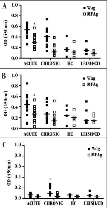
Serum immunoglobulin isotype reactivity in paracoccidioidomycosis patients. Serum from patients with both the acute and the chronic form of PCM showed higher reactivity towards all immunoglobulin isotypes (total IgG, IgM, IgA, IgG1 and
IgG2) when WAg was used than when MPAg was used (P < 0.05)
(Figures 1 and 2). Serum samples from patients with the acute form of PCM presented higher levels of total IgG (Figure 1A) and IgM
(Figure 1B) anti-Paracoccidioides brasiliensis antibodies than did
serum samples from patients with the chronic form of the disease,
when the MPAg was used (P < 0.05). On the other hand, IgA levels
specific to WAg were higher in the serum from chronic PCM patients
than in the serum from acute patients (Figure 1C) (P < 0.05).
There were no significant differences in the reactivity of serum IgG1 and IgG2 between patients with the acute and chronic forms of PCM, regardless of the antigenic preparation used (Figure 2). However, the reactivity of the IgG1 (Figure 2A) and IgG2
All serum samples were tested against whole (WAg)( )or sodium metaperiodate-treated WAg (MPAg)( )ofParacoccidioides brasiliensis;horizontal line (—) represents the median values of samples detected by ELISA. The optical density (OD) was measured at 450nm. LEISH: Leishmaniasis, CD: Chagas disease, *p < 0,05 vs chronic patients*p < 0,05 vs chronic patients (A and B), * p < 0.05 vs acute patients (C).
Figure 1 - Reactivity of total IgG (A), IgM (B) and IgA (C) in serum from patients with acute and chronic paracoccidioidomycosis. Serum from PCM patients, healthy controls and diseased controls were screened.
All serum samples were tested against whole antigen (WAg) ( ) or sodium metaperiodate-treated WAg (MPAg) ( ) of Paracoccidioides brasiliensis; horizontal line (—) represents the median values of samples detected by ELISA. The optical density (OD) was measured at 450nm. * p < 0.05 vs HC and LEISH/CD groups, LEISH: Leishmaniasis, CD: Chagas disease.
Figure 2 - Reactivity of IgG1 (A) and IgG2 (B) in the serum from PCM patients, healthy controls and diseased controls.
Figure
3
-
Recognition
profile
of
isotype-specific
IgG1
from
serum
of
PCM
patients
with
the
acute
fo
rm
(
A
, l
a
n
es
1
1
0
)
a
n
d
c
h
ro
n
ic
f
o
rm
(
B
, l
a
n
es
1
-
9
),
t
o
w
a
rd
s
fr
a
ct
io
n
a
te
d
a
n
ti
ge
n
ic
c
o
m
p
o
n
en
ts
of
Paracoccidioides brasiliensis
(W
Ag) by western blot assay
.
A
B
A
B
C
in the healthy controls or non-PCM diseased controls (Figure 2). Serum from patients with chronic PCM showed only low levels of specific IgG3 and IgG4 when either WAg or MPAg were used (data not shown).
Serum reactivity distinguished by western blotting.
Western blot analysis with Paracoccidioides brasiliensis WAg
DISCUSSION
Previous studies have shown the profile of serum
immunoglobulin isotypes that are reactive towards Paracoccidioides
brasiliensis antigens in acute and chronic forms of PCM, using
Paracoccidioides brasiliensis filtrate antigens or purified
recombinant proteins1 11. To our knowledge, this is the first study of
serum reactivity in PCM that has compared antigenic preparations
obtained from Paracoccidioides brasiliensis culture filtrates that
differed according to their carbohydrate content.
The use of serological tests for PCM diagnosis has been very limited, mainly due to the high frequency of cross-reactivity with other infections, such as American trypanosomiasis, leishmaniasis,
histoplasmosis and Jorge Lobo’s disease, among others18 19. In
order to improve the specificity, a purified 43kDa glycoprotein (gp43) was used in serological tests and showed high-specificity
results17 20. An additional study to evaluate the reactivity of gp43
in serological tests was performed after treatment of gp43 with metaperiodate to disrupt the carbohydrates in the molecule,
and this showed a reduction in nonspecific reactivity18. Despite
these findings, no study with disruption of carbohydrates from an
unpurified antigenic preparation of Paracoccidioides brasiliensis
has been performed. In the present study, antigens obtained from
Paracoccidioides brasiliensis culture filtrate (WAg) were treated with metaperiodate to obtain a carbohydrate-modified antigenic preparation (MPAg). WAg and MPAg were used to compare differences in the serum immunoglobulin isotype profile (total IgG, IgM, IgA, IgG1 and IgG2) in groups of patients with the acute and chronic forms of PCM, by means of ELISA.
The results presented here showed that serum from patients with either acute or chronic PCM had higher levels of total IgG and IgM reactivity towards WAg than against MPAg. However, only
the IgG anti-Paracoccidioides brasiliensis isotypes (and not the
IgM isotypes) were significantly increased in PCM patients, in comparison with the controls. Furthermore, acute PCM patients showed increased serum IgM and IgG levels only for MPAg. Several studies have demonstrated increased IgM production in early
Paracoccidioides brasiliensis infection2 3 6. IgG is now seen to
be higher in acute PCM patients than in chronic PCM patients and the present study is in agreement with other studies that showed that juvenile PCM patients had higher IgG anti-gp43 levels than
did patients with the adult form of the mycosis1 6.
Analysis of IgA production also showed differences between acute and chronic PCM patients. In contrast to the higher IgG and IgM isotype reactivity in acute patients, there was higher IgA reactivity in chronic patients, which was mainly anti-WAg antigenic
preparation. Together, these and previous observations7 suggest
that carbohydrate epitope removal leads to decreased levels of IgA. On the other hand, higher levels of IgA reactivity towards the
whole antigen could indicate lack of specificity for the reaction21.
In addition, the increased levels of IgA in the chronic PCM patients might relate to chronic mucosal antigenic stimulation, which is a characteristic of the chronic form of PCM. It is very well known that chronic inflammation of the mucosa stimulates production of IgA
antibodies1 9 10. In addition, the secretory IgA in saliva from PCM
patients may be indicative that it has a protective role regarding
antigen neutralization on mucosal surfaces15.
Previous studies showed that IgG1 antibodies were present,
frequently at high levels, in subacute and chronic PCM patients1 11.
Using both WAg and MPAg antigenic preparations, IgG1 and IgG2 subclass levels were found to be similar in the two clinical forms. However, a significant difference in IgG1 and IgG2 levels was detected when comparing PCM patients with healthy controls and non-PCM diseased controls. Isotype-specific IgG1 western blot analysis of
the reactivity towards fractionated Paracoccidioides brasiliensis
antigens indicated that acute patients consistently recognize a set of antigens that are of approximately 19, 27 and 31kDa, in contrast with recognition of 43 and 75kDa antigens, which was common to both the acute and the chronic form. Similar findings were described by
another author16, showing that a 38kDa antigen was recognized more
frequently and intensely by the serum of acute PCM than by the serum of chronic patients. Another study showed that components of the
Paracoccidioides brasiliensis with 27, 34 and 43kDa were reactive
towards IgG anti-Paracoccidioides brasiliensis antibodies13.
In conclusion, the present study suggests that the use of MPAg, obtained from treatment of WAg with sodium metaperiodate, could improve the serological test to distinguish between the chronic and acute forms of PCM, by testing the reactivity towards IgG and IgM. The measurement of total IgG and specific IgG1 or IgG2 was efficient in distinguishing between PCM patients and control individuals, using either WAg or MPAg. This study suggests that the detection of MPAg-specific IgG-antibodies reacting with low molecular weight antigens may be applied to the development of a better method for immunodiagnosis of paracoccidioidomycosis.
ACKNOWLEDGEMENTS
We thank Daniel Stockdell for language assistance.
REFERENCES
1. Baida H, Bicelli PJ, Juvenale M, Del Negro GM, Mendes-Giannini MJ, Duarte AJ, Benard G. Differential antibody isotype expression to the major Paracoccidioides
brasiliensis antigen in juvenile and adult form paracoccidioidomycosis. Microbes
and Infection 1:273-278, 1999.
2. Bernard G, Mendes-Gianini MJS, Juvenale M, Miranda ET, Duarte AJS. Immunosuppression in Paracoccidioidomycosis: T cell Hyporesponsiveness to two Paracoccidioides brasiliensis glycoproteins that elicit strong humoral immune response. The Journal Infectious Diseases 175:1263-1267, 1997. 3. Biagioni L, Souza MJ, Chamma LG, Mendes RP, Marques AS, Mota NGS, Franco M.
Serological of Paracoccidioidomycosis. II - Correlation between class-specific antibodies and clinical forms of the disease. Transactions of the Royal Society of Tropical Medicine Hygiene 78: 617-621, 1984.
4. Botteon FA, Camargo ZP, Bernard G, Coelho RF, Chamone DA, Itano EN.
Paracoccidioides brasiliensis - reactive antibodies in Brazilian blood donors.
Medical Mycology 40:387-391, 2002.
5. Brummer E, Castaneda E, Restrepo A. Paracoccidioidomycosis: in the update. Clinical Microbiology Review 6:89-117, 1993.
6. Bueno JP, Mendes-Giannini MJ, Del Negro GM, Assis CM, Takiguti CK, Shikanai-Yasuda MA. IgG, IgM and IgA antibody response for the diagnosis and follow-up of paracoccidioidomycosis: comparison of counterimmunoelectrophoresis and
complement fixation. The Journal Medical Veterinary Mycology35:213-217, 1997.
7. Chen YL, Ben KL, Cao XM. The terminal monosaccharide of antigenic determinant is involved in the binding of human sperm to specific monoclonal IgA antibody. Shi Yan Sheng Wu Xue Bao 29:101-107, 1996.
8. Del Negro GM, Garcia NM, Rodrigues EG, Cano ML, Aguiar MS, Lirio VdS, Lacaz CS. The sensitivity, specificity and efficiency values of some serological tests used in the diagnosis of paracoccidioidomycosis. Revista do Instituto de Medicina Tropical de São Paulo 33:277-280, 1991.
9. Fayette J, Dubois B, Vandenabeele S, Bridon JM, Vanbervliet B, Durant I, Banchereau J, Caux C, Briere F. Human dentritic cells skew isotype switching of CD-40-activated naive B cells towards IgA1 and IgA2. The Journal Experimental Medicine 185:1909-1918, 1997.
10. Franco M, Bagagli E, Cunha M, Chamma LG, Fecchio D. Paracoccidioides brasiliensis antigen batches from the same isolate show immunological and biochemical differences. Mycopathology 135:13-19, 1996.
11. Juvenale M, Del Negro GMB, Duarte AJS, Benard G. Antibody isotypes to a
Paracoccidioides brasiliensis somatic antigen in sub-acute and chronic form paracoccidioidomycosis. The Journal Medical Microbiology 50:127-134, 2001.The Journal Medical Microbiology 50:127-134, 2001. 12. Lacaz CS, Porto E, Martins JEC. Paracoccidioidomicose. In: Lacaz CS (ed)
Micologia Médica, 8th edição, Sarvier Editora, São Paulo, p. 248-261, 1991.
13. Martins R, Marquesa S, Alves M, Fecchio D, Franco MF. Serological follow-up ofSerological follow-up of patients with itraconazole using Dot-blot, ELISA and e Western-blot. Revista doRevista do Instituto de Medicina Tropical de São Paulo 39:261-269, 1997.
14. Mendes-Giannini MJ, Del Negro GB, Siqueira AM. Serodiagnosis. In: Franco MI, Lacaz CS, Restrepo A, Del Negro GB (eds) Paracoccidioidomycosis, CRC Press, Boca Raton, p. 345-363, 1994.
15. Miura CS, Estevão D, Lopes JD, Itano EN. Levels of specific antigen (gp43),Levels of specific antigen (gp43), specific antibody, and antigen-antibody complexes in saliva and serum of paracoccidioidomycosis patients. Medical MycologyMedical Mycology39: 423-428, 2001. 16. Panunto-Castelo A, Freitas-da-Silva G, Bragheto IC, Nartinez R, Roque-Barreira
MC. Paracoccidioides brasiliensis exoantigens: recognition by IgG from patients with different clinical forms of paracoccidioidomycosis. Microbes and Infection 5:1205-1211, 2003.
17. Puccia R, Schenkman S, Gorin PA, Travassos LR. Exocellular components ofExocellular components of
Paracoccidioides brasiliensis identification of a specific antigen. Journal Infection and Immunity 53:199-206, 1986.
18. Puccia R, Travassos LR. 43-kilodalton glycoprotein from Paracoccidioides brasiliensis: immunochemical reactions with sera from patients with paracoccidioidomycosis, histoplasmosis, or Jorge Lobo’s disease. Journal Clinical Microbiology 29:1610-1615, 1991.
19. Suzuki E, Toledo MS, Takahashi HK, Straus AH. A monoclonal antibody direct to terminal residue of B-galactofuranose of a glycolipid antigen isolated from
Paracoccidioides brasiliensis: cross-reactivity with Leishmania major and
Trypanosoma cruzi. Glicobiology 7:463-468, 1997.
20. Taborda CP, Camargo, ZP. Diagnosis of paracoccidioidomycosis by dot immnobindig assay for antibody detection using the purified and specific antigen GP43. Journal Clinical Microbiology 32:554-556, 1994.