R E V I E W
Open Access
Yellow fever in Africa and the Americas: a
historical and epidemiological perspective
Jean-Philippe Chippaux
1,2*and Alain Chippaux
3Abstract
Yellow fever was transported during the slave trade in the 15th and 16th centuries from Africa to the Americas where the virus encountered favorable ecological conditions that allowed creation of a sustainable sylvatic cycle. Despite effective vector control and immunization programs for nearly a century, yellow fever epidemics reemerged in many Latin American countries, particularly Brazil. The emergence or reemergence of vector-borne diseases encompasses many intricate factors. Yellow fever outbreaks occur if at least three conditions are fulfilled: the introduction of the virus into a non-immune human community, presence of competent and anthropophilic vectors and insufficiency of prevention and/or adequate management of the growing outbreak. On the other hand, two weapons are available to constrain yellow fever: vector control and immunization. In contrast, yellow fever is absent from Asia and the Pacific despite the presence of the vector and the susceptibility of human populations to the virus. Based on a review of the global history of yellow fever and its epidemiology, the authors deliver some recommendations for improving the prevention of epidemics.
Keywords:Yellow fever,Aedes aegypti,Haemagogussp., Sabethessp. Vector, Arbovirus, Epidemiology, Brazil, Latin America, Africa
Background
Brazil has experienced an exceptional yellow fever (YF) out-break since December 2016 (Table1). After the last major epidemic (1935–1940), sporadic cases were regularly re-ported from endemic states–mainly the Amazonian states
–with some incursions into those of the Southeast (Minas Gerais and São Paulo, respectively in 2002 and 2008) and South (Paraná and Rio Grande do Sul in 2008), until today (Fig. 1). Preceded by an upsurge of epizootics in monkeys since 2014 [1], the current epidemic resulted, from 1 De-cember 2016 to 8 May 2018, in 2050 confirmed cases of yellow fever including 681 deaths, indicating a case fatality rate of 33.2% (Fig. 2), while a further 1300 cases are still under investigation [2]. Despite a rapid and appropriate re-sponse, the epidemic spread to the east and south of the country, including areas generally considered non-endemic. This extension reproduces almost identically the course of all the epidemics observed in Brazil, including those
following the reappearance of Aedes aegypti in the 1970s, after an attempt to eliminate the vector [3,4]. However, in Brazil, this epidemic and the previous ones are marked by the role of wild vectors, Haemagogus sp. and Sabethessp., involved in the sylvatic cycle, while Ae-des aegyptiand A. albopictus, which induce epidemics in urban and peri-urban areas, respectively [3, 4], do not seem to be involved in the transmission of the virus at this stage [2].
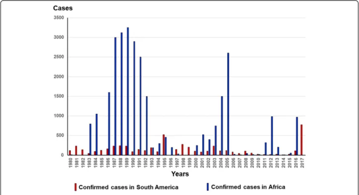
YF is an acute hemorrhagic hepatonephritis caused by an enveloped single-stranded RNA virus of approximately 12,000 base pairs belonging to the family Flavoviridae. It is transmitted by the bite of a mosquito belonging to the genusAedes(in Africa and the Americas) or to the genera Haemagogus and Sabethes (in America). Epidemics are more frequent and important in Africa than in the Ameri-cas (Fig.3) [5].
YF is native to Africa where it may have emerged around 3000 years ago [6]. It probably was introduced into the Americas during the beginning of the slave trade, and benefited from favorable ecological conditions, including the presence of sylvatic vectors which, although compe-tent, forced the virus to adapt by modifying its genome to * Correspondence:jean-philippe.chippaux@ird.fr
1UMR216, Mother and child facing tropical diseases, PRES Sorbonne Paris
Cité, Université Paris Descartes, Faculté de Pharmacie, Paris, France 2Centre de Recherche Translationnelle, Institut Pasteur, 28 rue du Dr Roux,
75015 Paris, France
Full list of author information is available at the end of the article
become a pathogen close to but distinct from that of Africa [7,8].
The main objective of this review is to describe the epi-demiological cycles of YF in Africa and South America, particularly Brazil, and their differences. The history of YF on the two continents, and its absence from Asia, are ele-ments that establish the conditions necessary for its main-tenance and development. Recent epidemiological studies seem to indicate characteristics that might be employed to anticipate the emergence of epidemics in major Brazilian cities and improve preventive measures.
History of yellow fever and origin of the virus
YF probably impacted the history – and the econ-omy – of Latin America more than those of Africa. Strongly linked to the development of the Americas, from its discovery by Europeans until the implementation of ef-fective control strategies in the middle of the twentieth century, YF gave rise to strong social and political
repercussions due, in particular, to the many deadly urban epidemics [9, 10]. It is certainly the recurrent YF epi-demics that dissuaded Napoleon Bonaparte from achiev-ing the conquest of the United States of America that he was preparing from then French Louisiana, with troops he had massed in the West Indies [11]. We can also evoke the scandal of the Panama Canal, the construction of which was delayed because of an epidemic of YF [12,13]. In addition, research on YF has led to conflicts between scientists because of their personal pride as well as coloni-alist and nationcoloni-alist positions [14].
The history of YF entails three main controversies–now resolved – namely its mode of transmission, geographical origin and the infectious agent responsible for the disease.
The first controversy concerned the transmission of the disease and was debated throughout the nineteenth cen-tury. The theory of contagion has long prevailed [12–14]. Like the transmission of cholera, proponents of this hy-pothesis defended, from the seventeenth century until the end of the nineteenth century, a transmission by water and/ or human contacts, then by“miasmas”. This theory–also named“aerism” –argued that the germ penetrated into the body via the respiratory system [15]. Changes in the con-cepts took place gradually between the end of the eight-eenth century–after the epidemics of Philadelphia in 1793, Cadiz in 1800 and Barcelona in 1821–1822, where the lack of direct contact between the patients excluded direct con-tamination between people–and the demonstration of the vectorial transmission in 1900 [15–17]. The “ non-conta-gion” was first demonstrated by several doctors in the French West Indies, notably masterfully by Lefort and his team [18]. It was on this basis that Beauperthuy, as early as 1854, suggested the transmission of YF by mosquito, which he illustrated by protecting healthy persons with a bed net Table 1Confirmed yellow fever cases and deaths in Brazil from
December, 2016 to 8 May, 2018 (from [2,131])
States Confirmed YF cases Deaths from confirmed YF
Pará 4 4
Tocantins 1 0
Goiás 1 1
Minas Gerais 1004 341
Espirito Santo 264 86
São Paulo 537 170
Rio de Janeiro 238 78
Distrito Federal 1 1
Total 2050 681
[19–21]. Following this new paradigm, Chervin proposed to abolish quarantining as the regular method for the pre-vention of YF [16]. Based on the filarial transmission model described by Manson in 1878, but without quoting the lat-ter nor Beauperthuy, although he certainly had access to their works [14], Finlay [22] supported the vectorial
transmission of the YF that was confirmed experimentally by Reed and his team in 1900–1902 [23–25]. Thus, three nations claim–with varying degrees of insistence–the dis-covery of the vectorial transmission of YF: France, Cuba and the USA in chronological order. The merit of the American Commission, led by Reed, was having shown that
Fig. 3Confirmed case reports in Africa and South America between 1980 and 2017 (based on WHO, weekly epidemiological record:http:// www.who.int/wer/en/–accessed on May 18, 2018)
the mosquito becomes infected during the first 3 days of the disease, when the viremia is sufficiently high, and that the sting contaminating the healthy subject should occur at least 12 days later to allow replication of the virus in the mosquito [26], which legitimized the authorship of the dis-covery [14].
The geographical origin of YF has also long been debated. The disease was considered to have originated from the Americas, discovered at the end of the fifteenth century by the first Spanish conquerors [12]. The first description in the New World rather than in the Old one resulted from circumstantial reasons: the Americas, actual colonies for settlements and economic exploitation, attracted much more consideration than Africa, which still consisted of trading posts essentially devoted to acquiring slaves con-tributing to the development of the Americas. Thus, many epidemics were reported in America and the West Indies from the middle of the seventeenth century. The mention of possible or probable cases before 1647, the date of the Guadeloupe epidemic, which is generally considered the first formally identified YF epidemic in history, led to the belief that YF was already present in America at the arrival of Spanish invaders. In fact, YF would have appeared for the first time in the West Indies 2 months after the battle of La Vega-Real that Christopher Columbus launched against the Amerindians on March 24, 1495, in Hispaniola, today known as the Dominican Republic [27]. However, the disease is mentioned under multiple names based on the very recognizable symptoms, by European navigators sail-ing along the African coast and the Canary Islands as early as 1494, regardless of the discovery of America [12]. It is therefore likely that the first American cases resulted from an introduction of the YF virus by the crews of Columbus between 1492 and 1495 coming from the Canary Islands
where the ships of Columbus made their last resupplying before the crossing of the Atlantic Ocean.
The arguments in favor of the African origin of YF prevail and this thesis has become the consensus. The frequency of epidemics and the adaptation of the virus to its hosts and vectors argue for an older presence in Africa, which molecu-lar biology today confirms by the greater genetic heterogen-eity of the virus in Africa [7,28–31].
Currently, there are seven genotypes: five in Africa and two in the Americas (Fig.4). African genotypes are charac-terized by their affinity for their respective vectors [32,33]. All the studies confirmed the African origin of the YF virus. The East African strain is the oldest, and probably diverged from an ancestral flavivirus about 3500 years ago [6, 34]. West African strains were separated from East African ones about 3 centuries before the alleged introduction of the virus into the Americas. American strains are closer to West African strains than the latter are related to the East African ones [6, 7, 34]. The virus has encountered in the Americas a competent mosquito that allowed permanent installment of a sylvatic cycle of the virus.
However, this does not explain why the disease was never mentioned in Europe before the fifteenth century, despite numerous and intense contacts between sub-Saharan Africa
–the land of YF’s origin–and Europe well before the dis-covery of America [12]. The most likely hypothesis is the ab-sence of a competent mosquito capable of establishing itself in a temperate zone. Subsequently, YF has been recurrent in Europe, as in North America, mainly during summer port epidemics, which are still not maintained in situ from 1 year to the next [27]. A greater number of epidemics in the Americas from the 17th to the nineteenth century in com-parison with Africa resulted from a combination of socio-economic, demographic and ecological circumstances that
favored the development of the disease in relation to Africa, where many human infections were probably limited by low population density and did not result in “conspicuous” epidemics.
While an African origin of YF is established, a new ques-tion arises: how did it cross the Atlantic Ocean? At the time of the slave trade, the duration of the journey between Afri-can and AmeriAfri-can coasts was longer than 1 month whereas the severe conditions of the crossing certainly did not favor the survival of the patients [35]. Incubation (3 to 6 days) and viremia (less than 10 days before the acquired immun-ity does not make it disappear) are too short for the virus to have been carried by slaves, if we admit the likelihood that some of them may have been infected at the time of their departure [36,37]. It is now accepted that the transfer was carried out via the mosquitoAedes aegypti, whose eggs survive for several months at desiccation [38], which en-sures transovarial transmission of the virus [39–41]. The circulation of the virus and the development of the epi-demics for which it is responsible are inseparable from the vectors.
Finally, the search for the pathogen has led to a long de-bate. Most of the works were performed in the Americas, mainly in Cuba, Brazil and Mexico, at least until the begin-ning of the twentieth century. This can be explained by the socioeconomic development of the Americas, particularly in the nineteenth century, the demographic changes that this region has experienced and the geopolitical influence it exerted on the world at that time. In addition, the lack of laboratories in Africa before the twentieth century did not favor research on the transmission and etiology of YF.
Brazilian scientists played a leading role. Lacerda iso-lated a fungus from the viscera and stool of patients [42]. Freire discovered a Cryptococcus that he believed respon-sible for YF and, after culture and attenuation of the yeast virulence, he experimented to obtain a vaccine [43]. In Rio de Janeiro, Havelburg found a coliform [44], the same year that Sanarelli in Montevideo identified another bacillus from Uruguayan and Brazilian patients [45]. In Cuba, Fin-lay and Delgado observed a bacterium, which they identi-fied as Micrococcus tetragenus Koch and Gaffky 1881, both in the vomit of patients, proboscis of the mosquito and blood from inoculated animals [46]. The microbiolo-gists proceeded by analogy, associating the pathogen with the seasonality of the disease, and inferring its adaptation to the environment in which it develops and/or relating the colors matching those of vomit, liver or skin.
A detailed review of potential candidates as etiological YF agents concluded that it was filterable, i.e., a pathogen not yet called a virus:“It is more than likely that the germs of yellow fever, as well as those of small pox, measles, hydro-phobia, etc. belong to a group of organisms, smaller than our bacteria and as yet unknown, awaiting discovery”[47]. The US Mission in Cuba led by Reed has shown that the
bacillus isolated by Sanarelli (Bacilus icteroides) was not the cause of YF but a secondary contaminant [25]. The French Mission in Brazil confirmed these observations and the fil-tering property of the YF pathogen [36].
The YF virus was isolated June 30, 1927, from the blood of a Ghanaian patient – who recovered – by Adrian Stokes, who contracted YF and died [48]. The 17D vaccine - still used today - was obtained by attenuation of this strain, called Asibi after the name of the Ghanaian pa-tient, a few years later by Theiler [49], which earned him the Nobel Prize in Medicine. A few months after the dis-covery by Stokes on December 21, 1927, another strain of the YF virus was isolated at the Institut Pasteur in Dakar [50], from François Miyeli a young patient from Rufisque, Senegal [51]. This strain was at the origin of the vaccine manufactured in Dakar (YF French Neurotropic Vaccine or FNV) [52]. Used throughout French-speaking Africa until 1982, the FNV was discontinued because of its adverse ef-fects [53,54].
Epidemiology of yellow fever in Africa
YF has been known in Africa since the end of the fifteenth century through sporadic cases and/or limited small epi-demics observed in the colonial counters of the West Afri-can coast. In East and South Africa, YF was probably rare or absent from coastal areas because of the lack of an ap-propriate vector.
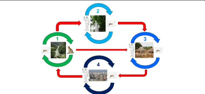
The epidemiology of YF in West and Central Africa was well described in the 1960s to 1980s [55–58] following a long cross-section follow-up of virus circulation during and outside of epidemic periods. It is possible, schematic-ally, to restrict the zones of virus circulation to three re-gions: a) the main one that harbors the endemic sylvatic cycle, i.e., the natural focus, b) a border zone of emer-gence, actually a latency area ensuring a transition be-tween the latter and c) the zone of epidemics, visible part of the iceberg, made up of regions inhabited by varying densities of human populations (Fig.5).
The endemic zone tends to spread, partly because of the transovarial transmission of YF in mosquitos [39–41] and, secondly, because of the anthropization of the environ-ment due to deforestation and population growth. Global warming could play an increasing role in the near future by changing environmental conditions to favor the vec-tors, in particular through alterations in the rain regime.
In-depth studies of viral infection in wild mammals and specific antibodies showed that vertebrates in general, in-cluding monkeys, play only a secondary role in the persist-ence and resilipersist-ence of the virus in the sylvatic cycle, because of the short duration of viremia and the resulting definitive immunity, which reflects an effective adaptation of the virus to its transient host [56,57,60–62]. Humans are no exception to this rule, whether they live in primary forest like the Pygmies of Central Africa or in villages close to natural foci, although their susceptibility to infec-tion is probably higher [63,64]. Wild vertebrates, no more than humans, cannot be considered a virus reservoir, a function that has essentially devolved to mosquitoes through the transovarial transmission of the virus. During estivation, the virus is maintained in the eggs of Aedes, thereby ensuring infection of the adults at their hatching at the beginning of the following season. On the other hand, monkeys–and possibly other small mammals–, es-pecially the young still immunologically naïve ones, amp-lify, reveal and propagate the virus from the first rains, thus perpetuating the sylvatic cycle [57].
In the epidemic zone, the virus does not circulate and the wild vector does not occur. The population shows a low immune status, except in immunized individuals, which in case of low immunization coverage explains the intensity of
the epidemic [53]. In urban and peri-urban areas, YF trans-mission is ensured by other Aedes species, first of all A. aegypti. The virus is spread in the municipality, amplified by the proliferation ofA. aegypti, where there are favorable conditions, lack of immunity among a large proportion of the inhabitants and, even today, health-system insufficiency based on the tripartite approach: mass vaccination, vector control, screening and treatment of cases placed under bed nets.
YF vaccination was made mandatory in 1941 in all French-controlled colonies. Facilitated by its stability and administration by scarification associated with the smallpox vaccine, mass immunization led to remarkably effective control of YF [5,53]. Its use was suspended in 1960 in chil-dren under 10 years because of the risk of serious adverse effects related to the use of the FNV manufactured in Dakar – although no case had been reported in a dozen years–then definitively interrupted in 1980. The disease reappeared and grew from 1965 in almost all French-speaking countries of sub-Saharan Africa. Since the late 1990s, in the face of renewed outbreaks of YF throughout Africa, sub-Saharan countries are progres-sively including the more tolerable 17D vaccine in the Ex-panded Program on Immunization by introducing routine YF vaccination in 9-month-old children together with the measles immunization supported by GAVI (Global Alli-ance for Vaccines and Immunization).
Epidemiology of yellow fever in the Americas
In South America, YF was mentioned at the same time as in Africa and in similar circumstances - during the slave trade
In Brazil, the first indisputable description of YF was made after the Pernambuco epidemic in Recife and Olinda in 1685 [65]. Legend has it that a boat from the Cape Verde Islands carried slaves with an unknown disease [66]. In fact, the boat had stopped in Guadeloupe, where YF was endemic if not epidemic [27], before reaching the Brazilian coast [67]. It is also possible that Cape Verdean mosqui-toes, and not Antillean patients, introduced the virus into Pernambuco. However, the infection of passengers by YF was refuted by Gouy [68] who diagnosed food poisoning by the spoiled meats consumed during the trip based on the kinetics and symptoms of the disease.
Anyway, the penetration of YF in America via Brazil is not excluded because of the considerable economic devel-opment of Brazil starting from 1554. The expansion of sugar plantations was favored by the large spaces available and the tax benefits granted by Portugal. On the one hand, the need for labor not met by indigenous natives was as-sumed by the massive influx of African slaves (3500 indi-viduals per year on average at the end of the sixteenth century). On the other hand, the crossing from Africa to Brazil was shorter than that to the West Indies or North America due to the proximity of both coasts and favorable winds, resulting in the multiplication of YF transport op-portunities [35,69,70]. In addition, the agricultural tech-niques favored the development ofA. aegyptiensuring the perpetuation and dispersal of the virus [69]. YF was re-ported in the ports of Pernambuco in 1640 and, perhaps, in other regions of Brazil [27,66]. It is therefore conceiv-able that the YF virus had been already endemic in Brazil since the sixteenth century and that the epidemic resulted from an indigenous strain emergence.
The endemic zone of YF is located in the Amazon and encompasses, to the west, Peru and Colombia, to the north the Guyana plateau (Venezuela, Guyana, Surinam and French Guiana), and to the northeast, Brazil. The de-scription of the sylvatic cycle was made on the occasion of the great 1942 epidemic in Brazil [71,72]. The circulation of the virus seems persistent, even if the detailed mecha-nisms were not yet discovered [73]. It is notable, for ex-ample, that specific immunity is maintained in monkeys from many parts of Brazil, even outside the Amazon, re-gardless of epidemic episodes, suggesting a regular infec-tion of the monkeys by the virus resulting in a lasting immunization in survivors [74]. While the general pattern remains fairly similar to the African one (Fig.5), there are some important differences. First, the American monkey is susceptible to the YF virus and many individuals die from it [75]. Thus, it not only plays the role of amplifying YF endemia as in Africa [73] but also reveals the risk of epidemics: periodic epizootics that occur near inhabited regions are used as a warning signal [76]. In addition, the vectors are different–at least within the jungle cycle–in whichHaemagogus janthinomysandH. leucocelaenusplay
an essential role while other species (including H. capri-cornii) or genera, notablyAedes, are little involved in the transmission [72, 77–83]. On the other hand, A. aegypti and A. albopictusrepresent a major risk of spreading YF in urban and peri-urban areas [84].
Despite common ecological conditions in the Amazon-ian region, there are two lineages of the YF virus: genotype 1 from Brazil (or possibly from the West Indies) and geno-type 2 from Peru [6,85–87]. A strong genetic heterogen-eity of the virus has been developing for some 30 years, increasing since the 2000s, probably as a result of human migrations that play a role of both gene mixing and viral dispersal [85,88,89].
The reemergence of YF in Brazil benefits from particu-larly favorable environmental conditions: high deforest-ation increasing the contact of human populdeforest-ations with the sylvatic virus, substantial migration between endemic and epidemic regions, poorly controlled urbanization fa-voring the multiplication of vectors, low immunization coverage in epidemic risk areas [90]. Finally, the role of the recent dengue and Zika epidemics, transmission of which is ensured by the same vectors, in diverting atten-tion and /or complicating the logistics of the health ser-vices, remains to be elucidated.
On the occasion of the Rio de Janeiro epidemic of 1900, the French scientific mission demonstrated the vertical transmission of the pathogen in mosquitos, and identified the former as small filtering organism. The first vaccine trials were undertaken [36,39,40]. At the end of the mis-sion, new health measures produced spectacular results.
The control ofA. aegyptistarted in the 1920s in most Latin American countries, reinforced by the use of DDT from 1947 [91]. Mass vaccination campaigns started after the discovery of the 17D vaccine in 1937. The elim-ination of the vector and immunization of a large part of the Brazilian population were considered major public health achievements and established an expectation of YF elimination in Brazil and perhaps more widely in Latin America. The persistence of the sylvatic transmis-sion of YF, or“jungle cycle”, and resurgence ofA. aegypti from jungle foci were unexpected [73,92].
The role of the vector and its cooperation with the virus
and, on the other hand, the human population lacks herd immunity.
These two species, in particular A. aegypti, favor the transport of the virus because of the resistance of their eggs to the desiccation [93] that leads to the resilience of the YF virus [94,95]. The spread of YF probably occurred at the same time as that ofA. aegypti. The short flying dis-tance of Aedes, especially A. aegypti and A. albopictus, leaves no alternative other than passive transport, espe-cially by the transportation of humans or goods [96–102].
The molecular phylogeny of A. aegypti strains con-firmed the African origin of the species although genetic variability reflected massive mixings due to multiple im-portations over time from all continents, and human intervention for controlling mosquitoes [103, 104]. In addition, genetic heterogeneity resulted in a high vari-ability in susceptibility to viral infection according to vector strains [105–108]. The original strain – now known asA. aegypti formosusas opposed to the subspe-ciesA. aegypti aegypti, which is the domestic and urban form of the species – is still present in African jungle where the larvae grow in tree trunks, and whose females are not anthropophilic [109–111].A. aegypti formosusis involved in the transmission of YF in nonhuman pri-mates within the sylvatic cycle [112]. It is not known whether the domestication ofA. aegypti, which led to its diffusion in America, was earlier or contemporaneous with its transport during the slave trade [111]. In either scenario, A. aegyptiadaptation to human populations in West Africa was early, which explains the many large urban epidemics in this part of the continent at the end of the eighteenth century [27]. On the other hand, in East Africa, A. aegypti does not seem to have adapted to the anthropization of the environment, as evidenced by the scarcity of urban epidemics up to the present day [113].
The colonization of Asia byA. aegyptiwas later, probably at the end of the nineteenth century when it was revealed as an urban vector of dengue fever [114]. Phylogenetic data suggest that the Asian strain originates from the Americas and not from East Africa as might have been expected, probably because of the poor adaptation of the East African strain to humans and its poor susceptibility to the YF virus [33,108,111]. In addition, the genetic homogeneity of Asian strains ofA. aegyptisuggests that the introduction was re-cent and accomplished by a limited number of entries [115]. However, although the current strain ofA. aegypti aegyptiis monophyletic [110], it has important local polymorphisms [116,117] that could explain, at least in part, the variability of the vector competence [32,105,107,118].
Absence of the YF from Asia and Oceania
The absence of YF from Asia and Oceania remains an en-igma. The population is susceptible to the virus as shown by Asian patients living in endemic areas (Africa or South
America).A. aegypti, although a recent arrival, is present in many places in Asia and Oceania. In addition, the abun-dant circulation of other arboviruses, dengue, chikun-gunya and Zika, the latter two coming from Africa like YF, indicates the possibility of YF extension in Asia. Recently, several human cases of YF introduced from Angola have been diagnosed in China [119], increasing YF outbreak fears. Several studies attempted to explain the absence of YF in Asia. The following main reasons were given: a) poor competency of the vector for the virus [32,105,108,118], b) poor adaptation of the vector to humans and competi-tion between vectors (Abrão and Fonseca, 2006, quoted in [120]), c) competition between Flavoviridae in the vector at the expense of YF and d) cross-reactivity with other Flavo-viridae, including dengue fever, in humans and other ani-mals [121,122]. Recent modeling of YF risk in Asia showed that two major factors limited the probability of YF expan-sion in Asia (“Asian hypothesis”) and dengue fever in Africa (“African hypothesis”) [123]. The first was based on cross-immunity between Flavoviridae–especially between dengue and YF, which could limit the risk of double infec-tion – and high viremia and severe clinical forms of YF [121,122]. The second was based on the competition be-tween A. aegyptiandA. albopictusas well as on their re-spective competencies in the transmission of dengue and YF to explain the absence of the latter in Asia and the rarity of the former in Africa [120]. Another explanation was the low probability of transporting the virus from Africa –or the Americas–to Asia and the Pacific. The slave trade to Asia was much smaller than the trade to the Americas. This reduces the probability – as to the transport of the virus into the Americas before air transport –by infected humans [124]. Mosquito transport did not occur, either from Africa or America, for reasons that have not yet been elucidated.
Rooting out and controlling of yellow fever in Brazil
Since its introduction in the seventeenth century, YF remained in the Amazonian region in the form of a jungle cycle that causes rural, peri-urban or urban epidemics at regular intervals. The sylvatic cycle is maintained by wild endemic vectors present before the introduction of YF in Brazil–belonging to the generaHaemagogusandSabethes –playing the role of virus reservoir, especially in the Ama-zon that is the refuge Ama-zone of the virus [3,79–83]. Exten-sion to peri-urban and urban areas involves A. albopictus and A. aegypti respectively, and follows the routes of hu-man migrations, particularly forest galleries [4].
The vector control carried out at the beginning of the twentieth century during the Rio de Janeiro epidemic was organized nationwide in 1928–29. It was reinforced by YF immunization in 1937 and DDT sprayings initiated 10 years later, which significantly reduced the burden of YF, that was considered as having been eliminated from Brazil in the 60s [91, 126]. However, the epidemics of dengue in 1963–64 then in 1968–69 marked the beginning of the re-infestation of the continent by A. aegypti, confirmed between 1971 and 1999 [127], which resulted in the return of YF and, more recently, the installation of other arboviruses (chikun-gunya and Zika, in particular) [2]. Several factors can ex-plain this phenomenon. The most important is, almost certainly the reinvasion of A. aegypti from isolated and poorly controlled Amazonian and Southern Brazilian forest localities, as a result of a relaxation of vector control and vaccination campaigns, as well as the deterioration of health systems in some remote areas [92, 103, 128]. An-other reason could be the insufficiency of vaccination coverage, which should have favored the extension of the disease beyond its natural limits, particularly in the regions of Brazil where vaccination was not recommended, e.g., in northeast and southern Brazil [90, 129]. Nevertheless, YF now seems well established in the form of a deeply rooted jungle cycle, as shown by the numerous epizootics re-ported in recent years (Fig.6). Finally, the sudden popula-tion growth and unplanned urbanizapopula-tion that results in
substandard housing, and inadequate water supply and waste management systems, at least partially explain the failure of preventive measures and the delay in the man-agement of cases [85, 128]. The vectorial competence of A. aegyptiandA. albopictusreinforce the risk of YF exten-sion in cities, especially in the overcrowded suburbs of major Brazilian cities [3,84].
The attempt to eliminate the YF during the twenty-first century, despite a relative failure, probably contained the risk of urban and peri-urban epidemics. For more than 50 years, Brazilian YF epidemics–and the most recent one was no exception–were sylvatic and involved wild vectors (Haemagogus andSabethes, as well as non-anthropophilic Aedes, i.e. other species than A. aegyptiandA. albopictus) and spared densely populated areas. So far, humans have been infected outside cities and their suburbs from multiple sylvatic foci [73]. However, the sylvatic cycle is expanding, and getting closer to the cities, infesting forest areas that could even be included in the conurbations of southern Brazil, notably in the states of Rio de Janeiro, São Paulo and Rio Grande do Sul, highlighting the risk of metropolitan ep-idemics. [80–84, 130, 131]. The epidemiological surveil-lance should be performed at two levels. On the one hand, the sentinel role of the monkeys that are the first to ex-press the reemergence of the YF, dying from the disease, must be favored [76]. On the other hand, the clinical diag-nosis of YF index cases, which may reveal an increased
risk of epidemic, must be reinforced. The“syndromic sur-veillance”approach recommended by US Centers for Dis-ease Control and Prevention incrDis-eases the sensitivity – and specificity–of first-line clinical detection [132]. This method speeds up the diagnostic process, which is employed to organize case management and contain the epidemic.
The return of YF has led to a considerable increase in the genetic diversity of its South American genotype I, leading to the appearance of a new viral line [89]. The reasons for this diversification and its consequences, particularly as to transmissibility and virulence, remain to be clarified. One of the pending issues is the need to assess the capacity of the YF virus currently present in Brazil to invade peri-urban and urban environments spread by the A. aegyptiandA. albopictuspopulations that abound [3,4].
Two strategies – certainly not exclusive – enable the containment of YF: vector control and immunization of populations at risk. The former is very effective but expen-sive. It requires a constant effort wherever the risk of vec-tor development is high. The recent epidemics of dengue, chikungunya and Zika in Brazil demonstrated the limits of this strategy [133]. However, integrated control – taking into account sanitation – and mutualized with other vector-borne diseases, is cost-effective and should be fa-vored [134,135]. Vaccination against YF is based on a se-lective strategy – routine mandatory vaccinations in endemic areas of Brazil, voluntary vaccinations elsewhere such as the east and south of the country, and large-scale vaccinations of populations threatened by an epidemic. In Brazil, vaccine coverage is irregular, with a drop in cover-age between 1990 and 2010, and insufficient in States where it is not compulsory [136], which requires making a considerable effort to catch up on vaccinations in the event of an epidemic. This defective coverage results from recurrent vaccine shortages and poor tolerance [137–139]. Once the causative agent of the YF was identified in 1927 [48], the vaccine was discovered 10 years later [49]. It is a live virus attenuated by repeated passages into chicken embryos (YF 17D), and genetically stable. It rep-licates in the vaccinated individuals and confers protec-tion for life [34, 138, 139]. The effectiveness of the vaccine in controlling YF has been emphasized above in relation to its epidemiological description of the in both Africa and the Americas. In the current Brazilian context, vaccination of at-risk populations, including those living outside endemic areas, appears to be a strategy of choice provided that at least 80% of the resident population is covered to obtain an effective herd immunity [139]. How-ever, global production–about 80 million doses annually
–is insufficient in some years to cover the needs, i.e. rou-tine newborn vaccination, immunization of travelers and protection of populations at risk of epidemic [54, 138]. The reduction of the vaccine dose administered –up to
one-fifth of the recommended dose–conferred sufficient immunity and would allow, in the event of an epidemic, to respond to the needs, although these results require confirmation in real-life situations [54,139–142].
Another important issue is the incidence of serious ad-verse events (SAE). In addition to vaccine hypersensitivity and anaphylactic shock, SAEs were categorized into two groups: YFV-associated neurotropic disease (YFV-AND), including encephalitis, myelitis, acute disseminated encephalo-myelitis, and Guillain-Barré syndrome, and YF-vaccine-associated viscerotropic disease (YDV-AVD) re-sponsible for multi-organ failure [139]. Risk factors are age (young children under 6 months and adults over 50), preg-nancy and immunodeficiency. Most SAEs concern primary vaccination [143, 144]. Based on meta-analyses in many countries with reliable pharmacovigilance, the incidence of SAEs was estimated to be 11.1–15.6 per million vaccinated persons, including 6.6 YFV-AND and YFV –AVD cumu-lated, with wide variations depending on the country [145]. These are linked both to the surveillance system and to the recruitment of vaccinated persons: in YF-free regions, the latter are most often travelers whose age is on average sig-nificantly higher than people from endemic countries where routine immunization targets young children. Des-pite the high incidence of SAEs, the risk-benefit profile re-mains extremely favorable for vaccination in endemic areas [139]. The mortality due to SAEs is below one per million vaccinated people [145], which must be compared with the 33% case fatality rate of YF in Brazil (Table1). In addition, it is likely that the risk of SAEs increases during large-scale vaccination campaigns due to reduced attention to risk fac-tors in vaccinated people and conditions of storage and vac-cine administration. The vaccinations carried out in Brazil and Argentina during the recent YF outbreak confirmed these data, showing an SAEs incidence ranging from 8.3 to 12.4 per million respectively, mainly involving people at risk, especially those over 50 years old [137,146]. Consider-ing the risks of urban epidemics and incidence of SAEs, it is urgent to develop a more tolerable vaccine. The vaccine strategy will need to be reconsidered to extend vaccine coverage to the whole country, including major urban metropolises.
Conclusion
The physiognomy of YF has reemerged abruptly over the last 50 years while the disease was likely to be controlled. Consequently, it is no longer a question of its eradication, if only because of the sylvatic foci present on two conti-nents, but rather of limiting the epidemic risk [147].
The reservoir of YF, both in Africa and the Americas, is the mosquito, especially the sylvatic species that maintain the jungle cycle. The short viremia and definitive immunity
However, they ensure amplification of the latter and, in the Americas in particular, are an alarm signal of viral reemer-gence that can result in human outbreaks able to extend into urban areas.
The history of YF, especially its installation in the Ameri-cas five centuries ago, taught us that the virus and its vector presented a remarkable capacity for environmental adapta-tion. The pair seem to react very quickly to the conditions that they face. While until 1960, the virus was spread by boat–limited to transport through mosquitos–which re-quired time and allowed the implementation of preventive or remedial measures, it is now rapidly spread via aircraft carrying infected humans, a scenario that is more difficult to control.
However, there are flaws in the transmission of the YF, as evidenced by its absence from Asia. The emergence or re-emergence of vector-borne diseases depends on complex, entangled biological, climatic, eco-logical, socio-economic and political factors [148]. Three conditions are necessary to produce YF epidemics: a) the introduction of the virus into a non-immune human com-munity, b) presence of competent and anthropophilic vec-tors and, c) insufficiency of prevention and/or adequate management. On the other hand, there are two weapons available to fight YF: vector control and immunization of human populations [147]. The first has the advantage of allowing the control of many other parasitic and viral dis-eases transmitted by arthropods, and may result in the mutualization of resources. It involves financial and eco-logical constraints that can limit its application and effect-iveness. Immunization is both cheaper and very effective, subject to strict and permanent application.
The objectives of the WHO are therefore to protect the at-risk population, prevent international spread and rap-idly contain epidemics. These objectives include a preven-tion component through appropriate urban development and an immediate response in the event of an YF outbreak by combining vector control against Aedes larvae and adults, and preventive mass immunization [149]. In Brazil, preventive vaccination is underway. However, it is neces-sary to reconsider the vaccination schedule. While waiting for a safer YF vaccine, vector control measures and epi-demiological surveillance should be strengthened to initi-ate emergency large-scale vaccination campaigns along two lines: monkey mortality [76,150] and syndromic sur-veillance in sentinel hospitals [132].
Public health strategies must combine both scientific and political criteria. The history of YF stressed, as many au-thors have already noted, that the opinions of scientists– sometimes giving rise to expertise followed by the author-ities–often came from assertions based on subjective argu-ments rather than evidence based on observations validated experimentally. Controversies conceal ideological cleavages, even political ones, which divert the public debate from its
objectives of prevention and management, make them lose their effectiveness, and involve useless and considerable ex-penses [15,16].
Authors’contributions
JPC and AC conceived and designed the review. JPC drafted the manuscript that was finalized by AC, and both authors approved the final version. They are guarantors of the paper.
Authors’information
AC, MD, is specialist in microbiology. He was director of Institut Pasteur de Bangui, Central African Republic, in the 60s, director of Institut Pasteur of Ivory Coast in the 70s, responsible of vaccine and blood product assessment in the French Government Regulatory Agency (Laboratoire National de la Santé) in the 80s, and head the Unit of the arboviruses and hemorrhagic fever viruses at Institut Pasteur Paris in the 90s. He is now retired and former President of the Société de Pathologie Exotique et filiales, Paris. JPC, MD, PhD, graduated in virology and medical entomology, is specialist in public health and epidemiology. He worked on Yellow Fever vectors in French Guiana during the early 80s.
1UMR216, Mère et enfant face aux infections tropicales; PRES Sorbonne Paris
Cité, Université Paris Descartes, Faculté de Pharmacie, Paris, France.2Centre
de Recherche Translationnelle, Institut Pasteur, 28 rue du Dr. Roux, 75,015 Paris, France.3Société de Pathologie Exotique, Hôpital Salpêtrière, BP50082,
75,622 Paris cedex 13; 18 rue Princesse, 75,006 Paris, France.
Ethics approval and consent to participate
Not applicable.
Consent for publication
Not applicable.
Competing interests
The authors declare that they have no competing interests.
Publisher’s Note
Springer Nature remains neutral with regard to jurisdictional claims in published maps and institutional affiliations.
Author details
1UMR216, Mother and child facing tropical diseases, PRES Sorbonne Paris
Cité, Université Paris Descartes, Faculté de Pharmacie, Paris, France.2Centre de Recherche Translationnelle, Institut Pasteur, 28 rue du Dr Roux, 75015 Paris, France.3Société de Pathologie Exotique, Hôpital Salpêtrière, BP50082, 75622 Paris cedex 13; 18 rue Princesse, 75006 Paris, France.
Received: 6 June 2018 Accepted: 14 August 2018
References
1. WHO. Yellow fever in Africa and the Americas, 2016. Wkly Epidemiol Rec. 2017;92(32):442–52.
2. Brasil. Ministerio da Saude. Febre Amarela. Situação Epidemiológica/Dados. Situação Epidemiológica no Brasil. http://portalms.saude.gov.br/saude-de-a- z/febre-amarela-sintomas-transmissao-e-prevencao/situacao-epidemiologica-dados(accessed 17 Apr 2018).http://www.saude.mg.gov.br/component/ search/?all=&exact=informe+epidemiol%C3%B3gico+da+febre +amarela&any=&none=&created=&modified=&area=stories(accessed 14 May 2018).https://www.paho.org/hq/index.php?option=com_topics&view= article&id=69&Itemid=40784&lang=en(accessed 14 May 2018).
3. Mondet B, Travassos da Rosa APA, Vasconcelos PFC. Les risques d’épidémisation urbaine de la fièvre jaune au Brésil par les vecteurs de la dengue:Aedes aegyptiet Aedes albopictus. Bull Soc Pathol Exot. 1996;89(2):107–13.
4. Mondet B. Epidémiologie de la fièvre jaune au Brésil. Bull Soc Pathol Exot. 2001;94(3):260–7.
5. Tomori O. Yellow fever in Africa: public health impact and prospects for control in the 21st century. Biomedica. 2002;22(2):178–210.http://www. redalyc.org/articulo.oa?id=84322213(accessed 18 Aug 2018
7. Mutebi JP, Rijnbrand RC, Wang H, Ryman KD, Wang E, Fulop LD, et al. Genetic relationships and evolution of genotypes of yellow fever virus and other members of the yellow fever virus group within the Flavivirus genus based on the 3′noncoding region. J Virol. 2004;78(18):9652–65. 8. Li Y, Yang Z. Adaptive diversification between yellow fever virus west
African and south American lineages: a genome-wide study. Am J Trop Med Hyg. 2017;96(3):727–34.
9. Patterson KD. Yellow fever epidemics and mortality in the United States, 1693-1905. Soc Sci Med. 1992;34(8):855–65.
10. Oldstone M. Viruses, Plagues, and history: past, present and future. New York: Oxford University Press; 2009. pp. 102–4.
11. Marr JS, Cathey JT. The 1802 saint-Domingue yellow fever epidemic and the Louisiana purchase. J Public Health Manag Pract. 2013;19(1):77–82. 12. Augustin G. History of yellow fever. Searcy & Pfaff ltd, New Orleans, USA; 1909. 13. Elton NW. Yellow fever in Panama; historical and contemporary. Am J Trop
Med Hyg. 1952;1(3):436–56.
14. Delaporte F. Histoire de la fièvre jaune. Paris: Payot; 1989.
15. Lódola S, Góis Jr E. Theories about the propagation of yellow fever: the scientific debate in the São Paulo press between 1895 and 1903. Hist Cienc Saude Manguinhos 2015;22(3):687–704.http://www.scielo.br/hcsm
(accessed 18 Apr 2018).
16. Ackerknecht EH. Anticontagionism between 1821 and 1867. Int J Epidemiol. 2009;38(1):7–21.
17. Fournier P. Zones humides et“aérisme”à l’époque moderne. In: Derex JM, editor. Zones humides et santé. Paris: GHZH; 2010. p. 9–23.
18. Lefort P. Mémoire sur la non contagion de la fièvre jaune. Fleurot et Cie, Gouvernements de la Martinique et de Guadeloupe. 1823.
19. Beauperthuy LD. 1854. Fiebre Amarilla. Gac Ofic Cumaná. 1854;57(23 de Mayo). 20. Beauperthuy LD. Recherches sur la cause du choléra asiatique, sur celle du
typhus ictérode et de fièvres de marécages. C R Acad Sci. 1856;14(13):692–3. 21. Beauperthuy de Benedetti R. Louis Daniel Beauperthuy et la méthode
scientifique, le rôle des moustiques dans la transmission de la fièvre jaune. Ann Hyg Langue Franc. 1966;2(6):25–32.
22. Finlay CJ. El mosquito hipoteticamente considerado como agente de transmisíon de la fiebre amarilla. An Acad Cienc Med Fis Nat Habana. 1881;18:147–69. 23. Reed W. Recent researches concerning the etiology, propagation, and
prevention of yellow fever, by the United States Army Commission. J Hyg (Lond). 1902;2(2):101–19.
24. Reed W, Carrol J, Agramonte A. The etiology of yellow fever - an additional note. JAMA. 1901;36:431–40.
25. Reed W, Carroll J, Agramonte A, Lazear JW. The etiology of yellow fever-a preliminary note. Public Health Pap Rep. 1900;26:37–53.
26. Chastel C. Le centenaire de la découverte du virus de la fièvre jaune et de sa transmission par un moustique (Cuba 1900-1901). Bull Soc Pathol Exot. 2003;96(3):250–6.
27. Béranger-Féraud LJB. Traité théorique et clinique de la fièvre jaune. Paris: Doin; 1891.
28. Deubel V, Digoutte JP, Monath TP, Girard M. Genetic heterogeneity of yellow fever virus strains from Africa and the Americas. J Gen Virol. 1986; 67(Pt 1):209–13.
29. Lepiniec L, Dalgarno L, Huong VT, Monath TP, Digoutte JP, Deubel V. Geographic distribution and evolution of yellow fever viruses based on direct sequencing of genomic cDNA fragments. J Gen Virol. 1994; 75(Pt 2):417–23.
30. Mutebi JP, Wang H, Li L, Bryant JE, Barrett AD. Phylogenetic and
evolutionary relationships among yellow fever virus isolates in Africa. J Virol. 2001;75(15):6999–7008.
31. von Lindern JJ, Aroner S, Barrett ND, Wicker JA, Davis CT, Barrett AD. Genome analysis and phylogenetic relationships between east, central and west African isolates of yellow fever virus. J Gen Virol. 2006;87(Pt 4):895–907. 32. Beaty BJ, Aitken THG. In vitro transmission of yellow fever virus by
geographic strains ofAedes aegypti. Mosq News. 1979;39(2):232–8. 33. Ellis BR, Barrett AD. The enigma of yellow fever in East Africa. Rev Med Virol.
2008;18(5):331–46.
34. Monath T, Cetron MS, Teuwen DE. Yellow fever vaccine. In: Plotkin SA, Orenstein WA, Offit PA, editors. Vaccines. 5th ed. Philadelphia: Saunders Elsevier; 2008. p. 959–1055.
35. Thomas H. The slave trade: the story of the Atlantic slave trade: 1440 - 1870. Touchstone, New York. 1999. Paris: French translation, Robert Lafont; 2006. 36. Marchoux E, Simond PL, Salimbeni A. Etudes sur la fièvre jaune, premier
mémoire. Ann Inst Pasteur. 1903;17:665–728.
37. Nassar ES, Chamelet EL, Coimbra TL, de Souza LT, Suzuki A, Ferreira IB, et al. Jungle yellow fever: clinical and laboratorial studies emphasizing viremia on a human case. Rev Inst Med Trop Sao Paulo. 1995;37(4):337–41.
38. Christophers SR.Aedes aegypti(L.) the yellow fever mosquito. In: Its life history, bionomics and structure. Cambridge, Great Britain: Cambridge University Press; 1960.
39. Marchoux E, Simond PL. La transmission héréditaire du virus de lafièvre jaunechez leStegomyia fasciata. C R Soc Biol (Paris). 1905;59:259–60. 40. Marchoux E, Simond PL. Etudes sur la fièvre jaune, deuxième mémoire. Ann Inst
Pasteur. 1906;25:16–40.
41. Fontenille D, Diallo M, Mondo M, Ndiaye M, Thonnon J. First evidence of natural vertical transmission of yellow fever virus in Aedes aegypti, its epidemic vector. Trans R Soc Trop Med Hyg. 1997; 91(5):533–5.
42. Lacerda JB. Mémoire présenté par M. de Quatrefages. Compt Rend Acad Sci. 1883;97(1):1708–9.
43. Freire D. Doctrine microbienne de la fièvre jaune et ses inoculations préventives. Imprimerie Nationale, Rio de Janeiro. 1885.
44. Havelburg W. Recherches expérimentales et anatomiques sur la fièvre jaune. Ann Inst Pasteur. 1897;11:515–22.
45. Sanarelli J. Etiologie et pathogénie de la fièvre jaune. Ann Inst Pasteur. 1897;11(7):433–513.
46. Finlay CJ, Delgado C. Del micrococo tetrágeno de la fiebre amarilla. Ann Acad Ciencias Med Fis Natur Habana. 1888;18:147–69.
47. Novy FG. The Etiology of Yellow Fever. Med News. 1898;73:326–331 and 360–9. 48. Stokes A, Bauer JH, Hudson NP. Experimental transmission of yellow fever to
laboratory animals. Am J Trop Med. 1928;8:103–64.
49. Theiler M, Smith HH. The use of yellow fever modified by in vitro cultivation for human immunization. J Exp Med. 1937;65:787–800.
50. Mathis C, Sellards AW, Laigret J. Sensibilité duMacacus rhesusau virus de la fièvre jaune. C R Acad Sci. 1928;186:604–6.
51. Ducloux M. François Miyeli (1911–1927 - 1973) et la souche française amarile. Bull Soc Med Afr Noire Lang Franc. 1975;20:174–9.
52. Sellards AW, Laigret J. Vaccination de l’homme contre la fièvre jaune. C R Acad Sci. 1932;194:1609–11.
53. Moreau JP, Girault G, Dramé I, Perraut R. Réémergence de la fièvre jaune en Afrique de l’Ouest : leçons du passé, plaidoyer pour un programme de contrôle. Bull Soc Pathol Exot. 1999;92(5):333–6.
54. Barrett ADT. Yellow fever live attenuated vaccine: a very successful live attenuated vaccine but still we have problems controlling the disease. Vaccine. 2017;35(44):5951–5.
55. Hamon J, Pichon G, Cornet M. La transmission du virus amaril en Afrique occidentale. Ecologie, répartition, fréquence et contrôle des vecteurs et observations concernant 1’épidémiologie de la fièvre jaune. Cah ORSTOM, sér Ent Med Parasitol. 1971;9(1):3–60.
56. Taufflieb R, Cornet M, Le Gonidec G, Robin Y. Un foyer selvatique de fièvre jaune au Sénégal oriental. Cah ORSTOM, sér Ent Med Parasitol 1973;11(3):211–220.
57. Germain M, Cornet M, Mouchet J, Herve JP, Robert V, Camicas JL, et al. La fièvre jaune selvatique en Afrique: données récentes et conceptions actuelles. Med Trop (Mars). 1981;41(1):31–43.
58. Cordellier R. L’épidémiologie de la fièvre jaune en Afrique de l’Ouest. Bull World Health Organ. 1991;69(1):73–84.
59. Hudson NP, Philip CB. Infectivity of blood during the course of experimental yellow fever. J Exp Med. 1929;50(5):553–99.
60. Chippaux-Hyppolite C, Chippaux A. Etude du réservoir animal dans le cycle de quelques arbovirus en Afrique centrale. I. Etude
immunologique chez divers animaux domestiques et sauvages. Bull Soc Pathol Exot. 1969;62(6):1034–45.
61. Chippaux A, Chippaux-Hyppolite C, Diederich-Decoux M. Etude du réservoir animal dans le cycle de quelques arbovirus en Afrique centrale. II. Virémie expérimentale chez les rongeurs sauvages avec les virus de la fièvre jaune et de West Nile. Bull Soc Pathol Exot. 1970;63(2):173–80.
62. Taufflieb R, Robin Y, Cornet M. Le virus amaril et la faune sauvage en Afrique. Cah ORSTOM. 1971;9(4):351–71.
63. Chippaux A, Chippaux-Hyppolite C. Immunologie des arbovirus chez des pygmées Babinga en République Centrafrique. Bull Soc Pathol Exot. 1965;58(5):820–33.
65. Rosa JF. Tratado Único da Constituição Pestilencial de Pernambuco. Pernambuco - Brasil 1694.http://objdigital.bn.br/objdigital2/acervo_digital/ div_manuscritos/mss1428710/mss1428710.pdf. Accessed 17 Aug 2018. 66. Franco O. História da Febre-Amarela no Brasil. Rio de Janeiro: Ministério da
Saúde; 1969.
67. dos Anjos BC. Single treaty on the pestilential constitution of Pernambuco: first description of the“males”by João Ferreira da Rosa in the XVII century. Tempo. 2016;8(1):11–36.
68. Gouy E. Histoire de la fièvre jaune au Brésil. Paris: Alfonse Dorenne; 1884. 69. McNeill JR. Yellow Jack and Geopolitics: environment, epidemics, and the struggles
for empire in the american tropics, 1650-1825. OAH Mag Hist. 2004;18(3):9–13. 70. Pétré-Grenouilleau O. Les Traites négrières. Essai d’histoire globale. Gallimard
coll. « Bibliothèque des Histoires », Paris; 2004. p. 201–5.
71. Taylor RM, Fonseca da Cunha J. An epidemiological study of jungle yellow fever in an endemic area in Brazil; epidemiology of human infections; investigation of vertebrate hosts and arthropod vectors. Am J Trop Med Hyg. 1946;26(6 Suppl 2):69.
72. Laemmert HW Jr, Ferreira LC, Taylor RM. An epidemiological study of jungle yellow fever in an endemic area in Brazil. Investigation of vertebrate hosts and arthropod vectors. Am J Trop Med Hyg. 1946;26(6 Suppl 2):23–69. 73. Moreira-Soto A, Torres MC, Lima de Mendonça MC, Mares-Guia MA, Dos
Santos Rodrigues CD, Fabri AA, et al. Evidence for multiple sylvatic transmission cycles during the 2016-2017 yellow fever virus outbreak, Brazil. Clin Microbiol Infect. 2018;
74. Kumm HW, Laemmert HW Jr. The geographical distribution of immunity to yellow fever among the primates of Brazil. Am J Trop Med Hyg. 1950;30(5):733–48. 75. Davis NC, Shannon RC. Studies on south American yellow fever: iii.
Transmission of the virus to Brazilian monkeys preliminary observations. J Exp Med. 1929;50(1):81–5.
76. Fernandes NCCA, Cunha MS, Guerra JM, Réssio RA, Cirqueira CDS, Iglezias SD, et al. Outbreak of yellow fever among nonhuman primates, Espirito Santo, Brazil, 2017. Emerg Infect Dis. 2017;23(12):2038–41.
77. Pajot FX, Geoffroy B, Chippaux JP. Écologie d’Haemagogus, janthinomysDyar (Diptera, Culicidae) en Guyane française. Cah ORSTOM. 1985;23(3):209–16. 78. Dégallier N, Travassos da Rosa AP, Vasconcelos PFC, Guerreiro SC, Travassos
da Rosa JF, Hervé JP. Estimation du taux de survie, de la densité relative et du taux d’infection d’une population d’Haemagogus janthinomysDyar (Diptera, Culicidae) ayant fourni des souches de fièvre jaune en Amazonie brésilienne. Bull Soc Pathol Exot. 1991;84(4):386–97.
79. Mondet B, Vasconcelos PF, Travassos da Rosa AP, Travassos da Rosa ES, Rodrigues SG, Travassos Rosa JF, et al. Isolation of yellow fever virus from nulliparousHaemagogus(Haemagogus)janthinomysin eastern Amazonia. Vector Borne Zoonotic Dis. 2002;2(1):47–50.
80. Cardoso JC, de Almeida MA, dos Santos E, da Fonseca DF, Sallum MA, Noll CA, et al. Yellow fever virus inHaemagogus leucocelaenusandAedes serratus mosquitoes, southern Brazil, 2008. Emerg Infect Dis. 2010;16(12):1918–24. 81. Alencar J, Mello CF, Barbosa LS, Gil-Santana HR, Maia DA, Marcondes CB,
et al. Diversity of yellow fever mosquito vectors in the Atlantic Forest of Rio de Janeiro, Brazil. Rev Soc Bras Med Trop. 2016;49(3):351–6.
82. Mucci LF, Cardoso RP Jr, de Paula MB, Scandar SAS, Pacchioni ML, Fernandes A, et al. Feeding habits of mosquitoes (Diptera: Culicidae) in an area of sylvatic transmission of yellow fever in the state of São Paulo, Brazil. J Venom Anim Toxins incl Trop Dis. 2015;21:6.https://doi.org/10.1186/ s40409-015-0005-z.
83. Mucci LF, Medeiros-Sousa AR, Ceretti-Júnior W, Fernandes A, Camargo AA, Evangelista E, et al.Haemagogus leucocelaenusand other mosquitoes potentially associated with sylvatic yellow fever in Cantareira State Park in the São Paulo metropolitan area, Brazil. J Am Mosq Control Assoc. 2016;32(4):329–32.
84. Couto-Lima D, Madec Y, Bersot MI, Campos SS, Motta MA, Santos FBD, et al. Potential risk of re-emergence of urban transmission of yellow fever virus in Brazil facilitated by competentAedespopulations. Sci Rep. 2017;7(1):4848. 85. Vasconcelos PFC, Bryant JE, da Rosa APAT, Tesh RB, Rodrigues SG, Barrett ADT. Genetic divergence and dispersal of yellow fever virus, Brazil. Emerg Infect Dis. 2004;10(9):1578–84.
86. Nunes MR, Palacios G, Cardoso JF, Martins LC, Sousa EC Jr, de Lima CP, et al. Genomic and phylogenetic characterization of Brazilian yellow fever virus strains. J Virol. 2012;86(24):13263–71.
87. Carrington CV, Auguste AJ. Evolutionary and ecological factors underlying the tempo and distribution of yellow fever virus activity. Infect Genet Evol. 2013;13:198–210.
88. de Souza RP, Foster PG, Sallum MA, Coimbra TL, Maeda AY, Silveira VR, et al. Detection of a new yellow fever virus lineage within the south American genotype I in Brazil. J Med Virol. 2010;82(1):175–85.
89. Mir D, Delatorre E, Bonaldo M, Lourenço-de-Oliveira R, Vicente AC, Bello G. Phylodynamics of yellow fever virus in the Americas: new insights into the origin of the 2017 Brazilian outbreak. Sci Rep. 2017; 7(1):7385.
90. Vasconcelos PF, Rodrigues SG, Degallier N, Moraes MA, da Rosa JF, da Rosa ES, et al. An epidemic of sylvatic yellow fever in the southeast region of Maranhao state, Brazil, 1993-1994: epidemiologic and entomologic findings. Am J Trop Med Hyg. 1997;57(2):132–7.
91. Severo OP. Eradication of theAedes aegyptiMosquito from the Americas. In “Yellow fever, a symposium in commemoration of Carlos Juan Finlay, 1955.Paper 6.http://jdc.jefferson.edu/yellow_fever_symposium/6(accessed 14 May 2018). 92. Kotsakiozi P, Gloria-Soria A, Caccone A, Evans B, Schama R, Martins AJ, et al. Tracking the return ofAedes aegyptito Brazil, the major vector of the dengue, chikungunya and Zika viruses. PLoS Negl Trop Dis. 2017;11(7):e0005653. 93. Reiter P.Aedes albopictusand the world trade in used tires, 1988-1995: the
shape of things to come? J Am Mosq Control Assoc. 1998;14(1):83–94. 94. Diallo M, Thonnon J, Fontenille D. Vertical transmission of the yellow fever
virus byAedes aegypti(Diptera, Culicidae): dynamics of infection in F1 adult progeny of orally infected females. Am J Trop Med Hyg. 2000;62(1):151–6. 95. Díaz-Nieto LM, Chiappero MB, Díaz de Astarloa C, Maciá A, Gardenal CN, Berón
CM. Genetic evidence of expansion by passive transport ofAedes(Stegomyia) aegyptiin eastern Argentina. PLoS Negl Trop Dis. 2016;10(9):e0004839. 96. Trpis M, Hausermann W. Dispersal and other population parameters
ofAedes aegyptiin an African village and their possible significance in epidemiology of vector-borne diseases. Am J Trop Med Hyg. 1986; 35(6):1263–79.
97. Trpis M, Häusermann W, Craig GB Jr. Estimates of population size, dispersal, and longevity of domesticAedes aegypti aegypti(Diptera: Culicidae) by mark-release-recapture in the village of Shauri Moyo in eastern Kenya. J Med Entomol. 1995;32(1):27–33.
98. Harrington LC, Scott TW, Lerdthusnee K, Coleman RC, Costero A, Clark GG, et al. Dispersal of the dengue vectorAedes aegyptiwithin and between rural communities. Am J Trop Med Hyg. 2005;72(2):209–20.
99. Liew C, Curtis CF. Horizontal and vertical dispersal of dengue vector mosquitoes,Aedes aegyptiandAedes albopictus, in Singapore. Med Vet Entomol. 2004;18(4):351–60.
100. Russell RC, Webb CE, Williams CR, Ritchie SA. Mark-release-recapture study to measure dispersal of the mosquitoAedes aegyptiin cairns, Queensland, Australia. Med Vet Entomol. 2005;19(4):451–7.
101. Maciel-de-Freitas R, Lourenço-de-Oliveira R. Presumed unconstrained dispersal ofAedes aegyptiin the city of Rio de Janeiro, Brazil. Rev Saude Publica. 2009;43(1):8–12.
102. Guagliardo SA, Barboza JL, Morrison AC, Astete H, Vazquez-Prokopec G, Kitron U. Patterns of geographic expansion ofAedes aegyptiin the Peruvian Amazon. PLoS Negl Trop Dis. 2014;8(8):e3033.
103. Mousson L, Dauga C, Garrigues T, Schaffner F, Vazeille M, Failloux AB. Phylogeography ofAedes(Stegomyia)aegypti(L.) andAedes (Stegomyia)albopictus(Skuse) (Diptera: Culicidae) based on mitochondrial DNA variations. Genet Res. 2005;86(1):1–11. 104. Moore M, Sylla M, Goss L, Burugu MW, Sang R, Kamau LW, et al. Dual
African origins of globalAedes aegyptis.L. populations revealed by mitochondrial DNA. PLoS Negl Trop Dis. 2013;7(4):e2175.
105. Tabachnick WJ, Wallis GP, Aitken TH, Miller BR, Amato GD, Lorenz L, et al. Oral infection ofAedes aegyptiwith yellow fever virus: geographic variation and genetic considerations. Am J Trop Med Hyg. 1985;34(6):1219–24. 106. Yébakima A, Charles C, Mousson L, Vazeille M, Yp-Tcha MM, Failloux AB.
Genetic heterogeneity of the dengue vectorAedes aegyptiin Martinique. Tropical Med Int Health. 2004;9(5):582–7.
107. Bernhardt SA, Simmons MP, Olson KE, Beaty BJ, Blair CD, Black WC. Rapid intraspecific evolution of miRNA and siRNA genes in the mosquitoAedes aegypti. PLoS One. 2012;7(9):e44198.
108. Tabachnick WJ. Nature, nurture and evolution of intra-species variation in mosquito arbovirus transmission competence. Int J Environ Res Public Health. 2013;10(1):249–77.
110. Brown JE, Evans BR, Zheng W, Obas V, Barrera-Martinez L, Egizi A, et al. Human impacts have shaped historical and recent evolution inAedes aegypti, the dengue and yellow fever mosquito. Evolution. 2014;68(2):514–25. 111. Powell JR, Tabachnick WJ. History of domestication and spread ofAedes
aegypti -a review. Mem Inst Oswaldo Cruz. 2013;108(Suppl 1):11–7. 112. Crawford JE, Alves JM, Palmer WJ, Day JP, Sylla M, Ramasamy R, et al.
Population genomics reveals that an anthropophilic population ofAedes aegyptimosquitoes in West Africa recently gave rise to American and Asian populations of this major disease vector. BMC Biol. 2017;15(1):16. 113. Mutebi JP, Barrett AD. The epidemiology of yellow fever in Africa. Microbes
Infect. 2002;4(14):1459–68.
114. Smith CE. The history of dengue in tropical Asia and its probable relationship to the mosquitoAedes aegypti. J Trop Med Hyg. 1956;59(10):243–51. 115. Wallis GP, Tabachnick WJ, Powell JR. Macrogeographic genetic
variation in a human commensal:Aedes aegypti, the yellow fever mosquito. Genet Res. 1983;41(3):241–58.
116. Scarpassa VM, Cardoza TB, Cardoso Junior RP. Population genetics and phylogeography ofAedes aegypti(Diptera: Culicidae) from Brazil. Am J Trop Med Hyg. 2008;78(6):895–903.
117. Louise C, Vidal PO, Suesdek L. Microevolution ofAedes aegypti. PLoS One. 2015;10(9):e0137851.
118. Black WC 4th, Bennett KE, Gorrochótegui-Escalante N, Barillas-Mury CV, Fernández-Salas I, de Lourdes Muñoz M, et al. Flavivirus susceptibility in Aedes aegypti. Arch Med Res. 2002;33(4):379–88.
119. Cui S, Pan Y, Lyu Y, Liang Z, Li J, Sun Y, et al. Detection of yellow fever virus genomes from four imported cases in China. Int J Infect Dis. 2017;60:93–5. 120. Wasserman S, Tambyah PA, Lim PL. Yellow fever cases in Asia: primed for
an epidemic. Int J Infect Dis. 2016;48:98–103.
121. Theiler M, Anderson CR. The relative resistance of dengue-immune monkeys to yellow fever virus. Am J Trop Med Hyg. 1975;24(1):115–7. 122. Xiao SY, Guzman H, da Rosa AP, Zhu HB, Tesh RB. Alteration of clinical
outcome and histopathology of yellow fever virus infection in a hamster model by previous infection with heterologous flaviviruses. Am J Trop Med Hyg. 2003;68(6):695–703.
123. Amaku M, Coutinho FA, Massad E. Why dengue and yellow fever coexist in some areas of the world and not in others? Biosystems. 2011;106(2–3):111–20. 124. Cathey JT, Marr JS. Yellow fever, Asia and the east African slave trade. Trans
R Soc Trop Med Hyg. 2014;108(5):252–7.
125. Tuboi SH, Costa ZG, Vasconcelos PF, Hatch D. Clinical and epidemiological characteristics of yellow fever in Brazil: analysis of reported cases 1998-2002. Trans R Soc Trop Med Hyg. 2007;101(2):169–75.
126. Wermelinger ED, Carvalho RW. Methods and procedures used inAedes aegypti control in the successful campaign for yellow fever prophylaxis in Rio de Janeiro, Brazil, in 1928 and 1929. Epidemiol Serv Saude. 2016;25(4):837–44. 127. Brathwaite Dick O, San Martín JL, Montoya RH, del Diego J, Zambrano B,
Dayan GH. The history of dengue outbreaks in the Americas. Am J Trop Med Hyg. 2012;87(4):584–93.
128. Guzmán MG, Kourí G. Dengue: an update. Lancet Infect Dis. 2002;2(1):33–42. 129. Romano AP, Costa ZG, Ramos DG, Andrade MA, Jayme Vde S, Almeida MA,
et al. Yellow fever outbreaks in unvaccinated populations, Brazil, 2008-2009. PLoS Negl Trop Dis. 2014;8(3):e2740.
130. Camargo-Neves VLF, Poletto DW, Rodas LAC, Pachioli ML, Cardoso RP, Scandar SAS, et al. Entomological investigation of a sylvatic yellow fever area in São Paulo state, Brazil. Cad Saude Publica. 2005;21(4):1278–86. 131. Cavalcante KRLJ, Tauil PL. Epidemiological characteristics of yellow fever in
Brazil, 2000-2012. Epidemiol Serv Saude. 2016;25(1):11–20. 132. Fortaleza CMCB, Rocha R, Aragão VDN, Almeida RAMB.“Syndromic
surveillance”and the reemergence of yellow fever in São Paulo state, Brazil, 2009. J Venom Anim Toxins incl Trop Dis. 2009;15(2):186–9.http://www. scielo.br/scielo.php?script=sci_arttext&pid=S1678-91992009000200002
133. Penna MLF. Um desafio para a saúde pública brasileira: o controle do dengue. Cad Saude Publica. 2003;19(1):305–9.
134. Lima EP, Goulart MOF, Rolim Neto ML. Meta-analysis of studies on chemical, physical and biological agents in the control ofAedes aegypti. BMC Public Health. 2015;15:858.
135. Fitzpatrick C, Haines A, Bangert M, Farlow A, Hemingway J, Velayudhan R. An economic evaluation of vector control in the age of a dengue vaccine. PLoS Negl Trop Dis. 2017;11(8):e0005785.
136. Shearer FM, Moyes CL, Pigott DM, Brady OJ, Marinho F, Deshpande A, et al. Global yellow fever vaccination coverage from 1970 to 2016: an adjusted retrospective analysis. Lancet Infect Dis. 2017;17(11):1209–17.
137. Martins Rde M, Pavão AL, de Oliveira PM, dos Santos PR, Carvalho SM, Mohrdieck R, et al. Adverse events following yellow fever immunization: report and analysis of 67 neurological cases in Brazil. Vaccine. 2014;32(49):6676–82. 138. Monath TP, Woodall JP, Gubler DJ, Yuill TM, Mackenzie JS, Martins RM, et al. Yellow
fever vaccine supply: a possible solution. Lancet. 2016;387(10028):1599–600. 139. Barrett AD, Monath TP, Barban V, Niedrig M, Teuwen DE. 17D yellow fever
vaccines: new insights. A report of a workshop held during the world congress on medicine and health in the tropics, Marseille, France, Monday 12 September 2005. Vaccine. 2007;25(15):2758–65.
140. Martins RM, Maia Mde L, Farias RH, Camacho LA, Freire MS, Galler R, et al. 17DD yellow fever vaccine: a double blind, randomized clinical trial of immunogenicity and safety on a dose-response study. Hum Vaccin Immunother. 2013;9(4):879–88.
141. Wu JT, Peak CM, Leung GM, Lipsitch M. Fractional dosing of yellow fever vaccine to extend supply: a modelling study. Lancet. 2016;388(10062):2904–11. 142. Visser LG, Roukens AH. Modelling a way out of yellow fever. Lancet. 2016;
388(10062):2847–8.
143. Thomas RE, Lorenzetti DL, Spragins W, Jackson D, Williamson T. The safety of yellow fever vaccine 17D or 17DD in children, pregnant women, HIV+ individuals, and older persons: systematic review. Am J Trop Med Hyg. 2012;86(2):359–72.
144. Seligman SJ, Cohen JE, Itan Y, Casanova JL, Pezzullo JC. Defining risk groups to yellow fever vaccine-associated viscerotropic disease in the absence of denominator data. Am J Trop Med Hyg. 2014;90(2):267–71.
145. Thomas RE, Lorenzetti DL, Spragins W, Jackson D, Williamson T. Active and passive surveillance of yellow fever vaccine 17D or 17DD-associated serious adverse events: systematic review. Vaccine. 2011;29(28):4544–55. 146. Biscayart C, Carrega ME, Sagradini S, Gentile A, Stecher D, Orduna T, et al.
Yellow fever vaccine-associated adverse events following extensive immunization in Argentina. Vaccine. 2014;32(11):1266–72.
147. WHO. Eliminate yellow fever epidemics (EYE): a global strategy, 2017-2026. Wkly Epidemiol Rec. 2017;92(16):193–204.
148. Reiter P. Global warming and mosquito-borne disease in USA. Lancet. 1996; 348(9027):622.
149. WHO. Yellow Fever - Brazil. Disease outbreak new, 9 March 2018:http://www. who.int/csr/don/09-march-2018-yellow-fever-brazil/en/(accessed 17 Apr 2018). 150. Almeida MAB, Cardoso JC, dos Santos E, da Fonseca DF, Cruz LL, Faraco
![Fig. 1 Confirmed human yellow fever cases in Brazil between 1980 and 2017 (from Sinan; GT-Arbo/UVTV/CGDT/DEVIT/SVS/MS [2])](https://thumb-eu.123doks.com/thumbv2/123dok_br/16029696.693958/2.892.86.435.164.374/confirmed-human-yellow-fever-cases-brazil-sinan-devit.webp)
![Fig. 4 Yellow fever genotypes (adapted from [30, 31, 85–87, 89, 113])](https://thumb-eu.123doks.com/thumbv2/123dok_br/16029696.693958/4.892.84.817.754.1070/fig-yellow-fever-genotypes-adapted.webp)
![Fig. 6 Municipalities with human cases and non-human primates: infected and immunized areas, Brazil, 2016/2017 [2]](https://thumb-eu.123doks.com/thumbv2/123dok_br/16029696.693958/9.892.87.813.632.1067/municipalities-human-cases-human-primates-infected-immunized-brazil.webp)
