Full paper published online: November 30, 2006 ISSN 1678-9199.
INTRASPECIFIC VARIATION OF Bothrops pubescens (COPE, 1869) VENOM IN URUGUAY (SERPENTES: VIPERIDAE)
MORAIS V. (1), BARAIBAR M. (2), CARREIRA S. (2)
(1) Laboratorio de Sueros, Departamento de Desarrollo Biotecnológico y Producción,
Instituto de Higiene, Montevideo, Uruguay; (2) Bioterio de Animales Ponzoñosos
(Serpentario), Convenio Facultad Ciências y Facultad Medicina, Instituto de Higiene,
Montevideo, Uruguay.
ABSTRACT: In Uruguay, there was no information about the variations degree in Bothrops pubescens venoms until the present work, in which we investigated
intraspecific venom variation using polyacrylamide gel electrophoresis (SDS-PAGE).
We found some differences in the venom protein profile; however, they were not
related to the parameters studied (geographic distribution, weight, sex, and captivity
time). Moreover, we distinguished two different groups in relation to band densities at
49 and 57 kDa. Specimens with predominant density in the 49kDa band tend to be
predominantly females. Weight distribution in this group extended for all the range
(150-1500 g) with an average weight of 720 g. The other group (57kDa predominant
band) showed restricted weight range (150-400 g) with an average weight of 280 g.
Cluster analysis was also performed. The variability observed in the venom profile
probably corresponds to genetic variations.
KEY WORDS: venom, Bothrops pubescens, Uruguay, antivenom production, electrophoretic pattern.
CORRESPONDENCE TO:
SANTIAGO CARREIRA, Bioterio de Animales Ponzoñosos (Serpentario), Convenio
Facultad Ciências y Facultad Medicina, Instituto de Higiene, A. Navarro 3051, CP
11600, Montevideo, Uruguay. Phone: 598 2 4871288 1047. Email:
INTRODUCTION
Venoms are complex mixtures of components which have a diverse array of actions
both on prey and human victims. These components are mainly biologically highly
active proteins and peptides whose primary function is to kill or immobilize prey and
also to assist in the digestion of that prey (5, 8).
Variation in snake venom composition was observed at the beginning of the 20th
century by Vital Brazil (2). Intraspecific level variation has also been reported and
associated with diverse factors, such as the snake geographic origin, sex and age (4,
11). Knowledge of venom variability would allow more efficacious treatment of
snakebite victims due to a more rational venom selection for effective antivenom
production (1, 9, 15, 16).
Proteomic tools make possible the study of individual venom composition and
variation (14). Some authors have examined venoms electrophoretic patterns as an
auxiliary tool for taxonomic and intraspecific variation studies (7, 10, 13, 14).
In recent years, the complex B. neuwiedi was divided into 7 species (12). Bothrops
neuwiedi pubescens was included in the synonymy of Bothrops pubescens. This new
systematic arrangement reduces the species distribution to Rio Grande do Sul
(Brazil) and Uruguay.
Bothrops pubescens is one of the species implicated in human envenomation in
Uruguay (3), where antivenom production includes venoms from different localities to
minimize geographic variation. It is the most important parameter for venom pool
design. However, investigation about differences in venom composition has not been
performed so far.
The aim of the present study was to investigate the existence of differences in B.
pubescens venoms based on whole protein profile and considering: geographic
distribution, weight, sex, and captivity time.
MATERIALS AND METHODS Animals
Thirty-nine B. pubescens specimens were captured from different localities in
Uruguay (Table 1) between 1995 and 2003 and kept in optimal captivity conditions
according to the national regulations established by CHEA (“Comisión Honoraria de
Experimentación Animal”). Feeding during captivity period consisted of mice (Mus
depends on the captured snake locality: north or south of Rio Negro, which divides
the country into two regions.
Venoms
Milking from both venom glands was performed under aseptic conditions. Venom
was immediately frozen until use.
SDS-PAGE
Sodium dodecyl sulphate polyacrylamide gel electrophoresis (SDS-PAGE) was
carried out according to the method of Laemmli (6). Venom samples (30 μg) were
loaded onto 15% acrylamide gel and stained with Coomassie Brilliant Blue.
Bicinchoninic acid method (Pierce, IL USA) was used to determine protein
concentration.
Statistical analysis
Chi-square test was used for sex and geographic comparisons. Weight and captivity
time data were contrasted by the Student’s t-test.
Gel bands evaluation was made using Quantity One software (Biorad, PA, USA).
For cluster analysis, matrix was generated using bands between 116 and 31 kDa.
The resulting absence-presence matrix (35 columns X 60 rows) was analyzed using
Statistica 5.0 (StatSoft, OK, USA). Tree diagrams were obtained using single linkage
and Euclidean distances.
RESULTS
All venoms showed similar band profiles. A 40kDa band was observed in seven
venoms only, but this variation did not correspond to any of the parameters studied
(Figure 1).
On the other hand, there was a visible difference in the distribution of protein
concentration, especially in protein bands with molecular weight above 20 kDa. A
relevant variation occurred in bands corresponding to 49 and 57kDa proteins (Figure
1). In some venoms, one of these protein bands was more concentrated, while in
others it was quite the opposite. The relation between this variation and the
parameters studied is shown in Table 2. This different distribution of protein
with geographic distribution and captivity time. Venoms with 49kDa predominant
protein band corresponded to female snakes and weight distribution in this group
extended for all the range (150-1500 g) with an average weight of 720 g, while the
other group (57kDa predominant band) showed restricted weight range (150-400 g)
with an average weight of 280 g.
The shortest distance in the cluster analysis was found between specimens 106 and
195 (Figure 2). On the other hand, we observed that specimens 106, 195, 196 and
179 were grouped by a distance of 2.0. The same was observed for the following
groups: 3, 198-136, 214-142, 102. Groups 106, 195, 179, 196, 180, 204, 199, 268,
111, 112, 49-3, 198, 163-136, 214, 142, 102, 331, 54 were observed between
distances of 2.2 and 2.4. No correlation was found between the resulting groups and
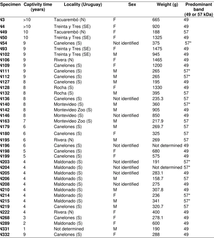
Table 1: Specimen data for the parameters studied.
Specimen Captivity time (years)
Locality (Uruguay) Sex Weight (g) Predominant
band (49 or 57 kDa)
N3 >10 Tacuarembó (N) F 665 49
N4 >10 Treinta y Tres (SE) F 920 49
N49 10 Tacuarembó (N) F 188 57
N50 10 Treinta y Tres (SE) F 1325 49
N54 9 Canelones (S) Not identified 375 57*
N93 9 Treinta y Tres (SE) F 1475 49
N102 9 Treinta y Tres (SE) M 945 49
N106 9 Rivera (N) F 1465 49
N109 9 Canelones (S) F 1200 49
N111 9 Canelones (S) M 265 57*
N112 9 Canelones (S) M 265 57*
N127 8 Canelones (S) M 195 49
N128 8 Rocha (S) F 1330 49
N132 8 Rocha (S) M 395 57
N136 8 Canelones (S) Not identified 235.3 57
N140 8 Montevideo (S) M 360 57*
N142 8 Montevideo Zoo (S) M 905 49
N146 8 Montevideo (S) Not identified 850 49
N163 7 Montevideo Zoo (S) M 217.9 57
N179 6 Canelones (S) M 269.7 57
N180 6 Canelones (S) F 325 57
N195 6 Rivera (N) M 269 57
N196 6 Canelones (S) Not identified Not determined 49
N198 5 Canelones (S) F 680 49
N199 5 Canelones (S) F 575 49
N203 4 Maldonado (S) Not identified 191 57*
N204 4 Maldonado (S) Not identified Not determined 57*
N205 4 Maldonado (S) Not identified 283.1 49
N206 4 Maldonado (S) M 158.7 57
N208 4 Maldonado (S) Not identified 275 49
N210 4 Maldonado (S) M 307.8 49
N214 4 Maldonado (S) F 236 57*
N215 4 Maldonado (S) M 341 57*
N219 4 Canelones (S) M 320.7 57
N222 4 Rivera (N) F 400 49
N268 3 Canelones (S) F 278.1 49
N289 2 Maldonado (S) F 600 49
N331 1 Not determined M 190 49
N332 9 Canelones (S) F 288 49
57*- Specimens with similar concentration in 49 and 57 kDa bands.
Groups 57* and 57 did not show significant differences in relation to the studied parameters.
Table 2: Statistical analysis between predominant band groups and the parameters
studied.
p value Correlation
Sex <0.01 Strong
Geographic variations 0.9 No
Captivity time 0.5 No
Weight <0.001 Very strong
Figure 1: SDS-PAGE (15%) of individual venom profiles. The arrow shows the 40kDa
band.
DISCUSSION
The existence of considerable variability at intraspecific level may be debatable and
perhaps reliant, to some extent, on the interpretation of similarity.
Our results show that individual variation in the protein profiles did not correspond to
the parameters studied. Relative quantity differences in 49 and 57 kDa bands are
strongly related to weight and must be considered for further protein identification
and analysis.
The groups proposed by the cluster analysis do not bring additional information.
Genetic variation could explain the analysis results.
Knowledge of intraspecific variations is very important for the design of reference
venom pool to obtain snake antivenom.
Traditionally, B. pubescens antivenom production in Uruguay has used venoms from
different localities to minimize geographic variations, although in Uruguay there are
not important geographic features or long distances.
According to our results, there are no evidences of geographic intraspecific variation.
Venoms from different localities of Uruguay may be unnecessary for antivenom
production. Further analysis, such as cross-neutralization assays, will be required to
confirm this point. This result is very relevant in relation to serpentarium management
and finally to economical cost of antivenom production.
ACKNOWLEDGMENTS
We are very grateful to Silvana Baletta for cooperation in venom extraction.
REFERENCES
1 BRAZIL, V. Do envenenamento ophidico e seu tratamento. São Paulo: Serviço
Sanitário do Estado de São Paulo, 1902. 27p.
2 BRAZIL, V. A serumtherapia do ophidismo em relação a distribuição geographica
das serpentes. Espécies venenosas americanas. Rev. Med. São Paulo. São
Paulo, 1907, 10, 10, 196-201.
3 CARREIRA S., MENEGHEL M., ACHAVAL F. Reptiles de Uruguay. Di. R. A. C.,
Facultad de Ciencias, Universidad de la República. Montevideo, 2005. 639 p.
4 CHIPPAUX JP., WILLIAMS V., WHITE J. Snake venom variability: methods of
study, results and interpretation. Toxicon, 1982, 29, 1279-303.
5 CHIPPAUX JP., GOYFFON M. Venoms, antivenoms and immunotherapy. Toxicon,
6 LAEMMLI UK. Cleavage of structural proteins during the assembly of the head of
bacteriophage T4. Nature, 1970, 227, 680-5.
7 MAGRO AJ., SILVA RJ. da, RAMOS PRR., CHERUBINI AL., HATAYDE R.
Intraspecific variations in the venom electrophoretic profile of recently captured
Crotalus durissus terrificus (Laurenti, 1768) snakes. J. Venom. Anim. Toxins,
2001, 7, 276-301.
8 MARCKLAND F. Snake venom and the hemostatic system. Toxicon, 1998, 36,
1749-800.
9 MONTEIRO R., DUTRA D., MACHADO O., CARLINI C., GUIMARAES J., BON C.,
ZINGALI R. Bothrops jararaca snake produce several bothrojaracin isoforms
following an individual pattern. Comp. Biochem. Physiol., 1998, 120B, 791-8.
10 PE T., LWIN NN., MYINT AA., HTWE KM., CHO KA. Biochemical and biological
properties of the venom from Russell`s viper (Daboia russelli siamensis) of
varying ages. Toxicon, 1995, 33, 817-21.
11 SCHEMBERG S. Geographical pattern of crotamine distribution in the same
rattlesnake subspecies. Science, 1959, 129, 1361-3.
12 SILVA V. da. Revisão sistemática do complexo Bothrops neuwiedi (Serpentes,
Viperidae, Crotalinae). Universidade de São Paulo, Instituto de Biociências,
2000, vol. 1: 134 p.; vol. 2: 241 p. [Thesis – Doctorate].
13 SOARES AM., ANZALONI LH.., FONTES MRM., SILVA RJ., GIGLIO JR.
Polyacrylamide gel electrophoresis as a tool for the taxonomic identification of
snakes from the Elapidae and Viperidae families. J. Venom. Anim. Toxins,
1998, 4, 137-42.
14 TAN N., PONNUDURAI G. A comparative study of electrophoretic patterns of
snake venoms. Comp. Biochem. Physiol., 1992, 102B, 103-9.
15 THEAKSTON RGD., PHILLIPS RE., WARREL DA., GALIGEDERA Y.,
ABEYSEKERA DT., DISSANAYAKE D., HUTTON R.A., ALOYSIUS DJ.
Failure of Indian (Haffkine) antivenom in treatment of Vipera russelli pulchella
(Russell’s viper) envenoming in Sri Lanka. Toxicon, 1989, 27, 82.
16 WARREL DA., PHILLIPS RE., THEAKSTON RGD.,GALIGEDERA Y.,
ABEYSEKERA DT., DISSANAYAKE D., HUTTON RA., ALOYSIUS DJ.
Neurotoxic envenomation by Indian krait (Bungarus caeruleus), Cobra (Naja
naja naja) and Russell’s viper (Vipera russelli pulchella) in Anuradhapura, Sri