ISSN 0100-2945 http://dx.doi.org/10.1590/0100-29452016048
SCIENTIFIC COMMUNICATION
FIRST REPORT OF Phytophthora nicotianae CAUSING ROOT
ROT OF SOURSOP IN NORTHEASTERN BRAZIL
1JAQUELINE FIGUEREDO DE OLIVEIRA COSTA2, IRAÍLDES PEREIRA ASSUNÇÃO3,
GAUS SILVESTRE DE ANDRADE LIMA4, MARIA DE FÁTIMA SILVA MUNIZ5,
EDNA DORA MARTINS NEWMAN LUZ6
ABSTRACT – In 2013, soursop trees showing symptoms of root rot were observed in a field in Maceió,
state of Alagoas, Brazil. It was isolated Phytophthora sp. which pathogenicity was confirmed in the host
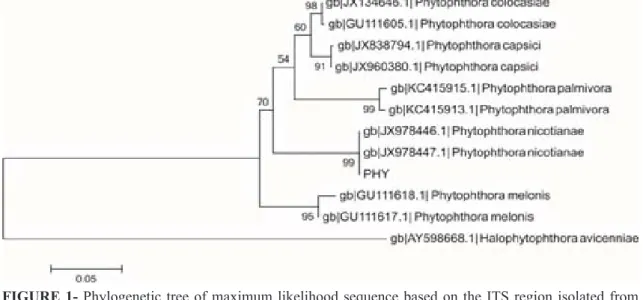
seedlings. Morphological and physiological characteristics in carrot-agar modified medium were consistent with Phytophthora nicotianae description. The PCR sequences products obtained with ITS1/ITS4 primers were compared to sequences of ribosomal DNA of Phytophthora species from the GenBank database ob-serving high identity with other P. nicotianae isolates. A phylogenetic tree was performed to compare the isolate with other sequences of P. nicotianae, which clustering has been verified with 99% of bootstrap,
confirming the morphophysiological studies. This is the first report of this pathogen on annonaceous plants in the Northeastern Brazil.
Index terms: Annona muricata, Phytophthora wilt, Oomycete, etiology.
PRIMEIRO RELATO DE
Phytophthora nicotianae
CAUSANDO PODRIDÃO
EM RAÍZES DE GRAVIOLEIRA NO NORDESTE DO BRASIL1
RESUMO – Em 2013, plantas de gravioleira exibindo sintomas de podridão em raízes foram observadas em um pomar localizado em Maceió, Alagoas, Brasil. Foi isolado Phytophthora sp., cuja patogenicidade foi confirmada em mudas do referido hospedeiro. Características morfofisiológicas do isolado em meio cenoura-ágar modificado apresentaram padrões idênticos àqueles descritos para Phytophthora nicotianae. As sequências dos produtos de PCR obtidos com os primers ITS1/ITS4 foram comparadas com sequências de DNA ribossômico de espécies de Phytophthora do GenBank observando-se alta identidade com outros isolados de P. nicotianae. Árvore filogenética foi construída para comparar o isolado com outras sequências
de P. nicotianae, cujo agrupamento foi verificado com 99% de bootstrap, confirmando os dados morfofisio -lógicos. Este é o primeiro relato deste patógeno em anonáceas no Nordeste do Brasil.
Termos para indexação: Annona muricata, murcha de Phytophthora, Oomiceto, etiologia.
1(Trabalho 086-15). Recebido em: 09-04-2015. Aceito para publicação em: 29-07-2015.
2MSc, doctoral student in Plant Protection, CAPES fellowship at the Centre for Agricultural Sciences of the Federal University of Alagoas, BR- 104 North, km 85, CEP 57100-000, Rio Largo-Alagoas. E-mail: jaquelinefigueredo@hotmail.com
3Associate professor at the Centre for Agricultural Sciences of the Federal University of Alagoas, 104 - BR North, km 85, CEP 57100-000, Rio Largo-Alagoas. E-mail:i_assuncao@hotmail.com
4Associate professor at the Centre for Agricultural Sciences of the Federal University of Alagoas, Brazil, BR – 104 North, km 85, CEP 57100-000, Rio Largo-Alagoas. E-mail:gaus@ceca.ufal.br
5Associate professor at the Centre for Agricultural Sciences of the Federal University of Alagoas, BR - 104 North km 85, CEP 57100-000, Rio Largo-AL. E-mail:mf.muniz@uol.com.br
The soursop (Annona muricata L.) is a tropical fruit tree, whose fruit is very appreciated for its food and nutritional properties, allied to its pleasant taste (CARDOSO et al., 2002) being considered the second most cultivated fruit by acreage and production in Brazil, losing only to the sugar apple (A. squamosa L.) (LEMOS, 2014). In Brazil, the Northeast region accounts for 80% of the culture production, Bahia being the leading producer, followed by Pernambuco, Ceará and Alagoas (IBGE, 2009).
T h e A n n o n a c e a e a r e s u s c e p t i b l e t o various diseases, highlighted by the Phytophthora
wilting which is caused by the Phytophthora spp. (JUNQUEIRA and JUNQUEIRA, 2014). In Brazil this disease is widespread in the states of São Paulo, Goiás, Minas Gerais and the Federal District, there is no confirmation on the productive regions from the North and Northeast (GRAMACHO et al., 2001; CARDOSO et al, 2002;. JUNQUEIRA and JUNQUEIRA, 2014). In São Paulo the species involved in the parasitism was identified as
Phytophthora nicotinae (= P. nicotinae van Breda de Haan var. parasitica (Dastur) Waterhouse), while in the Federal District and Goiás, the pathogen has not yet been identified at the species level (JUNQUEIRA; JUNQUEIRA, 2014).
The initial symptoms are manifested by leaf discoloration, which acquire a light green tonality. Then the leaves become chlorotic, wither and dry. At this stage of the infection, the roots exhibit necrosis. The lesions may progress toward the collar of the plant, reaching above the soil line. Plants of any age can be affected (CARDOSO et al., 2002; JUNQUEIRA; JUNQUEIRA, 2014). The disease incidence is increased by planting in poorly drained soils and also in areas with an excess of manure in the holes and with prolonged rainy periods, combined with temperatures below 22° C (JUNQUEIRA; JUNQUEIRA, 2014).
In Alagoas, root rot has been observed by producers of soursop, but there are no published data on the etiology of the disease. Thus, this study aimed to verify if the Phytophthora genus would be responsible for the appearance of symptoms and even to identify the species involved, through morphophysiological criteria and molecular analysis based on the sequencing of the internal transcribed spacer (ITS).
In 2013, soil samples were collected from the rhizosphere of soursop cv. Morada with symptoms of roots and collar rotting, followed by plant death from
a commercial orchard located in Maceió, Alagoas. Isolation was executed by the bait method employing a ripe fruit of pear cv. D’Anjou (ERWIN; RIBEIRO, 1996), which was placed on the surface of the soil, adding water according to indication of Luz et al. (2008). After three days, the fruit with lesions was removed from the ground and washed in tap water, proceeding to the isolation on PDA medium (potato dextrose agar), supplemented with Nystatin, PCNB (pentachloronitrobenzene), carbendazin (replacing the benomyl), rifampicin and ampicillin (MASAGO et al., 1977). The plates were incubated in the dark, at room temperature (26° C ± 2) and the developed colonies were transferred to Petri dishes containing PDA and, later preserved in sterilized distilled water (CASTELLANI, 1967) and kept at room tempera-ture, for subsequent studies.
The pathogenicity test was performed on soursop seedlings cv. Morada, five months old, cultivated in pots. The inoculation was done by stem injury, at about 5 cm above the ground with a scalpel (about 1 cm in length). In each hole was inserted a PDA culture disk of 5 mm diameter containing mycelia of the microorganism, taken from the periphery of the Petri plate culture, cultivated in the dark. Then, the site of inoculation was wrapped with a transparent PVC film. In the control group, only a disk of medium was placed. After the inoculation, the seedlings were kept in a greenhouse at room temperature (about 28°C). The symptoms were observed five months after inoculation, band* the
pathogen was reisolated on PDA selective medium as described above
To obtain asexual structures of the pathogen: mycelia disks (5 mm diameter) taken from the margins of the isolate colony, in modified carrot-agar medium-CA (RIBEIRO, 1978) cultivated at 25°C under alternate regime of light for three days, and were transferred to Petri dishes containing KNO3 0.01 M solution (ERWIN; RIBEIRO, 1996). After two days under the same incubation conditions, assessments of the structures were made.
For toospores** production, pairings of
Phytophthora sp. were made with isolates of P. palmivora (Butler) Butler from papaya (Carica papaya L.) of the two mating types: A1 (isolate no. 847) and A2 (isolated*** no. 839), assigned by the Section of Plant Pathology, CEPEC / CEPLAC, Ilhéus, BA, employing the sandwich technique (LUZ et al., 2008). The isolates were cultured in CA medium, and after five days of the cultures development, discs from the margin of the colonies
aqueous phase was transferred to a new tube and 400 µL of absolute ethanol was added.
The precipitated DNA was washed in 70% ethanol and dried at room temperature and then resuspended with 40 µL of TE (Tris-EDTA; 10 mM Tris-HCl, 1mM EDTA) + RNAse (10µg/mL). The DNA quality was visually estimated in 0.8% agarose gels, stained with ethidium bromide and observed under UV light. The material was stored at a temperature of -20°C.
The isolate used samples in the study had the region of ITS-rDNA amplified by PCR (polymerase chain reaction) using oligonucleotides ITS1 TCCGTAGGTGAACCTGCGG and ITS4 TCCTCCGCTTATTGATATGC. PCR reactions were performed with the buffer 10X, 50 mM MgCl2, 10 mM DNTP’s, 10 µM for each oligonucleotide, 1U of Taq DNA polymerase and1μL of DNA diluted (1:30). The final volume of the reactions was adjusted to 60 µL with autoclaved Milli-Q water. PCR reactions were performed in a thermocycler Applied Biosystems (2720 Thermal Cycler) under the following conditions: initial denaturation of 94° C and 34 cycles of 94°C for 1 min, 55°C for 30 sec, 72°C for 1 min and a final cycle of 10 min for 72°C.
After amplification, the sample was subjected to electrophoresis in 1.5% agarose gel, stained with ethidium bromide and viewed under UV light. The PCR product was purified using the GFX ™ PCR DNA Kit and Gel Band Purification Kit (GE Health -care), according to manufacturer’s recommendations and subsequently sent to sequencing at the Macrogen Inc. (Seoul, South Korea).
The sequence was first analyses with the BLASTn (Nucleotide Basic Local Alignment Search Tool algoritmo) for the preliminary identification of the species with the highest percentage of identity. The sequence was edited using the DNAMAN version 6 program (Biosoft Lynnon Corporation). For phylogenetic analysis, the sequence was initially aligned using MUSCLE (EDGAR, 2004) software available in the computer package MEGA 5 (Molecular Evolutionary Genetics Analysis) (TAMURA et al., 2011). The sequence alignment was the basis for phylogenetic reconstruction using a substitution model Kimura 2-parameter model, generating a phylogenetic tree of maximum likelihood. The reliability of the generated tree was obtained from 2,000 bootstrap replicates. Sequences
of other species of Phytophthora available at
GenBank (www.ncbi.nlm.nih.gov/) were included in the analyzes***. Phylogenetic analyses aimed to determine the taxonomic status of the isolate obtained in this study. The species Halophytophthora
were removed to be paired with other disc containing mycelium of the isolates A1, or A2 or from the isolate itself, interleaved by a disc of CA medium. After seven days at 20°C, in the dark, the oospores formation was observed.
Semipermanent slides with lactophenol were prepared to evaluate the morphology of the reproductive structures of the pathogen. Measurements were obtained from images captured by a digital camera (Olympus® IX2-SLP) coupled to an optical microscope (Olympus® CKX 41) using CellSens Standard software - Olympus 2010. Forty measurements were performed on each structure.
For physiological characterization, was evaluated the effect of different temperatures on the mycelia development of the isolate. In this way, mycelium discs with a diameter of 5 mm were collected from the colony edges on PDA medium at 25° C in the dark, with four days, they were transferred to the center of Petri dishes containing CA medium and incubated in the dark, with temperatures of 10, 15, 20, 25, 30 and 35° C for five days. Two perpendicular diameters from the colony of the isolate were measured, and the colony aspect was also observed. The experimental design was completely randomized with eight repetitions*.
To obtain the mycelial mass of the
Phythophtora isolate, three PDA discs containing the mycelium were transferred to Erlenmeyer flasks (capacity of 50 mL) containing 30 mL of Saccharose- Yeast extract- asparagine medium (saccharose 10 g, L-asparagine 2 g, yeast extract 2 g, KH2PO4 1 g, MgSO4.7H2O 0.1 g, ZnSO4.7H2O 0.44 mg, FeCl3.6H2O 0.48 mg, and MnCl2.H2O 0.36 mg) (ZAUZA et al., 2007). The culture was incubated for five days at 25 ± 1°C without agitation, under a 12 hour photoperiod.
For DNA extraction was used the Doyle and Doyle, (1987) protocol, where mycelium of the isolatewas** macerated with liquid nitrogen in a porcelain mortar with the aid of a pestle. The ground mycelium was transferred to a micro centrifuge tube with a 1.5 mL capacity. Then was added 1 mL of extraction buffer hexadecyltrimethylammonium bromide (CTAB) at 4% (4% CTAB, 1.4 M NaCl, 20 mM EDTA, 100 mM Tris-HCl, 1% PVP), 4 µL of β-mercaptoethanol and then, the tube was kept in a water bath at 65° C for 30 minutes. Subsequently, the sample was placed in a centrifuge at 12,000 rpm for 15 minutes. The supernatant was transferred to a new tube into which 600 mL of CIA (chloroform: isoamylalcohol 24: 1) was added and 40 µL of CTAB at 10%, heated at 65° C. After centrifugation, the
avicenniae (Gerr.-Corn. & Simpson) Ho & Jong (AY598668) was used as an out-group.
The isolate was pathogenic to soursop seedlings presenting necrotic lesions on the stem, but there was no chlorosis in the leaves or death of the plants up to five months after inoculation. Symptoms in the control plants were not observed. The reisola-tion of the oomycete was successfully obtained, thus confirming Koch’s postulates (AGRIOS, 2005).
The sporangia were ovoid, papillate, measuring 35.36 to 63.17 X 20.25 to 39.76 µm (average of 49.38 X 29.2 mM*), with length-breadth ratio of 1.7: 1. Rare chlamydospores were observed. The isolate is heterothallic forming aplerotic oospores, which measured 19.53 to 30.08 µm in diameter (average 25.04 µm) and amphigynous antheridia when paired with the mating type A1. These characteristics are consistent with those presented by Erwin and Ribeiro, (1996), to
Phytophthora nicotianae.
From the temperatures tested, there were only traces of mycelia growth at 10° C. In the temperatures of 15, 20, 25, 30 and 35° C, the mean diameter of the colonies was 4.8; 6.9; 8.9; 8.5 and 3.9 cm, respectively. The isolate produced cultures
FIGURE 1- Phylogenetic tree of maximum likelihood sequence based on the ITS region isolated from
Phytophthora nicotianae with Halophytophthora avicenniae as outgroup. The Phytophthora
PHY represents the isolated****from this study. The scale bar represents change of 0.05 of
bootstrap support values, 2000 repetitions**** are exhibit in the branches.
with a petaloid appearance, dense and cotton-like aerial mycelium. The cardinal temperatures for growth of the isolate studied are in accordance with the descriptions of Erwin and Ribeiro (1996), for the
P. nicotianae species.
The sequences of the PCR products obtained with the ITS1 / ITS4 primers compared to the ribosomal DNA sequences of Phytophthora species from the GenBank (http: www.ncbi.nlm.nih.gov) allowed species confirmation with 99% accuracy, as P. nicotianae (Figure 1). The identification of the
quoted species in black wattle (Acacia mearnsii De Wild.) was performed by molecular studies based on sequencing of regions of ITS** (SANTOS et al., 2005). According to Hyde et al. (2014), this is the most accurate method for identifying many species of Phytophthora.
Considering the observed morphophysiological criteria and molecular analysis based on sequencing of Its regions, the isolate of Phytophthora sp. obtained from soursop was identified as P. nicotianae. In Annonaceae, this is the first finding showing the oomycete as a causal agent of roots rot of the soursop in the Northeastern Brazil.
REFERENCES
AGRIOS, G.N. Plant pathology. 5th ed. San Diego: Academic Press, 2005. 922p.
CARDOSO, J.E.; VIANA, F.M.P.; FREIRE, F.C.O.; SANTOS, A.A. Doenças. In: CARDOSO, J.E. (Ed.). Graviola: fitossanidade. Brasília: Embrapa
Informação Tecnológica, 2002. p.11-21. (Frutas do Brasil, 20).
CASTELLANI, A. Maintenance and cultivation of the common pathogenic fungi of man in sterile distilled water: further researches. Journal of Tropical Medicine and Hygiene, Oxford, v.70, n.8, p.181-184, 1967.
DOYLE, J.J.; DOYLE J.L. A rapid DNA isolation procedure for small quantities of fresh leaf tissue. Phytochemical Bulletin, Oklahoma, v.19, n.1, p.11-15, 1987.
EDGAR, R.C.; MUSCLE: A multiple sequence alignment method with reduced time and space
complexity. BMC Bioinformatics, London, v.5,
p.1-19, 2004.
ERWIN, D.C.; RIBEIRO, O.K. Phytophthora
diseases worldwide. St. Paul, Minnesota: APS Press. 1996.
GRAMACHO, K.P.; BEZERRA, J.L.; JUNQUEIRA, N.T.V. Phytophthora sp. em espécies da família Anonaceae. In: LUZ, E.D.M.N.; SANTOS, A.F.;
MATSUOKA, K.; BEZERRA, J.L. (Ed.). Doenças
causadas por Phytophthora no Brasil. Campinas: Livraria e Editora Rural, 2001. p.91-99.
HYDE, K.D. et al. One stop shop: backbones trees for important phytopathogenic genera: I. Fungal Diversity, Chiang Mai, v.67, n.1, p.21-125, 2014.
IBGE – Instituto Brasileiro de Geografia e Estatística.
Censo agropecuário. Rio de Janeiro, 2009. 777p.
JUNQUEIRA, N.T.V.; JUNQUEIRA, K.P. Principais doenças de anonáceas no Brasil: descrição e controle.
Revista Brasileira de Fruticultura, Jaboticabal, v.36, n.1, p.55-64, 2014. Numero especial.
LEMOS, E.E.P. A produção de anonáceas no Brasil.
Revista Brasileira de Fruticultura, Jaboticabal, v.36, n.1, p.77-85, 2014. Numero especial.
LUZ, E.D.M.N.; SILVA, S.D.V.M.; BEZERRA, J.L.;
SOUZA, J.T.; SANTOS, A.F. Glossário ilustrado
de Phytophthora: técnicas especiais para o estudo de oomicetos. Itabuna: FAPESB, 2008.126p.
MASAGO, H.; YOSHIKAWA, M.; FUKADA, M.;
NAKANISHI, N. Selective inhibition of Pythium
spp. on a medium for direct isolation of Phytophthora
spp. from soils and plants. Phytopathology, St. Paul, v.67, n.3, p.425-428, 1977.
RIBEIRO, O.K. A source book of the genus
Phytophthora. Vaduz: J. Cramer, 1978. 417p. )
SANTOS, A.F.; LUZ, E.D.M.N.; SOUZA, J.T.
Phytophthora nicotianae: agente etiológico da
gomose da acácia-negra no Brasil. Fitopatologia Brasileira, Brasília, v.30, n.1, p.81-84, 2005.
TAMURA, K.; PETERSON, D.; PETERSON, N.; STECHER, G.; NEI, M.; KUMAR, S. MEGA 5: Molecular evolutionary genetics analysis using maximum likelihood, evolutionary distance, and
maximum parsimony methods. Molecular Biology
and Evolution, Oxford, v.28, n.10, p.2731-2739, 2011.
ZAUZA, E.A.V.; ALFENAS, A.C.; MAFIA, G.R. Esterilização, preparo de meios de cultura e fatores associados ao cultivo de fitopatógenos. In:
ALFENAS, C.A.; MAFIA, R.G. (Ed.). Métodos em
