Faculdade de Medicina de Lisboa
Brain-derived neurotrophic factor and adenosine
signalling on amyloid-β peptide induced toxicity:
impact on hippocampal function
André Jerónimo Santos
Doutoramento em Ciências Biomédicas
Especialidade em Neurociências
Faculdade de Medicina de Lisboa
Brain-derived neurotrophic factor and adenosine
signalling on amyloid-β peptide induced toxicity:
impact on hippocampal function
André Jerónimo Santos
Tese orientada por Professora Maria José Diógenes
Doutoramento em Ciências Biomédicas
Especialidade em Neurociências
As opiniões expressas nesta publicação são da exclusiva responsabilidade do seu
autor, não cabendo qualquer responsabilidade à Faculdade de Medicina de Lisboa
A impressão desta dissertação foi aprovada pelo Conselho Cientifico da
Faculdade de Medicina de Lisboa em reunião de 24 de Junho de 2014.
The scientific content of this thesis was included in the publications of the following original articles:
• Kemppainen S, Rantamaki T, Jerónimo-Santos A, Lavasseur G, Autio H, Karpova N, Karkkainen E, Staven S, Miranda HV, Outeiro TF, Diogenes MJ, Laroche S, Davis S, Sebastiao AM, Castren E, Tanila H (2012). Impaired TrkB receptor signaling contributes to memory impairment in APP/PS1 mice. Neurobiol Aging 33:1122 e1123-1139.
• Jerónimo-Santos A, Vaz SH, Parreira S, Rapaz-Lérias S, Caetano AP, Buée-Scherrer
V, Castrén E, Valente CA, Blum D, Sebastião AM, Diógenes MJ (2014). Dysregulation of TrkB receptors and BDNF function by amyloid-β peptide is mediated by calpain. Cereb Cortex. pii: bhu105. (Epub ahead of print).
• Jerónimo-Santos A, Batalha VL, Muller C, Hockemeyer J, Sebastião AM, Lopes LV
and Diógenes MJ (2014) Chronic blockade of adenosine A2A receptors prevents BDNF effect on hippocampal LTP. Neuropharmacology. 83: p. 99-106.
Other publications closely related to the content of this thesis:
• Diogenes MJ, Costenla AR, Lopes LV, Jerónimo-Santos A, Sousa VC, Fontinha BM, Ribeiro JA, Sebastiao AM (2011). Enhancement of LTP in aged rats is dependent on endogenous BDNF. Neuropsychopharmacology 36:1823-1836.
• Rodrigues TM* and Jerónimo-Santos A*., Outeiro TF, Sebastião AM,Diógenes MJ (2014). Challenges and promises in the development of neurotrophic factor-based therapies for Parkinson´s disease. Drugs Aging. 31: 239-261. *co-first authors
• Rodrigues TM, Jerónimo-Santos A, Sebastião AM,Diógenes MJ (2014). Adenosine A2A Receptors as novel upstream regulators of BDNF-mediated attenuation of
Figure index ...vii Acknowledgments ...ix Abbreviations list...xi Resumo...xv Abstract ...xvii 1. Introduction ...1 1.1. Neurotrophins ... 1 1.1.1. Neurotrophin release... 1 1.1.2. Neurotrophin receptors ... 3 1.1.3. Truncated TrkB receptors ... 3
1.1.4. TrkB-mediated signalling cascades ... 4
1.1.5. BDNF and long-term potentiation... 8
1.1.6. Facilitation of BDNF synaptic actions by adenosine ... 12
1.2. Calpain system ... 15
1.2.1. Calpains ... 15
1.2.2. Calpain substrates and cleavage specificity ... 18
1.2.3. Regulation of calpain activity... 19
1.2.4. Function in physiology and pathophysiology... 22
1.3. Alzheimer’s disease... 23
1.3.1. Amyloid-β peptides ... 26
1.3.2. Neurotoxicity mediated by Amyloid-β peptides ... 28
1.3.3. BDNF signalling in AD... 34 2. Aims ...37 3. Methods ...39 3.1. Materials ... 39 3.2. Amyloid-β peptides ... 39 3.3. BDNF ... 41
3.4. Animals and brain areas used ... 42
3.5. Primary Neuronal cultures and drug treatments ... 42
3.6. Human brain sample... 42
3.7. Western-blot ... 43
3.8. Cell death evaluation ... 44
3.9. N-sequencing... 45
3.11. Immunoprecipitation ... 47
3.12. Calpain in-vitro digestion ... 47
3.13. Acutely prepared hippocampal slices ... 47
3.14. Isolation of synaptosomes ... 48
3.15. [3H] Neurotransmitter release from hippocampal synaptosomes ... 48
3.16. Calculation of drug effects on GABA and glutamate release ... 49
3.17. KW-6002 treatment... 49
3.18. Ex-vivo electrophysiology recordings... 50
3.19. Input / Output curves ... 52
3.20. Statistical analysis ... 52
4. Dysregulation of TrkB receptors and BDNF function by Aβ peptide is mediated by calpain .53 4.1. Summary ... 53
4.2. Rational... 53
4.3. Aβ increases truncated TrkB protein levels ... 55
4.4. Aβ up-regulates TrkB-T1 mRNA levels... 59
4.5. Aβ induces a cleavage on TrkB-FL receptors... 60
4.6. Calpain mediates the Aβ-induced TrkB-FL cleavage... 61
4.7. TrkB-FL calpain cleavage site is located downstream the Shc binding site... 66
4.8. Calpain mediates detrimental effects of Aβ upon BDNF actions on GABA and Glutamate release. ... 71
4.9. Calpain mediates detrimental effects of Aβ upon BDNF actions on CA1 LTP... 75
4.10. Discussion ... 79
5. Impact of in-vivo chronic blockade of A2AR upon BDNF-mediated facilitation on LTP ...83
5.1. Summary ... 83
5.2. Rational... 83
5.3. Chronic blockade of adenosine A2AR prevents BDNF-induced facilitation of CA1 LTP which is not restored by acute A2AR activation... 84
5.4. Chronic A2AR blockade reduces protein and mRNA levels of TrkB-FL receptor without affecting BDNF levels... 87
5.5. Discussion ... 90
6. BDNF mediates neuroprotection against Aβ-induced toxicity in a mechanism independent on A2AR activation...93
6.1. Summary ... 93
6.2. Rational... 94
6.3. BDNF reduces cellular death induced by Aβ ... 95
6.4. Cellular death prevented by BDNF does not require A2AR activation ... 98
6.6. Discussion ... 103
7. General discussion and conclusion...107
8. Future perspectives ...109
9. References...111
10. Appendix...129
10.1. TrkB-ICD: putative conformation and localization. ... 129
Figure 1.1 – Regulation of BDNF release upon synaptic activity. ...2
Figure 1.2 – Schematic representation of TrkB receptors signalling pathways...7
Figure 1.3 – Modulation of glutamatergic synapse by BNDF ...11
Figure 1.4 – Cross-talk between TrkB and A2A receptors. ...14
Figure 1.5 – Schematic representation of µ-calpain structure. ...17
Figure 1.6 – Crystallographic structure of calcium-bound m-calpain enclosed by calpastatin. ...21
Figure 1.7 – Brain atrophy and hypothetical progression model in AD. ...25
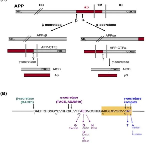
Figure 1.8 – APP processing and Aβ formation...27
Figure 1.9 – Simplified schematic representation of Aβ toxicity mechanisms...33
Figure 3.1 – Aβ1-42 andAβ25-35 peptides have fibrillary structures...41
Figure 3.2 – Recombinant BDNF purity...41
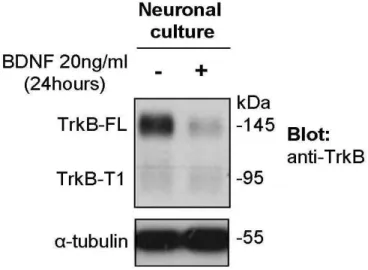
Figure 3.3 – Anti-TrkB antibody specificity...44
Figure 3.4 – Melting curves, amplification curves and Ct numbers from qPCR. ...46
Figure 3.5 – Electrophysiology recording configuration and LTP magnitude on both pathways. ...51
Figure 4.1 – Effect of Aβ 25-35 upon truncated and full-length TrkB receptors protein levels. ...56
Figure 4.2 – Effect of glial cells upon Aβ-induced changes in TrkB isoforms levels. ...57
Figure 4.3 – Effect of caspases inhibition upon Aβ-induced changes in TrkB isoforms levels...58
Figure 4.4 – Aβ peptide up-regulates mRNA levels of truncated TrkB-T1 and T2. ...59
Figure 4.5 – Aβ peptide induces a TrkB-FL receptor cleavage...61
Figure 4.6 – Aβ-induced cleavage of TrkB-FL is repressed by inhibitors of calpain-like activity. ...64
Figure 4.7 – Calpain cleaves rat and human TrkB-FL receptor. ...66
Figure 4.8 – Calpain inhibition does not block the Aβ-induced changes on TrkB mRNA levels...67
Figure 4.9 – Calpain cleaves TrkB downstream Shc binding site (Tyr 515). ...68
Figure 4.10 – Effect of Aβ upon BDNF-induced TrkB phosphorylation on Tyr 515. ...70
Figure 4.11 – Effect of Aβ upon BDNF-induced TrkB phosphorylation on Tyr 816. ...70
Figure 4.12 – Modulation of GABA and glutamate release by BDNF, Aβ1-42 and MDL28170...73
Figure 4.13 – Aβ inhibits BDNF effect upon neurotransmitter release in a calpain-dependent way. ...75
Figure 4.14 – Aβ decreases the effect of BDNF upon LTP, in a calpain-dependent way...78
Figure 4.15 – Reverse Aβ35-25 peptide does not affect BDNF effect upon LTP...78
Figure 4.16 – Schematic representation of Aβ-induced dysregulation of TrkB receptors...82
Figure 5.1 – Chronic treatment with KW-6002 abolishes the facilitatory effect of BDNF upon LTP...87
Figure 5.2 – Effects of KW-6002 are not reverted by treatment withdrawn or acute A2AR activation...88
Figure 5.3 – Protein and mRNA levels of TrkB-FL receptor are decreased in KW-6002 treated rats. ...89
Figure 6.1 – BDNF reduces the caspase-3 activation induced by Aβ...96
Figure 6.2 – BDNF reduces the αII-spectrin breakdown induced by Aβ. ...97
Figure 6.3 – A2AR does not influence BDNF-mediated reduction in Aβ-induced Caspase-3 activation ...99
Figure 6.5 – Effect of A2AR and BDNF upon Aβ-induced TrkB truncation ...102
Figure 10.1 – TrkB-ICD conformation ...129
Figure 10.2 – TrkB-ICD subcellular localization...130
Figure 10.3 – Amplification of TrkB-ICD by RT-PCR...132
Figure 10.4 – Analysis of selected E.coli colonies by RT-PCR...133
Em primeiro lugar gostaria de agradecer à minha orientadora, Maria José Diógenes, por todos os ensinamentos e sugestões que me deu, pela sua enorme simpatia e boa disposição constante e pela amizade e motivação que me transmitiu.
Gostaria também de mostrar o meu agradecimento à Professora Ana Sebastião e ao Professor Joaquim Alexandre Ribeiro pela receptividade e partilha de conhecimentos e pelas sugestões e discussão científica que me foram bastante úteis.
A todos os colegas do laboratório pelas ajudas em experiências, pela discussão científica e por proporcionarem um bom ambiente de trabalho com boa disposição.
Uma palavra especial de agradecimento à Rita por toda a sua paciência, pelos inúmeros conselhos, ajuda, carinho e apoio.
Um agradecimento ao Instituto de Medicina Molecular pelo acolhimento e bom ambiente, e não menos importante, gostaria de agradecer também à Fundação para a Ciência e Tecnologia por me ter financiado a bolsa de doutoramento (SFRH / BD / 62828 / 2009).
E finalmente, à minha família, em especial aos meus pais, pelo apoio incondicional e incentivo dado, nos bons e maus momentos.
5-FU 5-Fluoracil Aββββ Amyloid-beta A1R Adenosine A1 Receptor A2AR Adenosine 2A Rreceptor A2BR Adenosine 2B Receptor A3R Adenosine 3 Receptor
aCSF Artificial Cerebrospinal Fluid
AD Alzheimer’s Disease
ADA Adenosine Deaminase
ADDLs Aβ-derived Diffusible Ligands
ADK Adenosine Kinase
AFM Atomic Force Microscopy
AICD APP Intracellular Domain
AIF Apoptosis-Inducing Factor
ALLN N-Acetyl-Leu-Leu-Nle-CHO
AMP Adenosine 5'-Monophosphate
AMPA Alpha-Amino-3-Hydroxy-5-Methyl-4-Isoxazolepropionic Acid
AMPAR AMPA Receptor
ANOVA Analysis Of Variation
AOAA Aminooxyacetic Acid
Apaf-1 Apoptotic protease activating factor 1
APH1 Anterior Pharynx-defective 1
APOE Apolipoprotein E
APP Amyloid Precursor Protein
ATP Adenosine Triphosphate
BACE1 Beta-site APP Cleaving Enzyme 1
BAD Bcl-2-Associated Death promoter
BAK Bcl-2 homologous Antagonist/Killer
BBB Blood-Brain Barrier
Bcl-XL B-cell lymphoma-extra large
BDNF Brain-Derived Neurotrophic Factor
CA1 Cornu Ammonis area 1
CaMK Ca2+-Calmodulin-regulated Kinase
CaMKK Calcium/calmodulin-dependent protein kinase kinase
cAMP Cyclic Adenosine Monophosphate
CaMPDB Calpain for Modulatory Proteolysis Database
CANP Calcium-activated Neutral Proteinase
CAPN1 Calpain-1 catalytic subunit
CAPN2 Calpain-2 catalytic subunit
CAPNS1 Calpain small subunit 1
CAST Calpastatin
CDK5 Cyclin-dependent kinase 5
cDNA Complementary DNA
CRE cAmp-Responsive Element
CREB CRE binding protein
CRF Corticotropin-Releasing Factor CSF Cerebrospinal Fluid CTR Control CypD Cyclophilin D DAG Diacylglycerol DEVD-pNA Ac-Asp-Glu-Val-Asp-pNA DIV Days In-Vitro
DMSO Dimethylsulphoxide
DNA Deoxyribonucleic Acid
DTT 1,4-dithiothreitol
EC Enzime Classification
EDTA Ethylenediaminetetraacetic Acid
EF-hand Helix-loop-helix structural domain
EGF Epidermal Growth Factor
eIF4E Initiation Factor 4E
E-LTP Early LTP
EphB2 Ephrin type B receptor 2
ER Endoplasmic Reticulum
ERK Extracellular Signal-Regulated Kinase
FDG Fluorodeoxyglucose
fEPSP Field Excitatory Postsynaptic Potential
FL Full-length
G0/Gi Gi alpha subunit G protein
Gab1/2 Grb-associated binding protein
GABA Gamma-Aminobutyric Acid
GAP-43 Growth Associated Protein 43
GAPDH Glyceraldehyde 3-phosphate Dehydrogenase
GDP Guanosine 5'-Diphosphate
GFAP Glial Fibrillary Acidic Protein
GluR Glutamate receptor
GPCR G-Protein-Coupled Receptor
Grb2 Growth factor receptor-bound protein
GSK3 Glycogen Synthase Kinase 3
GTPase Guanosine-5'-triphosphate hydrolase
HBSS Hanks' Balanced Salt Solution
HEPES N-2-Hydroxyethylpiperazine-N'-2-Ethanesulfonic Acid
HFS High Frequency Stimulation
HRP Horseradish Peroxidase
IAPs Inhibitors of Apoptosis
ICD Intracellular Domain
IgG Immunoglobulin G
IP3 Inositol 1,4,5-triphosphate
IP3R IP3 Receptor
ISF Interstitial Fluid
IκB Inhibitor of kappa B
KHR Krebs-Henseleit Rinsing
KW6002 Istradefylline
LDH Lactate Dehydrogenase
LilrB2 Leukocyte immunoglobulin-like receptor B2
LL-37 Human Cathelicidin
L-LTP Late phase of LTP
LTD Long Term Depression
LTP Long-Term Potentiation
MAP Microtubule-Associated Protein
MAPK Mitogen-Activated Protein Kinase
MCI Mild Cognitive Impairment
MDL28170 N-[N-[(Phenylmethoxy)carbonyl]-L-valyl]-phenylalaninal
MEK Mitogen-activated protein kinase kinase
MG132 Z-Leu-Leu-Leu-CHO
mGluR Metabotropic Glutamate Receptor
MPTP 1-methyl-4-phenyl-1,2,3,6-tetrahydropyridine
mPTP Mitochondrial Permeability Transition Pore
MRI Magnetic Resonance Imaging
mRNA Messenger Ribonucleic Acid
NAIP-1 Neuronal Inhibitors of Apoptosis Protein 1
NF-κB Nuclear Factor Kappa-light-chain-enhancer of activated B cells
NGF Nerve Growth Factor
NMDAR N-Methyl-D-Aspartate Receptor
NT Neurotrophin
NTRK2 Neurotrophin Tyrosine Kinase 2
p75NTR P75 Neurotrophin Receptor
PBS Phosphate Buffered Saline
PC12 Pheochromocytoma cell line
PCR Polymerase Chain Reaction
PEN2 Presenilin enhancer 2
PET Positron Emission Tomography
PI3K Phosphatidylinositol 3-Kinase
PiB Pittsburgh compound B
PIP2 Phosphatidylinositol-4,5-bifosphate
PIP3 Phosphatidylinositol-3,4,5-trisphosphate
PKA Protein Kinase A
PKC Protein Kinase C
PKMζ Atypical Protein Kinase C Isoform
PLC Phospholipase C
PMSF Phenylmethylsulfonyl Fluoride
pNA p-nitroaniline
PPF Pair-pulse facilitation
ProBDNF Precursor of BDNF
PrPc Cellular Prion Protein
PS1 Presenilin-1
PSD Postsynaptic Density
PSEN Presenilin gene
PTB Phosphotyrosine Binding
PVDF Polyvinylidene Difluoride
qPCR quantitative Polymerase Chain Reaction
RAGE Receptor For Advanced Glycation End products
RhoGDI Rho GDP-dissociation inhibitor
RIPA Radioimmunoprecipitation Assay Buffer
RNA Ribonucleic Acid
ROS Reactive Oxygen Species
RSK Ribosomal S6 Kinase
RTK Receptor Tyrosine Kinase
RT-PCR Reverse Transcription-Polymerase Chain Reaction
SBDP Spectrin Breakdown Product
SCH-58261 A2AR antagonist
SDS-PAGE Sodium Dodecyl Sulfate-Polyacrylamide gel electrophoresis
SEM Standard Error Of The Mean
SH2 Src-Homology 2
siRNA small interfering RNA
SOS Son of Sevenless
TBS Theta-burst Stimulation
TBS-T Tris-buffered Saline-Tween 20
ThT Thioflavin T
TLR2 Toll-like Receptor 2
TM Transmembrane
TNF Tumor Necrosis Factor
tPA Tissue Plasminogen Activator
Trk Tropomyosin-related Kinase
TrkB-FL TrkB full-length
TrkB-T’ TrkB truncated (calpain-generated)
TrkB-T1 TrkB truncated isoform 1
TrkB-Tc TrkB truncated (total pool)
VGCC Voltage-gated Calcium Channels
Resumo
O factor neurotrófico derivado do cérebro (Brain-derived neurotrophic factor- BDNF) e o seu receptor de alta afinidade, TrkB-FL, desempenham um papel central no sistema nervoso, dado que promovem suporte trófico aos neurónios e que regulam a transmissão e plasticidade sinápticas.
A sinalização mediada pelo BDNF encontra-se diminuída na doença de Alzheimer (Alzheimer’s disease -AD), uma doença neurodegenerativa na qual ocorre acumulação do péptido beta amilóide (amyloid-beta -Aβ). Apesar dos mecanismos envolvidos na redução da sinalização mediada pelo BDNF na AD não serem totalmente conhecidos, o restabelecimento das acções do BDNF tem sido considerado como uma estratégia promissora para a terapêutica desta doença.
Na última década tornou-se claro que a maioria das acções sinápticas do BDNF, incluindo as acções na transmissão e plasticidade sinápticas e também na libertação de neurotransmissores, é dependente da activação dos receptores A2A da adenosina (A2AR).
Contudo, o uso de antagonistas dos A2AR tem sido apontado como uma possível estratégia
terapêutica para o tratamento da AD.
Dada a falta de evidências que clarifiquem os mecanismos envolvidos nas alterações da sinalização mediada pelo BDNF e o conhecimento de que a activação dos A2AR facilita a
maioria das acções sinápticas do BDNF, o objectivo principal desta tese foi estudar o impacto dos péptidos Aβ e dos A2AR na sinalização mediada pelo BDNF.
Este trabalho revelou que, em culturas primárias de neurónios corticais, o Aβ aumenta os níveis de mRNA dos receptores TrkB truncados, TrkB-T1 e TrkB-T2, sem afectar os níveis de mRNA dos receptores TrkB completos, TrkB-FL. Por outro lado, verificou-se que o Aβ aumenta os níveis proteicos do conjunto de receptores TrkB truncados e que diminui os níveis proteicos dos receptores TrkB-FL, por um mecanismo independente da proliferação glial e da activação de caspases. Foi ainda possível concluir que o Aβ induz a clivagem, mediada por calpaínas, dos receptores TrkB-FL, esta clivagem dá-se após o local de ligação da Shc e antes do início do domínio de cinase de tirosina, pelo que origina um novo receptor TrkB truncado T’), contendo o local de ligação à Shc, e um novo fragmento intracelular
(TrkB-intracellular domain- ICD), contendo a totalidade do domínio da cinase. No entanto, a presença
destes fragmentos, não mostrou afectar a fosforilação do receptor TrkB-FL induzida pela exposição ao BDNF. Interessantemente, foi possível detectar o fragmento TrkB-ICD em uma amostra, post-mortem, de cérebro humano. Mostrou-se também que a inibição das calpaínas previne as alterações dos níveis proteicos das isoformas do TrkB, induzidas pelo Aβ, sem afectar as alterações ao nível do mRNA do TrkB. Por outro lado, este trabalho revelou que o
BDNF exógeno reduz a activação da caspase-3 e das calpaínas induzida pelo Aβ, de uma forma independentemente dos A2AR.
Em fatias de hipocampo de ratos adultos, este trabalho mostrou que o Aβ diminui as acções do BDNF na plasticidade sináptica, nomeadamente na potenciação de longa duração (Long-term potentiation, LTP) na área CA1 do hipocampo, bem como no seu efeito sobre libertação de neurotransmissores (GABA e glutamato) de sinaptosomas. Notavelmente, o inibidor das calpaínas, MDL28170, mostrou restabelecer os efeitos do BDNF, na presença do péptido Aβ, tanto na plasticidade sináptica como na libertação de neurotransmissores.
Este trabalho permitiu ainda concluir que o bloqueio crónico dos A2AR, in-vivo, através
da administração de um antagonista selectivo (KW-6002), previne o efeito potenciador do BDNF na LTP, registada ex-vivo na área CA1 do hipocampo, e que diminui os níveis de mRNA e de proteína do receptor TrkB-FL, no hipocampo de rato.
Em suma, o presente trabalho revelou que o péptido Aβ induz a clivagem dos receptores TrkB-FL, mediada pelas calpaínas, e que bloqueia as acções mediadas pelo BDNF na plasticidade sináptica e na libertação de GABA e glutamato por um mecanismo dependente da actividade das calpaínas. Se por um lado, o efeito do BDNF na plasticidade sináptica é perdido aquando da inibição crónica dos A2AR, o efeito protector desta neurotrofina contra a toxicidade
induzida pelo Aβ mostrou-se independente da activação dos A2AR.
Palavras-Chave:
Doença de Alzheimer; neurodegeneração, neurotrofinas, potenciação de longa duração, libertação de neurotransmissores, TrkB, KW-6002, istradefylline, neuroprotecção
Abstract
Brain-derived neurotrophic factor (BDNF) and its high-affinity full-length receptor, TrkB-FL, play a central role in the nervous system by providing trophic support to neurons and by regulating synaptic transmission and plasticity.
BDNF signalling is impaired in Alzheimer’s disease (AD), a neurodegenerative disorder characterized, among other features, by the accumulation of the amyloid-β (Aβ) peptide. Although the mechanisms implicated in the reduction of BDNF signalling in AD were not clarified, the reestablishment of BDNF actions is considered as a promising strategy for AD treatment.
In last decade it became clear that most of synaptic actions of BDNF, including the ones upon synaptic transmission, plasticity or upon neurotransmitter release, are fully dependent on adenosine A2A receptors (A2AR) activation. However, evidences indicate that A2AR antagonists
can prevent the deficits in AD animal models.
Given the lack of data clarifying the mechanisms behind the changes on BDNF signalling, namely changes on TrkB receptors, and the knowledge that A2AR activation
facilitates most of BDNF synaptic actions, the main goal of this project was to study the impact of Aβ peptides and A2AR on BDNF signalling.
This work revealed that in rat primary neuronal cultures Aβ selectively increases mRNA levels for the truncated TrkB-T1 and TrkB-T2 isoforms without affecting TrkB full-length (TrkB-FL) mRNA levels. Moreover, Aβ increases protein levels of total pool of truncated TrkB receptors (TrkB-Tc) and decreases TrkB-FL protein levels. This effect is explained by the Aβ-induced calpain-mediated cleavage on TrkB-FL receptors, downstream of Shc binding site, which results in the formation of a new truncated TrkB receptor (TrkB-T’) and a new intracellular fragment (TrkB-ICD), which is also detected in post-mortem human brain samples. In hippocampal slices it was observed that Aβ impairs BDNF function in a calpain-dependent way, upon modulation of GABA and glutamate release from hippocampal nerve terminals, and upon modulation of long-term potentiation (LTP). Finally, the exogenous BDNF strongly reduces the Aβ-induced activation of caspase-3 and calpain in neuronal cultures, an effect not affected by A2AR agonist or antagonist.
Moreover, for the first time it was shown that chronic in vivo blockade of A2AR by a
selective antagonist, prevents the facilitatory action of BDNF upon ex-vivo CA1 hippocampal LTP and decreases both mRNA and protein levels of the TrkB-FL receptor in rat hippocampus.
In conclusion, the present work shows that Aβ induces a TrkB-FL cleavage mediated by calpain and impairs BDNF-mediated effects in synaptic plasticity and neurotransmitter release in a calpain-dependent way. While the BDNF action upon synaptic plasticity is abolished under
chronic in vivo A2AR blocking conditions, the protective actions of this neurotrophin against Aβ
toxicity were found to be dependent on A2AR activation.
Keywords:
Alzheimer´s disease, neurodegeneration, neurotrophins, long-term potentiation, neurotransmitter release, TrkB, KW-6002, istradefylline, neuroprotection
1. Introduction
1.1.
Neurotrophins
Neurotrophins (NTs) are a closely related group of secreted proteins that promote growth, survival and differentiation of developing neurons and provide trophic support and regulate synaptic plasticity in mature neurons [1]. The first neurotrophin was discovered in 1949, by Rita Levi-Montalcini. After a transplantation of a rat sarcoma tumour into chicken embryos she observed an increased growth and a hypertrophy of sensory and sympathetic neurons [2]. This observation led to the postulation that the tumour was able to release a soluble factor which induced the abnormal neuronal growth and differentiation. Later, with the collaboration of Stanley Cohen, the soluble factor was isolated and named as nerve growth factor (NGF). These findings were rewarded with Nobel Prize in physiology and medicine in 1986. After the discovery of NGF, more neurotrophins were identified in vertebrates, namely the Brain-derived neurotrophic factor (BDNF), Neurotrophin-3 (NT3) and NT-4 [3-5].
1.1.1. Neurotrophin release
Neurotrophins are initially synthesised as a precursor form (pro-neurotrophin) and secreted as homodimeric proteins [6, 7]. Pro-neurotrophins can be subsequently cleaved intracellularly by furin, or extracellularly by plasmin to produce the mature form (neurotrophins). Intracellular pro-neurotrophins can be released after the cleavage of the pro- domain (released as a mature neurotrophin), or can be released as an unprocessed pro-neurotrophin [6]. The pro-pro-neurotrophins and mature pro-neurotrophins preferentially activate different type of receptors, p75NTR and Trk, respectively, which triggers different signalling pathways producing opposite cellular responses. Neurotrophins can be constitutively released, due to the spontaneous fusing of vesicles with plasma membrane, or can be released in a regulated-way dependent on neuronal activity. In particular, high frequency synaptic activity, such as theta-burst stimulation (TBS), increases the synaptic levels of mature BDNF by either increasing its release and the extracellular plasmin-dependent cleavage of pro-BDNF into mature BDNF. In opposition, low frequency stimulation, which induces synaptic depression, increases the release of pro-BDNF which remains uncleaved at the synapse (see Figure 1.1) [6, 8].
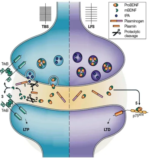
Figure 1.1 – Regulation of BDNF release upon synaptic activity.
Left: In response to theta-burst stimulation (TBS), tissue plasminogen activator (tPA) and proBDNF are released into synaptic cleft. 1) Then, tPA cleaves plasminogen producing the active protease plasmin. 2) Plasmin cleaves proBDNF producing the mature BDNF. 3) BDNF then binds to its high-affinity receptor (TrkB-FL) triggering multiple intracellular signalling pathways which in turn contribute to long-term potentiation (LTP). Right: During low-frequency stimulation (LFS) proBDNF is secreted into the synapse and remains uncleaved in the synapse. Uncleaved proBDNF binds to its high-affinity receptor (p75NTR) and
1.1.2. Neurotrophin receptors
The biological actions of neurotrophins are mediated by the activation of their cognate tropomyosin-related kinase receptor (TrkA, TrkB or TrkC) and by the activation of the common p75 neurotrophin receptor (p75NTR), which has been shown to modulate the affinity and selectivity of Trk activation [9]. Trk receptors are members of receptor tyrosine kinase (RTK) superfamily and promote neuronal survival and plasticity, while p75NTR is a member of tumour necrosis factor (TNF) receptor superfamily and can promote neuronal death under certain circumstances [1]. TrkA is the cognate receptor for NGF [10, 11], while TrkB was identified as the receptor for BDNF and NT-4/5 [12, 13], and TrkC as the receptor for NT-3 [14].
The Trk receptors and p75NTR receptor can function synergistically, antagonistically or independently of each other [8]. The mature neurotrophins bind with high affinity to Trk receptors, and p75NTR may act synergistically as a co-receptor [15]. In opposition, the pro-neurotrophins bind with high affinity to the p75NTR with Sortilin acting as a co-receptor. The effects of neurotrophins upon neuronal survival, differentiation and synaptic plasticity are mediated by the Trk receptors system [16], while the opposing effects of pro-neurotrophins, such as cell death and decreased synaptic function, are mediated by p75NTR and Sortilin complex [17-19].
All Trk receptors share a significant sequence homology and a conserved domain organization. The extracellular region of Trk receptors are composed by a leucine rich domain flanked by two cysteine rich regions. Under those domains, and prior to the transmembrane region, there are two immunoglobulin-like domains which define the ligand binding specificities of the receptor [20, 21]. Intracellularly, the Trk receptors are composed by a juxtamembrane sequence that includes the Shc binding site, a tyrosine kinase domain and a C-terminal tail containing the phospholipase C gamma (PLCγ) binding site [21].
Considering the focus of the present thesis, henceforth only BDNF and its receptor TrkB will be explored in more detail.
1.1.3. Truncated TrkB receptors
The TrkB gene (NTRK2) can originate a full-length TrkB receptor (TrkB-FL) and, by an alternative splicing mechanism, it also can originate truncated receptors (TrkB-T1, TrkB-T2 and TrkB-T-Shc) [22-24]. The TrkB-T1 and TrkB-T2 have a unique short C-terminal tail (T1 with 11 aminoacid residues and T2 with 9 aminoacid residues) [22], while the TrkB-T-Shc is a human brain-specific isoform which lacks the tyrosine kinase domain but contains the Shc
binding site. The truncated TrkB receptors cannot activate the canonical signalling pathways of full-length receptors, since they lack the intracellular kinase domain [24]. Additionally, truncated TrkB receptors can inhibit the BDNF effects by acting as dominant negative inhibitors of the TrkB-FL receptors [23, 25-27]. Indeed, multiple studies have shown that increased levels of truncated TrkB receptors have a negative impact in neuronal function and survival in both artificial and pathological conditions [28-30].
While the TrkB-FL is the most abundant isoform expressed in the early developmental period, in the post-natal period, and throughout aging, the truncated TrkB-T1 receptor became the most predominant TrkB isoform expressed in forebrain [31, 32]. TrkB-T1 can be expressed by neurons and astrocytes depending on the brain region. Accordingly, TrkB-T1 is highly expressed in astrocytes from pre-frontal cortex and subcortical white matter, but it is not present in astrocytes of the cerebellum and motor and visual cortex [33].
One possible biological role of TrkB-T1 is to regulate extracellular levels and localization of BDNF in the brain. When extracellular BDNF is abundant, TrkB-T1 binds and sequesters the available BNDF, and it is internalized along with its ligand. After BDNF and TrkB-T1 internalization, the BDNF can be degraded in lysosomes or can be sorted to another cellular location and be released by exocytosis [34, 35]. Although the in-vivo function of truncated receptors remains unknown, it was shown that TrkB-T1 deficient mice have increased anxiety in association with morphological abnormalities in dendrites of basolateral amygdale neurons. The same study showed that the depletion of TrkB-T1 can also partially rescue the BDNF haploinsufficiency phenotype, further suggesting that TrkB-T1 at physiological levels may regulate and attenuate TrkB-FL signalling [36]. Despite the lack of intracellular kinase domain, some studies have shown that TrkB-T1 receptor activates distinct signalling cascades in astrocytes. In fact, TrkB-T1 alone can promote Ca2+ release from the endoplasmic reticulum (ER) in astrocytes, through the activation of G-protein and PLCγ, with consequent inositol-1,4,5-triphosphate (IP3) formation (see Figure 1.2) [37]. Moreover, TrkB-T1 can bind to Rho GDP dissociation inhibitor I (RhoGDI1) and regulate actin cytoskeleton and glial morphology by modulating RhoGTPase activity [38].
1.1.4. TrkB-mediated signalling cascades
The binding of BDNF to TrkB receptor homodimmers, activates the intrinsic tyrosine kinase domain of the receptor promoting an auto-phosphorylation of specific tyrosine residues located in the intracellular domain of TrkB [39, 40]. In particular, the binding of BDNF to TrkB results in a fast phosphorylation of 5 tyrosine residues of the receptor, within seconds to minutes. These phosphorylated residues include 3 tyrosines in the kinase activation loop of
TrkB (Y701, Y705 and Y706) which regulate the kinase activity, and 2 tyrosines residues (Y515 and Y816) responsible for the activation of signalling cascades [41]. Phosphorylation of Y515 of TrkB (or equivalent residues in other Trks receptors) forms an adaptor binding site that couples the receptor to phosphatidylinositol-3 kinase (PI3K) and mitogen-activated protein kinases (MAPK) signalling pathways, while phosphorylation of Y816 recruits and initiates PLCγ signalling pathway (see Figure 1.2) [42].
PI3K / Akt signalling pathway:
When phosphorylated at Y515, TrkB receptors recruit Src homology 2 domain containing (Shc) adaptor protein through its phosphotyrosine-binding (PTB) domain [43]. In turn, Shc protein associates with Growth factor receptor-bound protein 2 (Grb2), Grb-associated binding protein (Gab1/2) and son of sevenless (SOS), culminating in the transient activation of small GTPases, such as Ras. Active Ras stimulates signalling through c-Raf/MEK/ERK (MAPK pathway) and class I PI3 kinase (PI3K) pathway (see Figure 1.2) [44]. Activated PI3K phosphorylates phosphatidylinositol-4,5-bisphosphate (PIP2), producing the second messenger phosphatidylinositol-3,4,5-trisphosphate (PIP3), which in turn stimulates the serine-threonine kinase Akt.
The signalling through the PI3K and Akt pathway are necessary and sufficient for the survival of certain populations of neurons [45]. The Akt kinase modulates the function of several substrates involved in the regulation of cell survival and growth. Akt phosphorylates and inactivates several pro-apoptotic proteins, such as procaspase-9 and Bcl2-associated death promoter (BAD), as well as Forkhead 1 transcription factor [46, 47]. Activated Akt can also inactivate GSK-3β, a kinase which has been implicated in neuronal apoptosis and inhibition of axon growth [48, 49]. On the other hand, Akt activates transcription factors that regulate the expression of anti-apoptotic proteins, such as cyclic AMP response element-binding protein (CREB) and nuclear factor-κB (NFκB) [50].
Ras/MAPK pathway:
As mentioned above, the binding of Shc adaptor protein to the phosphorylated Y515 of TrkB activates the Ras protein. Activated Ras stimulates the MAPK/ERK kinase (MEK) which in turn activates the extracellular signal regulated-kinases (ERK). The ERK/MAPK signalling cascade activates transcription factors such as CREB [51], which in turn control the expression of several proteins implicated in survival, growth and differentiation of neurons. ERK activates, by phosphorylation, the ribosomal s6 kinase (RSK), which in turn further activates transcription factors, such as CREB, c-Fos and NF-κB [52-54]. Additionally, BDNF enhances protein translation in neurons through the Erk/MAPK pathway, by phosphorylating eukaryote initiation factor 4E (eIF4E) and its binding protein (eIF4E-binding protein-1) [55].
PLCy pathway:
Phosphorylated Y816 of TrkB directly recruits PLCγ1 which in turn is phosphorylated and activated by the TrkB kinase domain. Activated PLCγ1 hydrolyses PIP2 and generates
inositol triphosphate (IP3) and diacylglycerol (DAG) [56]. While DAG activates DAG-regulated protein kinase C (PKC) isoforms, the IP3 promotes the release of Ca2+ from intracellular stores, such as ER, through activation of IP3 receptor (IP3R). The increase in cytosolic Ca2+ activates diverse enzymes, including Ca2+-regulated PKC isoforms and Ca2+ -calmodulin-dependent kinases (CaMKII, CaMKK and CaMKIV). PLCγ pathway is crucial for synaptic plasticity, since mice with point mutations on Y816 of TrkB, but not on Y515, have impaired long-term potentiation. The PLCγ also promotes the activation of CREB transcription factor through CaMKIV, and point mutation on Y816 strongly impairs CREB activation [57].
Figure 1.2 – Schematic representation of TrkB receptors signalling pathways.
BDNF binds to the extracellular region of TrkB receptor, which is equal in both TrkB truncated T1 and full-length (FL) receptor. After the binding of homodimeric BDNF to homodimeric TrkB-FL, the kinase domain of the receptor became active and trans-phosphorylates the receptor in the tyrosine 515 and 816 (also named as Y484 and Y785, respectively, when the TrkB signal peptide is ignored for the aminoacid counting). Phosphorylated tyrosine 515 recruits the binding of Shc adaptor protein, which in turn lead to the activation of Ras/MEK/ERK (MAPK pathway) and PI3K/Akt signalling pathway. PLCγ signalling pathway is triggered after the binding of PLCγ to phosphorylated tyrosine 816 of TrkB. The signalling mediated by TrkB-FL promotes transcriptional programs that regulate synaptic plasticity, neuronal differentiation, growth and survival and motility. TrkB-T1 lacks the tyrosine kinase domain, however, evidences show that its activation can trigger Ca2+ waves in astrocytes. Figure adapted from [58]
1.1.5. BDNF and long-term potentiation
Long-term potentiation (LTP) is a long-lasting increase in synaptic strength induced by some patterns of synaptic stimulation, such as high-frequency stimulation (HFS) or TBS, and it is commonly accepted as the neurophysiological basis for learning and memory [59, 60]. In opposition, long-term depression (LTD) is a long-lasting depression on synaptic transmission after a period of low-frequency stimulation of the synapse. Thus, synaptic plasticity is the ability of synapses to change their efficacy depending on their activity.
The LTP on Cornu Ammonis 1 (CA1) area in the hippocampus is dependent on NMDAR, a glutamate receptor permeable to Ca2+ ions, in which the pore is usually occluded by Mg2+ at a resting membrane potential. During high-frequency stimulation the synaptic release of glutamate depolarizes the post-synaptic terminal, through an AMPAR-dependent Na+ influx, and allows the removal of Mg2+ and the consequent activation of the NMDAR by the glutamate in the synapse. The NMDAR-mediated influx of Ca2+ at the post-synaptic terminal activates Ca2+-dependent proteins, such as CaMKII, which trigger intracellular cascades necessary for LTP induction. The increased cytosolic Ca2+ on post-synaptic terminal also triggers the AMPARs trafficking to post-synaptic density (PSD), resulting in a greater post-synaptic response to glutamate [61]. LTP is classically divided into early LTP (E-LTP) and late LTP (L-LTP). E-LTP requires modifications in existing proteins, whereas L-LTP is only induced by strong stimulation and requires de-novo proteins synthesis and structural modifications on synapses [62]. Despite years of intensive investigation on hippocampal LTP induced by HFS, only recently it was proven that learning actually induces LTP in the hippocampus of behaving animals [63, 64].
The first evidence that neurotrophins are important for synaptic function arise in early 1990s, when it was discovered that exogenous BDNF or NT-3, but not NGF, enhances synaptic activity on neuromuscular synapses [65]. Later, it was found that BDNF or NT-3, but not NGF, increases the basal synaptic transmission in hippocampal CA1 area [66]. The finding that BDNF might also have a role in hippocampal LTP came from experiments preformed in a BDNF-deficient mice. In this mice model, a significant impairment on hippocampal LTP magnitude was detected [67]. Interestingly, LTP was restored after reintroduction of BDNF gene in CA1 area by a virus-mediated gene transfer [68], or by exogenous addition of BDNF [69]. Several other works have been demonstrating the central role of BDNF in synaptic plasticity. Accordingly, intrahippocampal infusion of BDNF in living rats was shown to elicit long-term synaptic potentiation [70]; the application of exogenous BDNF to hippocampal slices from young mice enhanced the L-LTP induced by tetanic stimulation, which in the absence of BDNF only elicit a short-term potentiation (E-LTP) [71]; the scavenger of endogenous BDNF by
soluble TrkB-IgG fusion protein or by specific antibodies resulted in a reduced hippocampal LTP magnitude [71, 72] not seen for NT-3 or NT-4 [72]. The TrkB or BDNF null mice have severe phenotype and die between birth and weaning age, hampering the study of these proteins in LTP in an adult stage. However, a conditional TrkB knockout mouse, where the gene deletion was restricted to the forebrain neurons in the post-natal period, showed impaired LTP on CA1 hippocampal synapses, and impaired learning behaviour in the adult stage, without gross phenotypical aberrations [73]. Together, these evidences clearly showed that endogenous BDNF is required for normal LTP and learning, and that exogenous BDNF can induce or facilitate the LTP expression.
Other studies have provided mechanistic clues through which BDNF and TrkB activation facilitates LTP at glutamatergic hippocampal synapses (see Figure 1.3). Endogenous BDNF is released from glutamatergic synapses, in a Ca2+-dependent way, in response to stimulus used to induce LTP, such as TBS (see Figure 1.1) [74, 75]. Released BDNF can facilitate LTP at excitatory CA1 synapses by increasing presynaptic release of glutamate, and by amplifying the postsynaptic response to this neurotransmitter [76]. In particular, BDNF increases the Ca2+-dependent release of glutamate in cortical and hippocampal nerve terminals (synaptosomes) [77, 78] and in cultured hippocampal neurons [79]. The presynaptic stimulation of glutamate release by BDNF is mediated by a MAPK-dependent phosphorylation of the synaptic vesicle protein synapsin-I. In mice lacking the synapsin the effect of BDNF upon glutamate release is strongly attenuated [80]. On the other hand, BDNF, through its post-synaptic TrkB receptor, stimulates tyrosine kinase Fyn, which in turn phosphorylates the NMDAR and increases its activity [81, 82]. In cultured neurons, BDNF further modulates glutamatergic synapse at postsynaptic level, by increasing the levels and the trafficking of AMPAR to membrane [83, 84]. Moreover, exogenous BDNF, at nanomolar concentration, depolarizes and excites hippocampal and cortical neurons just as quickly and effective as glutamate at a micromolar concentration [85]. In fact, BDNF induces a fast neuronal depolarization, in a TrkB dependent-way, by activating Na+ channel NaV1.9 allowing the influx
of sodium ions [85, 86]. Consequently, the BDNF-induced depolarization activates voltage-gated Ca2+ channels (VGCC) evoking Ca2+ transients which are detectable in the dendrites and spines of hippocampal neurons, but not at presynaptic sites [87]. In this way, BDNF cooperates with NMDAR during LTP induction by promoting an additional influx of Ca2+ in the post-synaptic terminal. Thus, pairing a brief application of BDNF in dendrites and a weak burst of synaptic stimulation, elicit a fast and robust induction of LTP [87].
Interestingly, a recent study showed that acute or gradual increases in BDNF elicit distinct signalling and neuronal effects. While a gradual increase in BDNF concentration (slow perfusion rate) selectively facilitates LTP in hippocampal slices, a rapid increase in BDNF concentration (fast perfusion rate) increases the synaptic basal transmission instead [88]. This
study highlighted the importance of the kinetics of TrkB activation, and explained, in part, some conflicting results in the literature, regarding the pre- and post-synaptic effects of BDNF.
On top of BDNF-mediated fast changes in synaptic efficacy, BDNF has also a crucial role in maintenance of late-phase LTP (L-LTP), synaptic consolidation and long-term memory storage [89, 90]. BDNF synthesis is found to be increased in hippocampal neurons 2-4 hours after L-LTP-inducing stimuli, such as strong TBS [91, 92]. Unlike E-LTP, the L-LTP expression is dependent on protein synthesis. Surprisingly, application of exogenous BDNF is able to rescue L-LTP in the presence of protein synthesis inhibitors [93]. This perplexing result was recently demonstrated to be dependent on PKMζ, an atypical PKC isoform present in brain [94]. Weak TBS normally induce an E-LTP, which last less than 2 hours, and fail to elicit an L-LTP. However, when pairing BDNF perfusion and weak TBS, it produces a reliable L-LTP in CA1 area of hippocampus [93]. Moreover, mice lacking tissue plasminogen activator (tPA), a protease involved in the conversion of pro-BDNF into BDNF, have a selective deficit in L-LTP expression without affecting E-LTP in hippocampus [95]. Moreover, perfusion of BDNF in tPA null mice prevented the L-LTP impairment [93]. Indeed, evidences suggest that BDNF may trigger L-LTP by regulating local dendritic protein translation and concomitantly increasing synthesis of LTP-associated proteins, such as Arc, GluR1, CaMKII, PSD-95 among others. BDNF can also regulate actin cytoskeletal dynamics which are required for structural changes of synapses and L-LTP formation [96].
Figure 1.3 – Modulation of glutamatergic synapse by BNDF.
BDNF can enhance the transmission and plasticity of glutamatergic synapses. Presynaptic activation of TrkB-FL by BDNF increases the glutamate release. Postsynaptic activation of TrkB-FL increases the postsynaptic response to glutamate by distinct mechanisms: 1) TrkB induces the phosphorylation of NMDAR, through Fyn kinase, increasing its activity; 2) TrkB depolarizes the postsynaptic terminal by promoting the influx of cations through transient receptor potential channels (TRPC) which might facilitate the Ca2+ entry through NMDAR and voltage-gated channels and 3) TrkB modulates AMPAR expression and trafficking to the postsynaptic membrane. Figure adapted from [97].
1.1.6. Facilitation of BDNF synaptic actions by adenosine
Adenosine is a ubiquitous nucleotide that acts as an extracellular signalling molecule. Adenosine is a neuromodulator that regulates synaptic activity, by modulating the presynaptic neurotransmitter release, by depolarizing or hyperpolarizing the postsynaptic neuron or even by regulating glial cells activity. Overall, adenosine acts as a depressant of excitatory glutamatergic transmission and reduces excitability [98]. Extracellular adenosine can derive from the direct release of intracellular adenosine by equilibrative nucleoside transporters (ENT), or from catabolism of extracellular adenosine triphosphate (ATP) [99]. Adenosine can be released constitutively, or in an active-dependent manner through a calcium-dependent presynaptic release [100]. Extracellular adenosine has a short half-life time, since it is quickly reuptaked or converted to inosine or to adenosine monophosphate (AMP) by adenosine deaminase (ADA) or by adenosine kinase (ADK), respectively [99].
Extracellular adenosine exerts its actions through activation of four distinct G protein-coupled receptors (GPCR) namely, A1, A2A, A2B and A3 receptors. Adenosine A1 receptors
(A1R) are prevalent in the brain, being highly expressed in the cortex, cerebellum, hippocampus
and spinal cord. A1R and A2AR are high affinity receptors, with a Kd of 70 and 150nM,
respectively [101]. A1R are coupled to inhibitory G-proteins (G0/Gi) which inhibit synaptic
transmission by inhibiting cyclic adenosine monophosphate (cAMP) production [102]. In opposition, the A2AR are coupled to stimulatory Gs-proteins and potentiate synaptic
transmission by increasing cAMP production [101, 102]. A2AR are mainly expressed in
olfactory bulb and striatum, being also present in hippocampus at lower levels [103, 104]. Both A2AR and A1R can be present in the same synapse, and activated simultaneously by adenosine
[99]. Regarding the adenosine A2BR and A3R, both have low affinity for adenosine (Kd = 5 and
6µM, respectively) and are weakly expressed in CNS [98].
In addition to the modulatory actions of adenosine upon neurotransmitter release and synaptic plasticity, adenosine also modulates the actions of other modulators, such as neurotrophins [99]. The first direct evidence for the cross-talk between adenosine and neurotrophins, arose from the observation that adenosine, or a A2AR agonist, trans-activates
TrkA or TrkB receptors in PC12 cells or hippocampal neurons, respectively, in the absence of neurotrophins [105]. Nevertheless, the A2AR-mediated transactivation of Trk receptors has
different aspects when comparing to the conventional Trk activation by the respective neurotrophins. In particular, A2AR activation promotes the phosphorylation of an immature,
non-glycosylated, sub-population of Trk receptors associated mainly with Golgi membranes. In addition, this Trk transactivation is only detectable after 3 hours of A2AR activation, while the
classical phosphorylation of Trk receptors by the cognate neurotrophin, occurs in the mature, fully-glycosylated, receptors within seconds to minutes [106, 107].
In functional experiments, multiple evidences have been shown that A2AR activation is
necessary for synaptic effects of BDNF in hippocampus. Indeed, in hippocampal slices from young rats, exogenous application of BDNF increases basal synaptic transmission only when a previous depolarization stimulus is made, an effect blocked by an A2AR antagonist. Moreover,
pre-synaptic stimulation, or activation of A2AR by a selective agonist or by adenosine, triggered
the excitatory action of BDNF upon synaptic transmission, in a process dependent on PKA signalling [108, 109]. Thus, it is concluded that BDNF effects upon synaptic transmission require an activity-dependent presynaptic release of adenosine and consequent A2AR activation.
In addition, in hippocampal slices from adult rats, where both levels and actions of A2AR are
increased, the addition of exogenous BDNF spontaneously increases basal synaptic transmission, an effect fully blocked by an A2AR antagonist [109]. Interestingly, in
ADK-deficient mice, which have increased levels of extracellular adenosine, the spontaneous increase in hippocampal synaptic transmission induced by BDNF is observed even in young animals, an effect not present in age-matched wild-type mice. In opposition, the BDNF-induced spontaneous increase in hippocampal synaptic transmission present in adult wild-type mice, is not detected in age-matched ADK-overexpressing mice, which have lower adenosine levels [110]. Furthermore, the genetic deletion of A2AR abolished the excitatory effects of BDNF upon
synaptic transmission in mice [111]. Similarly to hippocampus, it was also found that A2AR and
PKA activation are required for the excitatory BDNF effects upon transmission in neuromuscular junction [112]. In addition to synaptic transmission, the effects of BDNF upon synaptic plasticity, both in LTP and LTD, are also fully dependent on A2AR activation, since
both effects are lost when extracellular adenosine is removed or when PKA or A2AR are
inhibited [108, 111, 113-116]. Together, these results indicate that A2AR activation, and
subsequent PKA signalling, are essential to trigger the BDNF excitatory effects upon synaptic plasticity and transmission. Since that exogenous BDNF spontaneously facilitates LTP induction and expression in hippocampal slices from young animals, it suggests that the release of adenosine and its precursor ATP induced by the high-frequency stimulation is enough to activate A2AR and trigger the BDNF actions [99].
Recently, it was shown that A2AR activation promotes the translocation of TrkB-FL to
lipid rafts domains in the membrane. Importantly, high-frequency stimulation resulted in increased levels of TrkB in lipid rafts, an effect abolished by the removal of endogenous extracellular adenosine. Thus, adenosine, through A2AR activation, promotes an
activity-dependent insertion of TrkB in lipid rafts , facilitating the phosphorylation of TrkB-FL and the BDNF-mediated actions [117].
Furthermore, the effects of BDNF upon GABA and glutamate release in synaptosomes are also dependent on A2AR activation (unpublished data from our lab). Conversely, the effects
of BDNF upon neuronal branching [118], or upon GABA uptake at nerve endings [119], do not depend on tonic activation of A2AR (see Figure 1.4). However, pharmacological activation of
A2AR in synaptosomes enhances the inhibitory effect of BDNF upon GABA uptake [119].
The findings that many synaptic actions of BDNF are dependent on A2AR activation
may open new therapeutic possibilities to boost the BDNF effects in neurodegenerative diseases where its signalling is known to be impaired. Multiple evidences showed that BDNF administration into the brain produces substantial benefits in in-vitro and in-vivo models of neurodegeneration. However, the translation of this approach to patients has been hampered by difficulty of BDNF to cross the blood-brain barrier (BBB) and by the poor bioavailability and stability of BDNF, which has a short half-life time in biological fluids [120]. Thus, small-molecules that modulate A2AR activity, or its signalling, might constitute a way to promote
BDNF synaptic effects in situations where BDNF signalling is compromised. Moreover, in opposition to the indiscriminate administration of BDNF into the brain, the approach of A2AR
modulation has the advantage of only stimulating BDNF effects on neuronal subpopulations which co-express A2AR, TrkB and BDNF, such as hippocampus or cerebral cortex, both regions
affected in Alzheimer’s disease (AD) [105, 121].
Figure 1.4 – Cross-talk between TrkB and A2A receptors.
BDNF actions upon synaptic transmission in hippocampus and neuromuscular junction, as well as, synaptic plasticity in hippocampus are fully dependent on A2AR activation. Figure adapted from [99]
1.2.
Calpain system
Proteases are a class of enzymes that catalyse the hydrolysis of peptide bounds. Depending on mechanism of catalysis, proteases are currently classified into 6 subgroups: aspartic, glutamic, cysteine, metalloproteases, serine, and threonine proteases. Cysteine proteases, also known as thiol proteases, use a nucleophilic cysteine thiol in the active site to attack the carbonyl-carbon of the amide bond and hydrolyse the peptide bond [122]. In the protease classification system of MEROPS database [123], the pure cysteine proteases are currently divided into 10 clans accordingly to their evolutionary relationship, tertiary structure and sequence motifs around catalytic site. Each clan of proteases can be further divided into several families based on their sequence homology. Calpains, papain and cathepsins are all members of clan CA, however, papain and cathepsins belong to the C1 family and are synthetized as inactive proenzymes with N-terminal propeptides, while calpains belong to the family C2 and are not synthetized as classical proenzymes [124].
1.2.1. Calpains
Calpains (EC 3.4.22.17), previously named as Ca2+-activated neutral protease, are cytosolic proteases ubiquitously expressed in mammals and have the peculiarity of being activated by a Ca2+-induced conformational change [125]. Calpains were discovered 50 years ago, in 1964, when a Ca2+-dependent protease activity was found in soluble fractions of rat brain at neutral pH [126]. The prototypical members of calpain family are the µ-calpain and m-calpain. These proteases differ in the Ca2+ concentration required for their activation in-vitro. The µ-calpain requires 3-50µM of Ca2+ for half-maximal activity, while m-calpain requires 400-800µM of Ca2+ [127]. Both proteases are heterodimers consisting in an 80 kDa catalytic large subunit (calpain-1 in µ-calpain and calpain-2 in m-calpain), associated with a common 30-kDa small regulatory subunit, CAPNS1 (see Figure 1.5). In 1984 the catalytic large subunit of µ-calpain (µ-calpain-1) was cloned, and its primary sequence revealed that it contained 4 domains. The Domain II of calpain-1 is the catalytic domain and is similar to papain-like thiol proteases, while the domain IV is similar to calmodulin-like Ca2+-binding proteins. These findings suggest that calpains are evolutionary derived from the fusion of a thiol protease with a Ca2+-binding protein [128].
Until now, it was found 15 human isoforms of calpain large subunit based on human genome sequence, being these homologs classified as ubiquitous or tissue-specific [129]. For instance, the mRNA of calpain-3a and calpain-8a are mainly found in muscle, while calpain-6 is found in placenta and calpain-11 in testis [127]. Although the protease domain (domain II) is
conserved in calpain family, there are members that lack, or have different, domains resulting in atypical calpains that may not dependent on Ca2+ (eg: Calpain-3) or may not have regulatory subunits. Moreover, there are also a calpain member (Calpain-6), that lacks the essential cysteine residue of the catalytic triad and may not even have proteolytic activity [129]. Nevertheless, atypical calpains have a biological function, and recent evidences showed that the non-catalytic calpain-6 regulates microtubule dynamics in cultured cells and regulates skeletal muscle development in mice [130].
The prototypical calpain-1 and calpain-2 (the large subunits of µ- and m-calpain, respectively) are ubiquitously expressed in mammalian cells and are the most abundant and characterized isoforms in the brain [131]. The calpain small regulatory subunit (CAPNS1) are also expressed in the brain [132]. It was shown that calpain-2 mRNA levels were 15-fold higher than calpain-1 levels in whole mice brain homogenate, whereas the distribution of calpain-1 mRNA was uniform throughout the brain, calpain-2 mRNA was enriched in certain neuronal populations including hippocampal and cortical pyramidal neurons [133]. Subcellular localization of calpains is widely attributed to soluble (cytosolic) fraction; however, recent studies showed that calpains are also associated with different subcellular compartments. In fact, evidences obtained from neuroblastoma cells, neuronal cultures and rat cortex, showed that µ-calpain is present at mitochondrial intermembrane space, placing it in proximity to its mitochondrial substrates and to Ca2+ released from mitochondrial stores [134]. During ischemic neuronal injury, the intra-mitochondrial µ-calpain can cleave and activate mitochondrial pro-apoptotic proteins, such as apoptosis inducing factor (AIF), which in turn mediate neuronal death signalling [135]. The m-calpain was also found in the nucleus of cultured neurons, where this nuclear calpain regulates Ca2+-dependent signalling by cleaving the CaMKIV [136].
The crystal structure of Ca2+-free m-calpain revealed that the catalytic site located in domain II (in large subunit), is not assembled in the absence of Ca2+, suggesting that Ca2+ may trigger conformational changes necessary to form a functional active site. Moreover, in opposition to classical proenzymes (eg: papain) whereas the N-terminal propeptide blocks the active site, the structure of calpain-2 revealed that the N-terminal anchor (Domain I) does not occupy the active site but inhibits its assembly. In addition, the structure indicated that the N-terminal anchor regulates the calpain affinity to Ca2+ by interacting with the small regulatory subunit (CAPNS1) [137]. The N-terminus of Domain I is autolysed during initial Ca2+-induced calpain activation. Although calpain autolysis reduces the requirement for Ca2+, it is not a prerequisite for its activation [124, 127, 138]. Furthermore, the domain III of the large subunit contains C2 subdomains that are implicated in conformational changes during Ca2+ binding and may be involved in binding to membrane phospholipids [139, 140]. The carboxy-terminal domain IV of calpain large subunit is a calmodulin-like domain and contains five Ca2+-binding EF-hand motifs, in which the fifth motif promotes the dimerization and binding to the small
subunit CAPNS1 [141]. In addition, the 30-kDa CAPNS1 has two distinct domains. The N-terminal domain (domain V) contains a hydrophobic sequence of aminoacids that may interact with plasma membrane [142, 143]. During calpain activation this domain is autolysed [144]. The Domain VI of CAPNS1 also contains a penta EF-hand domain and is very similar to the domain IV of the large subunit (see Figure 1.5). The biological function of CAPNS1 is not fully understood, and early evidences show that this small subunit is not required for catalytic activity of the large subunit [136]. Nevertheless, in-vitro experiments showed the small subunit acts as a chaperone and assists in the folding of the catalytic large subunit [136].
Either the large subunit of m-calpain (calpain-2) or the small subunit CAPNS1 are essential for mammalian life, since the genetic deletion of these proteins cause early embryonic lethality in mice [145, 146]. Interestingly, knockout mice for the large subunit of µ-calpain (calpain-1) are viable and fertile, despite showing a reduced platelet function [147].
Figure 1.5 – Schematic representation of µ-calpain structure.
The 80-kDa large subunit of µ-calpain (CAPN1 or calpain-1), or m-calpain (CAPN2 or calpain-2), contains four domains (DI to DIV), while the 30-kDa small subunit (CAPNS1) contains two domains (DV and DVI). CAPNS1 associates with CAPN1, or with CAPN2, to form a heterodimer (µ-calpain and m-calpain, respectively). Domain I is autolysed upon calpain activation. The Domain IIa and IIb constitute the protease core, and contain the catalytic triad residues (shown with ovals). C2-like domain (DIII) is involved in binding to phospholipids and Ca2+, and influences the calcium-induced activation of calpain. The domain IV and VI contain five calcium-binding EF hands motifs (shown with ovals), and associate to form the heterodimeric calpain. Domain V is a glycine-rich domain and may interact with cell membranes. The m-calpain large subunit, CAPN2, is structurally similar to CAPN1. The figure was adapted from [148].
1.2.2. Calpain substrates and cleavage specificity
The substrate specificities of the conventional calpains (m- and µ-calpain) are very similar. Calpains usually cleaves the substrates in inter-domain regions, producing large fragments rather than small peptides [127]. Thus, in opposition to digestive enzymes, the limited hydrolysis of calpains indicates their function as modulatory proteases [149]. The crystallography structure of Ca2+-bound m-calpain showed that the active site of this protease is deeper and narrower than others papain-like proteases. The constrains imposed by the cleft size indicate that the substrate should be in an extended conformation to fit the cleft, and may explain why calpains usually cleave unstructured inter-domain regions [150]. Although the protease domain (domain II) is highly conserved in calpain family, the substrate specificities of non-conventional calpains differ from the conventional µ- and m-calpain [151]. These differences suggest that the cleavage in the active site may depend on interactions between the substrate and other calpain interfaces. In opposition to other cysteine proteases, such as caspases, the cleavage mediated by calpains is not merely determined by the aminoacids sequence in the substrate. For calpains, the cleavage site is strongly determined by the conformation of the substrate rather than its primary aminoacid sequence [127]. Studies of bioinformatics have been attempted to predict calpain cleavage sites based on known cleavage sites determined experimentally. The most advanced predicting tool uses a machine learning process instead of the standard sequence analysis algorithms, such as the position-specific scoring-matrix method [152]. However, the accuracy of calpain cleavage site prediction still needs further improvements [153].
Calpains cleave a wide range of substrates. Accordingly to Calpain for Modulatory Proteolysis Database (CaMPDB – www.calpain.org) there are currently 97 experimentally confirmed mammalian calpain substrates and more than 1.000 computationally predicted mammalian substrates [154]. The confirmed calpain substrates include several proteins, such as:
1) cytoskeletal proteins (eg: integrins, cadherin, microtubule-associated proteins MAP1 and
MAP2, neurofilament 1 and 2, glial fibrillary acidic protein - GFAP, spectrin, tau); 2) signal transduction proteins (CaMKIV, epidermal growth factor receptor kinase, protein kinase A and C, GSK3β, IP3R, calcineurin, IκB, protein tyrosine phosphatase 1B); 3) apoptotic controllers (Apaf-1, AIF, Bax, Bid, Bcl-XL, BAK, caspase-3, 7, 8, 9, 12 and 14); 4) transcription factors (p53, c-jun, c-fos) and 5) synaptic proteins (APP, metabotropic and ionotropic glutamate receptors, dynamin-1, GAP-43, PSD-95), among others [155]. Interestingly, calpains often produce fragments with a reduced, enhanced or even different activity than the original substrate. For instance, a recent study showed that, in hippocampal neurons, an