Biomonitoring Atlantic deep waters through the
assessment of shark biomarkers
Luís Miguel Fonseca Alves
Biomonitoring Atlantic deep waters through the
assessment of shark biomarkers
Luís Miguel Fonseca Alves
Dissertação para obtenção do Grau de Mestre em Biotecnologia Aplicada
Dissertação de Mestrado realizada sob a orientação do Doutor Marco Lemos, do Doutor João Correia e da Doutora Sara Novais
II
Title: Biomonitoring Atlantic deep waters through the assessment of shark biomarkers
Título: Biomonitorizar as águas profundas do Atlântico através da análise de biomarcadores de tubarão
Copyright © Luís Miguel Fonseca Alves
A Escola Superior de Turismo e Tecnologia do Mar e o Instituto Politécnico de Leiria têm o direito, perpétuo e sem limites geográficos, de arquivar e publicar este trabalho de projeto através de exemplares impressos reproduzidos em papel ou de forma digital, ou por qualquer outro meio conhecido ou que venha a ser inventado, e de a divulgar através de repositórios científicos e de admitir a sua cópia e distribuição com objetivos educacionais ou de investigação, não comerciais, desde que seja dado crédito ao autor e editor.
III
Agradecimentos
Chegou ao fim mais uma etapa na minha formação académica e pessoal, depois de incontáveis horas de trabalho e dedicação. A conclusão deste projeto não seria possível sem a colaboração, disponibilidade e boa vontade das pessoas que passo a nomear e às quais quero prestar os meus sinceros agradecimentos.
Aos meus três orientadores, de uma maneira geral mas muito sentida. Todos eles me transmitiram conhecimentos valiosos e mostraram disponibilidade para me ajudar sempre que necessário.
Ao Doutor Marco Lemos, por me obrigar a dar o máximo, e depois mais um pouco. Obrigado acima de tudo por ter aceitado o desafio de um rapaz que queria estudar tubarões, quando todos lhe diziam ser impossível.
Ao Doutor João Correia pelo entusiasmo contagiante, pela confiança que sempre depositou em mim e por me ter mostrado, ainda antes de ser meu orientador, que era possível estudar tubarões em Portugal.
À Sara Novais, por uma orientação sempre presente. Obrigado pelo apoio durante as horas passadas no laboratório e pelo encorajamento. Foi já com muito respeito por ti que iniciei esta aventura e é com muita alegria e orgulho que a esse sentimento junto agora o da amizade.
À professora Susana Mendes pela ajuda indispensável na realização e interpretação do tratamento estatístico dos resultados.
Ao Tiago Bento, por me ajudar em situações de desespero e pelo conhecimento transmitido. Aprendi muito durante as minhas visitas à Lota.
Ao Nuno Costa, cuja contribuição foi essencial para a realização deste projeto. Espero que esta nossa colaboração se mantenha em aventuras futuras.
IV
A todos os membros do Grupo de Investigação de Recursos Marinhos (GIRM) por me terem recebido e tratado tão bem, mesmo quando deixei os laboratórios e corredores a cheirar a entranhas de tubarão.
À minha família, especialmente aos meus pais, por me terem ajudado a concretizar este projecto e por acreditarem nas minhas capacidades.
À Solange pela paciência e pelo exemplo de que com dedicação tudo se faz. Obrigado por me acalmares nas horas de aflição e por me dares razões para sorrir todos os dias, mesmo quando as coisas correm menos bem. Obrigado por seres quem és e por me ajudares a ser quem sou.
V
Resumo
Os ecossistemas marinhos estão continuamente a ser sobrecarregados com contaminantes derivados de atividades humanas resultando numa dimunuição dos recursos marinhos. A exposição crónica a contaminantes, como metais pesados e poluentes orgânicos persistentes (POPs), pode afetar negativamente o ambiente marinho e, eventualmente, também os seres humanos. Grandes predadores pelágicos, como tubarões, são particularmente afetados pela poluição, principalmente através de processos de bioacumulação e biomagnificação.
A fim de resolver o problema acima mencionado, são necessários estudos de avaliação de risco ambiental para prever os padrões de contaminação e evitar efeitos adversos que muitas vezes só são visíveis quando é tarde demais para tomar ações preventivas.
Análises de concentração de químicos fornecem-nos informações sobre o nível de contaminação no ambiente; no entanto, podem não ser suficientes para entender como os organismos estão a ser afetados e há uma necessidade de relacionar essas quantificações com parâmetros biológicos. A avaliação de parâmetros bioquímicos, como a atividade enzimática, pode fornecer uma visão mais sensível e precisa sobre os níveis de contaminação. Tubarões como Prionace glauca são predadores de topo e portanto extremamente importantes nos ecossistemas marinhos. A sua grande distribuição, juntamente com o fácil acesso a amostras, fornecidas por barcos de pesca comercial, tornou-as um alvo favorável para utilização em ensaios toxicológicos.
Este estudo teve como objetivo avaliar o potencial de P. glauca como uma espécie sentinela para pesquisas de monitorização de poluição, através do desenvolvimento e da aplicação de biomarcadores apropriados. As amostras de tecidos foram recolhidas de vinte tintureiras na costa de Portugal, a bordo de um barco comercial de pesca de espadarte. Níveis de POPs, assim como parâmetros bioquímicos relacionados com destoxificação, stress oxidativo e funções neuronais, foram medidos. A caracterização prévia da atividade das colinesterases no músculo e cérebro de P.
glauca foi feita, já que não havia dados disponíveis sobre esta matéria. Esta
caracterização foi essencial devido à existência de três classes de ChE conhecidas em peixes, acetilcolinesterase (AChE), butirilcolinesterase (BChE) e propionilcolinesterase (PChE), todas bastante suscetíveis a agentes anticolinérgicos, e outros contaminantes, tornando-as biomarcadores relevantes em estudos de monitorização de poluição. Os
VI
resultados obtidos indicaram que o cérebro de P. glauca aparenta possuir ChEs atípicas, revelando propriedades mistas de AChE e BChE e, que o músculo aparentemente possui maioritariamente AChE. A exposição in vitro a chloropyrifos-oxon provocou inibição de ChE das tintureiras em ambos os tecidos, com o cérebro sendo o tecido mais sensível e, por isso, o mais adequado para a detecção de compostos anticolinérgicos no ambiente. Este estudo indica que a actividade de ChE em tintureiras tem potencial para ser usada como um biomarcador sensível e fiável em programas de biomonitorização marinha.
O fígado apresentou níveis mais elevados de POP, quando comparado com músculo. Foram encontradas correlações positivas e negativas entre os parâmetros de contaminação e de stresse oxidativo.
Este estudo destaca a importância da caracterização de Che antes de a usar como um biomarcador em estudos ecotoxicológicos, e demonstra o grande potencial de P.
glauca como espécie modelo e como sentinela de poluição marinha, através do uso de
biomarcadores adequados.
Palavras chave: Poluição; Ecotoxicologia; Biomarcadores; Stress oxidativo;
VII
Abstract
Marine ecosystems are being continuously loaded with contaminants derived from human activities resulting in a decline of the marine resources. The chronic exposure to contaminants like heavy metals and persistent organic pollutants (POPs) can cause negative impacts to marine environment and, eventually, to humans. Big pelagic predators, like sharks, are particularly affected by pollution, mainly through bioaccumulation and biomagnification.
Given the aforementioned problem, environmental risk assessment studies are needed to predict contamination patterns and prevent injurious effects that are often only visible when it is too late to take preventive actions.
Chemical analysis of the pollutants’ concentrations provide information about the contamination level in the environment, but may not however be enough to fully understand how organisms are being affected, and there is a need to link these measurements with biological endpoints. Assessment of biochemical endpoints, like enzymatic activity, can provide a more sensible and accurate view on the effects of contamination and risk levels. Sharks like Prionace glauca, are apex predators and therefore extremely important parts of marine ecosystems. Their large distribution along with the fairly easy access to samples, provided by commercial fishing boats, has made them a favorable target for use in biomonitoring studies.
This study aimed to evaluate the potential of P. glauca as a sentinel species for pollution monitoring surveys, through the development and application of suitable biomarkers. Tissue samples were collected from twenty blue sharks of the coast of Portugal aboard a commercial swordfishing boat. POPs concentrations, as well the levels of biochemical parameters related with detoxification, oxidative stress and neuronal functions, were measured. As a first part of this work, a characterization of the cholinesterases present in muscle and brain tissues of P. glauca was made, as there was no data available on this matter. This characterization was essential due to the existence of three known types of ChE in fish, acetilcholinesterase (AChE), butyrylcholinesterase (BChE) and propionylcholinesterase (PChE), all very susceptible to anticholinergic agents, and to other contaminants, making them relevant biomarkers in pollution monitoring studies. The results suggest that the brain of P. glauca seems to contain atypical ChEs, revealing mixed properties of AChE and BChE, and that the muscle tissue seems to contain mostly AChE. In vitro exposures to chloropyrifos-oxon
VIII
inhibited blue shark’s ChE in both tissues, the brain being the most sensitive tissue and therefore the most suitable for detection of exposure to low concentrations of anticholinergic compounds in the environment. This study indicates that ChE activity in blue sharks has the potential to be used as a sensitive and reliable biomarker in marine biomonitoring programs.
Liver tissue presented higher POP levels, when compared to muscle. Both positive and negative correlations were found between physiological parameters and POPs accumulation levels. DNA damage was the main consequence of contamination and the inhibition of ChE activity the most strongly correlated effect.
This study highlights the relevance of ChE characterization before using it as a biomarker in ecotoxicology and biomonitoring studies, and demonstrates the great potential of P. glauca to be used as a model species and as a sentinel of marine pollution, through the use of suitable biomarkers of effect.
Keywords: Pollution, Ecotoxicology, Biomarkers, Oxidative stress, Acetylcholinesterase, persistent organic pollutants; Sharks
IX
Table of contents
Resumo --- V Abstract --- VII List of Figures --- XI List of Tables --- XIII List of Abbreviations --- XV
Chapter 1. General Introduction --- 17
1.1 Pollution of marine ecosystemns – an overview --- 19
1.2 Ecotoxicology and biomarkers: Nature’s early-warning signals --- 20
1.3 Introduction --- 23
1.4 Material and methods --- 26
1.5 Test organisms --- 29
Chapter 2. The potential of cholinesterases as tools for biomonitoring studies with sharks: biochemical characterization in brain and muscle tissues of Prionace glauca --- 31
2.1 Introduction --- 33
2.2 Material and methods --- 35
2.2.1. Test organisms --- 35 2.2.2. Chemicals --- 35 2.2.3. Tissue preparation --- 36 2.2.4. Cholinesterase characterization --- 36 2.2.4.1. Substrates --- 36 2.2.4.2. Inhibitors --- 37
2.2.5. In vitro effects of chlorpyrifos-oxon on ChE activity --- 37
2.2.6. Statistical analysis --- 37
2.3. Results --- 38
2.3.1. Cholinesterase characterization --- 38
2.3.2. In vitro effects of chloropyrifos-oxon --- 40
2.4. Discussion --- 42
2.4.1. Cholinesterase characterization --- 42
2.4.2. In vitro effects of chloropyrifos-oxon --- 44 Chapter 3. Biochemical responses in Blue sharks (Prionace glauca) exposed to
persistent organic pollutants (POPs) – an Atlantic survey ---
X
3.1. Introduction --- 50
3.2. Material and methods --- 52
3.2.1. Organisms --- 52
3.2.2. POPs chemical analysis --- 52
3.2.3. Tissue preparation --- 53
3.2.4. Protein quantification --- 54
3.2.5. Oxidative stress parameters --- 54
3.2.5.1. Lipid peroxidation --- 54
3.2.5.2 DNA damage --- 54
3.2.5.3. Glutathione S-transferase activity --- 55
3.2.5.4. Superoxide dismutase activity --- 55
3.2.5.5. Catalase activity --- 56
3.2.5.6. Glutathione reductase activity --- 56
3.2.5.7. Glutathione peroxidase activity --- 56
3.2.6. Total glutathione activity --- 56
3.2.7. Cholinesterase activity --- 57
3.2.8. Statistical analysis --- 57
3.3. Results --- 58
3.3.1. POPs chemical analysis --- 58
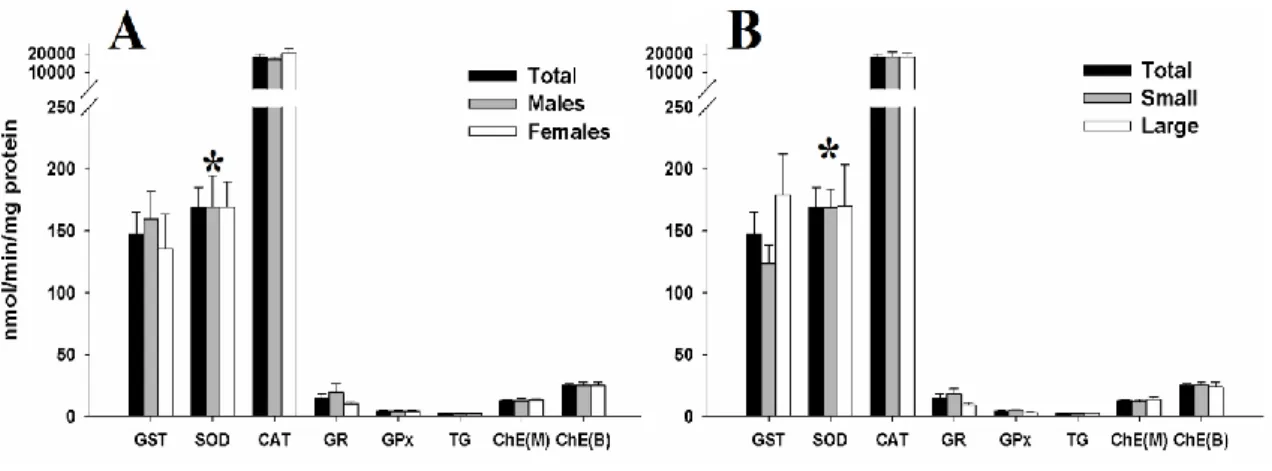
3.3.2. Oxidative damage --- 61
3.3.3. Detoxification/antioxidant and neurotoxicity related enzymes --- 61
3.3.4. Multivariate analysis --- 63
3.4. Discussion --- 66
Chapter 4. General discussion and concluding remarks --- 71
XI
List of Figures
Chapter 1. General introduction
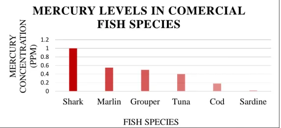
Figure 1 - Relationship between ecological relevance, time of response and the different levels of biological organization after stress exposure --- 21 Figure 2 - Main enzymatic mechanisms involved in reactive oxygen species neutralization (adapted from Howcroft et al., 2009) --- 25 Figure 3 - Levels of mercury in some commercial fish species, according to the 2010 US Food and Drug Administration report (1990-2010) --- 27 Figure 4 - Drawing of a blue shark by A. López, “Tokio”, as seen in ICCAT Manual (2009) --- 28
Chapter 2. The potential of cholinesterases as tools for biomonitoring studies with sharks: biochemical characterization in brain and muscle tissues of Prionace glauca
Figure 5 – Cholinesterase substrate preferences in the brain (A) and muscle (B) of
Prionace glauca --- 38 Figure 6 – Effect of the inhibitors eserine (A), BW284C51 (B) and iso-OMPA (C) on Prionace glauca cholinesterase (ChE) activities in brain and muscle tissues (expressed as mean values ± standard error) using acetylthiocholine as substrate - 40 Figure 7 – Cholinesterase (ChE) activity values and percentage of activity inhibition (expressed as mean values ± standard error) in the brain (A) and muscle (B) of Prionace glauca exposed in vitro to chlorpyrifos-oxon --- 41
Chapter 3. Biochemical responses in Blue sharks (Prionace glauca) exposed to persistent organic pollutants (POPs) – an Atlantic survey
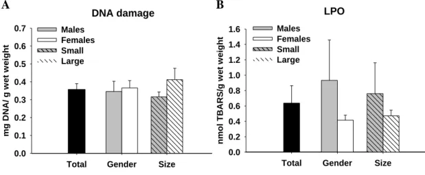
Figure 8 – Oxidative damage measured in the liver of juvenile blue sharks (Prionace glauca) by means of A) DNA damage levels and B) Lipid peroxidation (LPO) levels --- 61 Figure 9 - Distribution of enzymatic activity values of the total juvenile blue sharks (Prionace glauca) samples (n= 20) when comparing to values of: A – male and female group of individuals; B – small and large group of individuals --- 62 Figure 10 - Triplot of axes 1 and 2 of the Canonical Correspondence Analysis (CCA) on contaminants and oxidative stress parameters data --- 65
XIII
List of Tables
Chapter 2. The potential of cholinesterases as tools for biomonitoring studies with sharks: biochemical characterization in brain and muscle tissues of
Prionace glauca
Table 1 - Values of the Michaelis–Menten constant (Km), maximal velocity (Vmax) and the catalytic efficiency (Vmax/Km) of Prionace glauca cholinesterases for the three tested substrates --- 39 Table 2 – In vitro inhibition concentrations (IC50) of chlorpyrifos-oxon in
Prionace glauca, depending on gender and size of the organisms --- 42
Chapter 3. Biochemical responses in Blue sharks (Prionace glauca) exposed to persistent organic pollutants (POPs) – an Atlantic survey
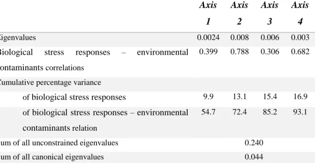
Table 3 - Lipid percentage and concentrations of quantified POPs (ng/g wet weight) in muscle and liver from 20 blue sharks (Prionace glauca) captured in the Atlantic Sea --- 59 Table 4 - Correlation analysis (Pearson Correlation) between all oxidative stress parameters assessed in this study. The pair of variables with significant positive or negative correlation coefficients (p < 0.05) are underlined and highlighted in bold --- 63 Table 5 - Eigenvalues for CCA axes and correlation coefficients between environmental factors and CCA ordination axes --- 64
XV
List of abbreviations
AChE – acetilcholinesterase ANOVA – analysis of variance ATCh – acetylthiocholine iodide. BChE – butyrylcholinesterase
BTCh = S-butyrylthiocholine iodide. CAT – catalase
CCA – canonical correspondence analysis CDNB – 1-chloro-2,4-dinitrobenzene DDTs – dichlorodiphenyltrichloroethanes DNA – Deoxyribonucleic acid
DTPA – diethylene triamine pentaacetic acid EDTA – ethylenediamine tetraacetic acid GPx – glutathione peroxidase
GR – glutathione reductase GSH – glutathione
GST – glutathione S-transferase GSSG – glutathione disulfide H2O2 – hydrogen peroxide
ICCAT – international commission for the conservation of atlantic tunas IOC – Intergovernmental Oceanographic Commission
LPO – lipid peroxidation
NADPH – nicotinamide adenine dinucleotide phosphate-oxidase NRC – National Research Council
O2•− – superoxide radical OH• – hidroxyl radical PCFs – perfluorochemicals PCBs – polychlorinated biphenyls PCDFs – polychlorinated dibenzofurans PCDDs – polychlorinated dibenzo-p-dioxins PCFs – perfluorochemicals PChE – propionylcholinesterase POPs – persistent organic pollutants
XVI
PPM – parts per million
PTCh – Propionylthiocholine iodide. ROS – reactive oxygen species SOD – superoxide dismutase TG – total glutathione
UNESCO – United Nations Educational, Scientific and Cultural Organization WHO – World Health Organization
Chapter 1.
19
1.1.
Pollution of marine ecosystems – an overviewComprising over 71% of our planet’s surface, the oceans are home to a great percentage of the World’s biodiversity (IOC/UNESCO et al., 2011). According to Lalli and Parsons (1993), besides having an essential role in the regulation of the planet’s climate, the oceans are also responsible for over 35% of the primary production of the planet. Furthermore, the importance of marine waters is not only ecological but also economical, with 60% of the total economic value of the biosphere being attributed to the oceans (Costanza et al., 1997).
The environment is continuously burdened with xenobiotic contaminants released by urban communities and industries and, most of the times, the final destination for many of these contaminants is the aquatic environment, either due to direct discharges or to hydrologic and atmospheric processes (Stegeman and Hahn, 1994; Van der Oost, et al., 2003). Human activities are responsible for the great majority of the contamination of marine environments, and consequently, for the decline of their resources (Derraik, 2002; Matthiessen and Law, 2002).
Xenobiotic compounds like persistent organic pollutants (POPs), organochlorine pesticides (DDTs), and heavy metals pose a serious threat to both marine fauna and humans because they are highly toxic and persistent (Gramatica and Papa, 2007). Being strongly bound to particulate material and accumulating in sediments, they are taken up by benthic organisms, entering this way the marine food web, becoming concentrated as the trophic levels rise (Kalay et al., 1999; Farombi et al., 2007; Storelli et al. 2011). Bioaccumulation happens when the intake of a substance by an organism is faster than its excretion. Since these contaminants accumulate in the tissues, predators tend to amass the contaminants present on their prey. In marine environments, bioaccumulation occurs mainly in fish, especially in large long-lived predators like sharks (Gomes et al. 2004).
Along with bioaccumulation, persistent hydrophobic chemicals may accumulate in aquatic organisms through different mechanisms, like the direct uptake from water by gills or skin (bioconcentration) and via uptake of suspended particles (ingestion) (Van der Oost et al., 2003). Because of the aforementioned facts, although the presence of a xenobiotic compound in a segment of an aquatic ecosystem may not indicate, per se, a deleterious effect, its accumulation/biomagnefication can eventually cause injurious effects (Franke et al., 1994; Tillitt et al., 1992; Wang, 2002; Dautremepuits et al., 2004).
20
Some of the most well studied POPs include polychlorinated biphenyls (PCBs), perfluorochemicals (PFCs), polychlorinated dibenzo-p-dioxins (PCDDs) and polychlorinated dibenzofurans (PCDFs), all of them having negative effects on the living organisms present in the water such as growth impairment and effects resulting from endocrine disruption (Jones and de Voogt, 1999; Storelli et al. 2011). PCBs and DDTs usually form complex mixtures, and their presence is a clear indication of anthropogenic pollution (Storelli et al., 2005). PCDDs and PCDFs, unlike the remaining POPs, are not intentionally produced by industries but are unwanted by-products of industrial and thermal processes (Stevens et al., 2000).
Heavy metals like lead (Pb), nickel (Ni), cadmium (Cd), and mercury (Hg) can all be found in aquatic environments and once accumulated by marine organisms, can induce the formation of reactive oxygen species, causing excessive oxidative stress which can lead to damage on important macromolecules like proteins, lipids or DNA (Bryan, 1976; Mart et al., 1982; Stohs and Bagchi, 1995; Canli and Atli, 2003). The organic form of Hg can bind to sulfhydryl groups of fish enzymes and proteins and can easily cross both the placental and blood brain barrier (Boening, 2000; Castro-Gonzalez and Mendez-Armenta, 2008; Guzzi and La Porta, 2008). High and ecologically alarming concentrations of these metals have been found in marine organisms, particularly in those higher in the food chain, like sharks (Branco et al., 2007; Barrera-García et al., 2013; de Carvalho et al., 2014).
1.2. Ecotoxicology and biomarkers: Nature’s early-warning signals
Ecotoxicology aims to understand how different xenobitotics can disturb the ecosystems to be able to protect it and prevent those effects from becoming permanent (Newman, 2008). This is not a simple task given the numerous xenobiotics that coexist in the environment, along with biotic and abiotic stress factors (eg. temperature and predation), which can have combined and complex effects (Calabrese, 1991; Lemos et al., 2010). In order to efficiently understand how xenobiotics affect organisms in the ecosystems, ecotoxicologists cannot rely solely in environmental levels of contaminants and need have the need to develop and apply reliable and accurate tools for pollution biomonitoring studies (Bucheli and Fent, 1995; Newman, 2008).
21
Figure 1 - Relationship between ecological relevance, time of response and the different levels
of biological organization after stress exposure.
In order to detect contamination before it hinders a community or even an entire ecosystem, and because effects at higher hierarchical levels are preceded by variations in biological processes at lower levels of organization (Fig. 1), early-warning signalling biomarkers can be created (Bayne et al., 1985). In an environmental context, these early-warning signals, also known as biomarkers, can help scientists to better understand which contaminants the organism has been exposed to, in which tissues they have been accumulated and if they are causing a toxic effect at critical targets (McCarthy and Shugart, 1990).
Numerous candidate biomarkers can be used in order to assess if an organism has been exposed to a pollutant and to evaluate the effects caused by environmental pollutants on aquatic ecosystems. Many studies have successfully used the activity of biotransformation enzymes involved in detoxification mechanism (Gorbi et al., 2004), oxidative stress parameters like lipid peroxidation (Barrera-García et al., 2012), hormones involved in reproductive and endocrine mechanisms (Koenig, 2012) and neuromuscular enzymatic activities (Solé et al., 2008) as biomarker of effect to contamination.
A pollutant stress situation usually provokes a cascade of biological responses and, initially, McCarty et al. (1991) stated that each of those responses could serve as a
22
biomarker. A few years later, in 1996, Van Gastel and Van Brummelen proposed a definition that is widely accepted today, stating that a biomarker can be “any biological
response to an environmental chemical at the subindividual level, measured inside an organism or in its products” (eg. blood, liver, urine, faeces, etc.), demonstrating an
deviation from the normal status, that cannot be detected in the intact organism.
Biomarkers may be grouped in different classes, but their classification can vary depending on the way they are used (Suter, 1993). A generally accepted description is the one used by the National Research Council (NRC) (1987) and WHO (1993), according to which biomarkers of exposure are used to detect and measure a xenobiotic or its metabolites, or the product of an interaction between a xenobiotic a target molecule or cell within an organism; biomarkers of effect allow to measure alterations in an organism (biochemical, physiological, etc.) that are associated with health impairment; lastly, biomarkers of susceptibility were described as indicators of the ability of an organism to respond to the effects of exposure to a xenobiotic (eg. genetic factors) which affect the vulnerability to that contamination. Increased activities of antioxidant enzymes and detoxification agents, oxidation of proteins and lipids, DNA damage and alterations in hormonal pathways, among others, have the potential to be used as biomarkers (Winston and Di Giulio, 1991; Filho, 1996; Elia et al., 2006; Labrada-Martagón et al., 2011).
Biomarkers can be useful tools in several steps of the risk assessment process allowing an evaluation of effect, exposure and hazard caused by the contaminant, providing means to monitoring the environmental quality of the ecosystems (Van der Oost et al., 2003). Particularly in aquatic ecosystems, fish species have attracted considerable interest for studies assessing biological and biochemical responses to environmental contaminants (Powers, 1989). Certain species can operate as sentinels, demonstrating the presence of bioavailable contaminants and how they affect the organisms, giving us insights on long-term effects on the health of populations or the integrity of the ecosystem (McCarthy and Shugart, 1990). Although a great deal of variation among fish species can occur, concerning both the basic physiological features and the responsiveness of certain biomarkers, fish have been the group par excellence for pollution monitoring in aquatic systems (Lopes et al., 2001; Van der Oost et al., 2003; Dautremepuits et al., 2004) since they are abundant and vastly distributed in all aquatic environments, being very susceptible to environmental pollutants. Even though most of the general biomarker criteria appear to be directly transferable to certain fish
23 biomarkers, monitoring fish species should be chosen taking some practical considerations: the test species must be part of the exposed community and have an important ecological role in the ecosystem (Stegeman et al., 1992; Suter, 1993).
Van der Oost and co-workers (2003), based upon the work made by Stegeman et al. (1992), proposed the following criteria to evaluate the strength and weaknesses of fish biomarkers objectively: 1) the methodology to assess the quantity/activity of the biomarker should be reliable; 2) the biomarker response should be sensitive to stressors; 3) the baseline data of the biomarker should be clear and the impacts of confounding factors to biomarker response should be well understood; 4) the underlying mechanism of the relationships between biomarker response and pollutant exposure should be well-known; 5) the toxicological significance of the biomarker should be established.
Since it remains hard to accurately predict bioaccumulation in fish, even with highly sophisticated models, analyses of tissue contaminant levels are required.
1.3. Organisms’ responses to contamination
When an exogenous harmful compound enters a living organism, a detoxification process is initiated, comprised by two main phases: phase I and phase II (Liska, 1998; Chen, 2012). During phase I, or functionalization phase, the organism first response is to facilitate the excretion of the contaminant and, in order to do that, a functional group is introduced in the chemical structure of the lipophilic xenobiotic, usually by oxidizing or hydrolysing it (Schlichting et al., 2000; Chen, 2012). A common Phase I oxidation involves conversion of a C-H bond to a C-OH. Although this process assists the organism to eliminate hazardous compounds, it can sometimes convert a nontoxic molecule into a toxic one, generating reactive oxygen species (ROS) that are injurious and cause stress to the cells (Di Giulio et al., 1989). In phase II, the second phase of detoxification, the metabolites generated in phase I are conjugated with small molecules like, for instance, glutathione (GSH) (Jakoby and Ziegler, 1990). This process increases the molecular weight of these xenobiotics and increases their polarity, thus making them less harmful and easier to excrete (Liska, 1998) (Fig.2).
It is not easy to define stress, but for this work purposes, the definition proposed by Brett in 1958 will be used, where he states that stress is “a physiological state
produced by an environmental factor that extends the normal adaptive responses of the animal, or disturbs the normal functioning to such an extent that the chances for survival are significantly reduced”. Oxidative stress specifically, or oxygen toxicity, is
24
defined as injurious effects due to cytotoxic reactive oxygen species (ROS) (Di Giulio et al., 1989; Halliwell and Gutteridge, 1999; Winzer, 2001).
Cells are constantly suffering some degree of oxidation, either by anaerobiosis, spontaneous mutagenesis and/or self defense mechanisms (Imlay, 2003; Valko et al., 2007). The oxygen metabolism in mitochondria originates a great amount of ROS, such as superoxide radical (O2•−) and hydrogen peroxide (H2O2) (Halliwell and Gutteridge,
2001). Due to this, when animals practice some kind of intense physical activity, like hunting prey or escaping predators, or try to eliminate harmful xenobiotics, higher rates of ROS production occur (Bejma and Ji, 1999; Best et al., 1999; Ji, 1999). The reduction products of molecular oxygen are capable of reacting with critical cellular macromolecules, potentially leading to enzyme inactivation, lipid peroxidation, DNA damage and, ultimately, cell death (Winston and Di Giulio, 1991; Lucas and Szweda, 1998; Ambrosio and Tritto, 1999; Semenza, 2000).
Many pollutants, or the metabolites deriving from them, can induce oxidative stress by increasing the production of ROS, disturbing the natural balance in the organism (Lackner, 1995; Pandey et al., 2003; Di Giulio and Meyer, 2008). These pollutants can be detoxified, and the effects of ROS minimized, through the activity of several enzymes and cofactors (Fig. 2). Variations in activity rates of biotransformation and antioxidant enzymes are extremely sensitive biomarkers of effect, and xenobiotics have been proven to cause alterations on these enzymes (Bucheli and Fent, 1995).
25
Figure 2 - Main enzymatic mechanisms involved in reactive oxygen species neutralization
(adapted from Howcroft et al., 2009). GST – glutathione S-transferase; SOD – superoxide dismutase; CAT – catalase; LPO – lipid peroxidation; GPx – Glutathione peroxidise; GR – glutathione reductase; GSH – reduced glutathione; GSSG – oxidized glutathione.
The metalloenzyme superoxide dismutase (SOD) is usually one of the most important agents in the process of preventing and counteracting the negative effects of ROS, being responsible for the transformation of O2•− into H2O2. The role of this
enzyme is so important that it can be found in all aerobic organisms studied so far (Stegeman et al., 1992). The H2O2 molecules resulting from the action of SODcan still
cause harmful effects and need to be removed from the organisms.
Catalase (CAT) and glutathione peroxidase (GPx) are two enzymes responsible for the elimination of H2O2 and preventing its accumulation. Catalase is a
hematin-containing enzyme that facilitates the elimination of H2O2 by metabolizing it to
molecular oxygen and water. Catalase specifically reduces H2O2, unlike some
peroxidases that can reduce different peroxides (Stegeman et al., 1992; Filho, 1996). CAT uses one of H2O2 molecule as donor in the reduction another, while peroxidases
like GPx need to use other reductant molecules. Glutathione peroxidase aids in the conversion of H2O2 to H2O, involving a concomitant oxidation of reduced glutathione
(GSH) to its oxidized disulfide form (GSSG).
The enzyme glutathione reductase (GR), as its name suggests, is involved in the transformation of the GSSG to the reduced form (GSH) (Worthington and Rosemeyer,
26
1974). GSH can act directly to neutralize oxidizing molecules and also as a cofactor used by GST, increasing the water solubility of xenobiotic compounds, facilitating their excretion (Egaas et al., 1995; Halliwell and Gutteridge, 2001; Livingstone, 2001; Valko et al., 2007).
Aside from the enzymes involved in oxidative stress responses, pollutants can also affect the activity of enzymes like acetylcholinesterase (AChE). Acetylcholinesterase is part of a family of enzymes that catalyze the hydrolysis of the neurotransmitter acetylcholine into choline and acetic acid, an essential process for both neuronal and motor capabilities (Pohanka, 2011). ChE inhibition is a serious impairment as it can cause behaviour alterations and even death to the organisms (Fulton and Key, 2001),
Because of their high sensitivity to organophosphate and carbamate pesticides, and also to other contaminants often present in marine ecosystems, the measurement of ChE activity has already been widely used in pollution monitoring studies as a biomarker of effect (Galgani et al., 1992; Payne et al, 1996; Kirby et al., 2000; Fulton and Key, 2001; Chambers et al., 2002; Van der Oost et al., 2003; Galloway et al., 2002; Arufe et al., 2007). Since usually there is more than one type of ChE in different fish tissues, to use these enzymes in ecotoxicology and biomonitoring studies, it is often necessary to perform a previous characterization of these enzymes in the target sampling tissue (Sturm et al., 1999a; Sturm et al., 2000; Kirby et al., 2000; Solé et al., 2008). The three known types of ChE in fish are acetilcholinesterase (AChE), butyrylcholinesterase (BChE) and propionylcholinesterase (PChE) (Solé et al.; 2010). AChE is essential to neuronal function but the roles of BChE and PChE, often named pseudocholinesterases, are still unclear (Chambers et al., 2002).
1.4. Sharks as tools for biomonitoring studies
Sharks are elasmobranchs, an ancient group of fish, existing for more than 400 million years (Ballantyne, 1997). These animals have always attracted the interest of scientists and oceanographers, like the Portuguese King Carlos I who reigned between 1889-1908 and published a pioneering study of portuguese sharks, including deepwater species not known to science at the time (Bragança, 1904).
Scientists estimate that over 90% of all marine predators have been lost and the decline of these animals can cause potentially irreversible effects in the ecosystems, by increasing prey populations and altering the normal interactions between the mentioned
27 prey and other members of the ecosystem (Schindler et al., 2002; Myers et al., 2007). The majority of shark species are at the top of their respective food chains and, as all large predators, play a very important role in the oceans, by maintaining the ecosystems in balance.
Being apex predators with long life spans, these animals are more susceptible to environmental contamination via bioaccumulation and biomagnification through the food web (Al-Yousuf et al., 2000; Escobar-Sánchez et al., 2011). A very good example of the aforementioned is the higher concentration of mercury that sharks tend to have in their bodies when compared to other fish species (figure 3), usually in the form of monomethylmercury, the most toxic form of the metal, which accounts for more than 95% of organic mercury in fish muscle tissues (Bloom, 1992; Porcella, 1994; Hueter et al., 1995). Extensive accumulation of heavy metals and POPs makes sharks’ immune system particularly affected, leading to a population decrease (Safe, 1994, Ross et al. 1995; Jones and de Voogt, 1999). Additionally, as a result of the previously stated, the consumption of shark meat makes it a potential entrance route of heavy metals and other pollutants into the human food web (Castro-Gonzalez and Mendez-Armenta, 2008).
Figure 3 - Levels of mercury in some commercial fish species, according to the 2010 US Food
and Drug Administration report (1990-2010).
Given their susceptibility to pollutant accumulation, their wide distribution and their importance to the ecosystems, sharks are ideal candidates to be used in marine pollution monitoring studies.
There are over 470 known species of sharks in the world and about 30 can be found in Portuguese waters (Compagno, 1999). The blue shark (Prionace glauca L.1758) is the most frequently caught shark species in the planet, with an
under-0 0.2 0.4 0.6 0.8 1 1.2
Shark Marlin Grouper Tuna Cod Sardine
MER C UR Y C ON C EN TRA TI ON (PP M) FISH SPECIES
MERCURY LEVELS IN COMERCIAL
FISH SPECIES
28
estimation of 20 million individuals caught annually as target or by-catch by fishing fleets such as the Portuguese longline swordfish fishing fleet (Bonfil, 1994; Santos et al., 2002; Stevens, 2009). A typical blue shark (Figure 4) has a slender body that can measure over 350 cm. These animals hunt mainly squids and small teleost fish, but they also consume other small sharks, birds, and whale carcasses (Compagno, 1999).
Figure 4 - Drawing of a blue shark by A. López, “Tokio”, as seen in ICCAT Manual (2009).
Being known for making long migrations, usually due to prey distribution, water temperature and reproduction, these animals segregate by size and gender (Stevens, 1976; Nakano and Seki, 2003; Aires-da-Silva et al., 2009; Stevens et al., 2010; Campana et al., 2011).
Pollutants quantification in these animals have already been done by several authors, mainly focusing on heavy metals (Branco et al., 2004; 2007; De Carvalho et al., 2014) and POPs (Storelli and Marcotrigiano, 2001; Storelli et al., 2005; 2006; 2011; Strid et al., 2007). Other sharks have been object of the same type of studies (Maz-Courrau et al., 2012; Shanshan et al., 2013), but contamination levels alone can’t explain the effects caused by xenobiotics (Van der Oost et al., 2003).
Recently some authors have establish correlations between contaminant trace elements body burden and detoxification mechanisms and antioxidant responses in blue sharks (Barrera-García et al., 2012; 2013), with the results indicating that this species has the potential to be used as biomonitor. Studying enzymes like cholinesterase is also of interest when trying to understand the consequences of pollutant contamination but to characterize the different forms of cholinesterases present in each tissue is imperative. Up until now the characterization of ChE in sharks have only been done for the muscle of Scyliorhynus canicula and Galeus melastomus (Solé et al., 2008).
29
1.5. Aims of the study
The main purpose of this work was to assess if Prionace glauca could be used as a sentinel species in pollution monitoring studies recurring to biomarker analysis. Potential biomarkers, involved in detoxification, oxidative stress response and neurological functions were evaluated as potential tools for use in environmental effect assessment.
To fulfil the main objective, the following tasks were completed:
1) Characterization of cholinesterases’ activity in muscle and brain tissues of P.
glauca in order to use it as a biomarker of effect in future studies (Chapter 2);
2) Measurement of contaminant body burden (persistent organic pollutants) in muscle and liver tissues of P. glauca and relate them with physiological responses (Chapter 3);
3) Development and optimization of sample preparation procedures and biomarker protocols to better address the blue shark as a sentinel species for marine pollution assessment (Chapters 2 and 3).
Chapter 2.
The potential of cholinesterases as tools for
biomonitoring studies with sharks: biochemical
characterization in brain and muscle tissues of
Prionace glauca
33
The potential of cholinesterases as tools for biomonitoring studies with sharks: biochemical characterization in brain and muscle tissues of Prionace glauca
Abstract
Cholinesterases (ChE) are a family of enzymes that play an essential role in neuronal and motor functions. Because of the susceptibility of these enzymes to anticholinergic agents and to other contaminants, their activity is frequently used as biomarker in pollution monitoring studies. The three known types of ChE in fish are acetilcholinesterase (AChE), butyrylcholinesterase (BChE) and propionylcholinesterase (PChE), for which the presence in each tissue tends to differ between species, thus demanding enzyme characterization before using them as biomarkers. Sharks, mostly acting as apex predators, help maintain fish population’s balance, performing a key role in the ecosystem. Blue sharks (Prionace glauca) are one of the most abundant and heavily fished sharks in the world, thus being good candidate organisms for ecotoxicology and biomonitoring studies. The present study aimed to characterize the ChE present in the brain and muscle of the blue shark using different substrates and selective inhibitors, and to assess the in vitro sensitivity of these sharks’ ChE to chlorpyrifos-oxon, a metabolite of a commonly used organophosphorous pesticide, recognized as a model anticholinesterase contaminant. The results suggest that the brain of P. glauca seems to contain atypical ChEs, displaying mixed properties of AChE and BChE, and that the muscle tissue seems to contain mainly AChE. In vitro exposures to chloropyrifos-oxon inhibited blue shark’s ChE in both tissues, the brain being the most sensitive tissue and therefore the most suitable for detection of exposure to low concentrations of anticholinergic compounds in the environment. This study indicates that ChE activity in blue sharks has the potential to be used as a sensitive and reliable biomarker in marine biomonitoring programs.
Keywords: biomarker, blue shark, pollution, chlorpyrifos-oxon
2.1. Introduction
Cholinesterases (ChE) are a family of enzymes that catalyze the hydrolysis of the neurotransmitter acetylcholine into choline and acetic acid, an essential process for both neuronal and motor capabilities (Pohanka, 2011). The measurement of ChE
34
activity is widely used in pollution monitoring as a biomarker of effect, mainly due to their high sensitivity to anticholinergic chemicals, such as organophosphate and carbamate pesticides, (Kirby et al., 2000; Fulton and Key, 2001; Chambers et al., 2002; Van der Oost et al., 2003; Galloway et al., 2002; Arufe et al., 2007) but also to other contaminants often simultaneously present in marine environments (Galgani et al., 1992; Payne et al., 1996).
Additionally, ChEs are among the less variable biomarkers, making them suitable for environmental pollution biomonitoring studies (Solé et al., 2008).
Currently there are three known types of ChE in fish: acetilcholinesterase (AChE), butyrylcholinesterase (BChE) and propionylcholinesterase (PChE) (Sturm et al., 1999a; Sturm et al., 2000; Kirby et al., 2000; Solé et al., 2008). AChE is a key enzyme of the nervous system, and a well-accepted biomarker of exposure to neurotoxic chemicals, existing predominantly in brain tissue but also,for example, in muscle, liver and blood (Stien et al., 1998; Bresler et al., 1999; Burgeot et al., 2001; Lionetto et al., 2004; Solé et al., 2008). BChE and PChE, the other types of cholinesterases frequently named pseudocholinesterases, have also been found, for example, in plasma, liver and muscle (Sturm et al., 1999a, Sturm et al., 2000; Kirby et al., 2000; Chambers et al., 2002; Solé et al.; 2010). The roles and physiological functions of pseudocholinesterases are still not completely understood but in humans they seem to be involved in detoxification processes, cell regeneration, lipid metabolism, neurogenesis, and neural development (Mack and Robitzki, 2000).
Activity levels, and types of ChE present in each tissue, vary among species (Chuiko, 2000). Given the lack of information on the forms of ChE present in several organisms, studies reporting screening for AChE may, in fact, be dealing with other ChE types that may have distinct sensitivities to contaminants. Different types of cholinesterases can be distinguished using both the aid of different substrates and specific inhibitors (Silver, 1974).
In recent years, fish have become very useful for the quality assessment of aquatic environments, acting as bioindicators of environmental pollution (Lopes et al., 2001; Dautremepuits et al., 2004).
Like all top of the food chain animals, sharks play a very important role in the oceans population dynamics, by maintaining other fish populations in balance. Thus, if the number of predators is significantly altered, the consequences for the ecosystem structure, functioning, and resilience can be significant (Paine, 1996; Duffy, 2002;
35 Baum and Worm, 2009). According to Bonfil (1994) and Stevens (2009), blue sharks are one of the most abundant and heavily fished sharks in the world, with an estimated 20 million individuals caught annually as target or by-catch species, making them suitable organisms for ecotoxicology and biomonitoring studies. To our knowledge, and to date, ChE of shark species have only been characterized in the muscle tissue of
Scyliorhinus canicula and Galeus melastomus (Solé et al., 2008).
This research had three main goals: 1) to characterize the ChE present in the brain and muscle of blue shark (Prionace glauca), using different substrates and specific inhibitors, in order to optimize tissue selection and experimental procedure; 2) to assess the in vitro sensitivity of these sharks’ ChE to chlorpyrifos-oxon, a metabolite of a vastly used organophosphorous pesticide, recognized as a model anticholinesterase contaminant; and 3) to address the blue shark ChE as a tool for future biomonitoring studies in marine ecosystems.
2.2. Materials and methods
2.2.1. Test Organisms
Muscle and brain tissues of eight juvenile blue sharks (P. glauca) were sampled aboard a commercial fishing boat on November 2013, off the Atlantic coast of Portugal at 36°43'11.2"N 13°09'30.0"W. The organisms used in this study consisted of four males and four females ranging 105 to 157 cm and 113 to 167 cm, respectively. Tissues from each shark were collected immediately after capture and landing on the vessel, after which all samples were stored on ice until they were deep-frozen in the lab at −80° C for further biochemical measurements.
2.2.2. Chemicals
The substrates acetylthiocholine iodide (ATCh), S-butyrylthiocholine iodide (BTCh) and propionylthiocholine iodide (PTCh), as well as the inhibitors eserine hemisulfate, 1,5-bis[4-allyl dimethyl ammonium phenyl] pentan-3-one dibromide (BW284C51), tetra[monoisopropyl]pyrophosphortetramide (iso-OMPA), were purchased from Sigma–Aldrich (St. Louis, MO, USA). Chlorpyrifos-oxon (CPF-oxon) was obtained fromGreyhound Chromatography (Birkenhead, Merseyside, UK).
36
2.2.3. Tissue preparation
Brain and muscle tissues from each shark were homogenized in potassium phosphate buffer (0.1 M, pH 7.2) in a 1:5 proportion. The homogenates were centrifuged at 3000 g, for 3 minutes (4º C), and the supernatant of each sample was transferred to new microtubes and stored at -80º C.
2.2.4. Cholinesterase characterization
Before the enzymatic assays, the total protein concentration in the supernatant was quantified according to the Bradford method (Bradford, 1976), adapted from BioRad’s Bradford microassay set up in a 96 well flat bottom plate and using bovine γ-globuline protein standard. The ChE activity of each muscle and brain sample was determined in quadruplicates in the previously diluted supernatant (final protein concentration of 0.8 mg/mL) by the method proposed by Ellman (1961) adapted to microplate (Guilhermino et al., 1996). For the determinations, 250 μl of the reaction solution [30ml potassium phosphate buffer (0.1M, pH 7.2), 1 ml of reagent 5,5-dithiobis-(2-nitrobenzoic acid) 10mM (DTNB) and 200 μl of substrate] was added to 50 μl of the diluted supernatant. The absorbance was measured every 20 seconds for 5 min at 414 nm (25º C).
All spectrofotometric measurements were performed using a microplate reader Synergy H1 Hybrid Multi-Mode (BioTek® Instruments, Vermont, USA).
2.2.4.1 Substrates
Cholinesterases substrate preferences were assessed in both muscle and brain tissues by determining the enzyme activity at 12 increasing concentrations of the substrates ATCh, BTCh, and PTCh: 0.01, 0.02, 0.04, 0.08, 0.16, 0.32, 0.64, 1.28, 2.56, 5.12, 10.24 and 20.48 mM. Cholinesterases activity in the presence of these substrates was determined as described in the previous section, with 200 μl of each substrate being dissolved in the reaction buffer (Howcroft et al., 2011). Blank reactions were made for each substrate concentration using the same volume of potassium phosphate homogenization buffer (0.1 M, pH 7.2) instead of sample.
37
2.2.4.2 Inhibitors
Eserine sulfate, BW284C51, and iso-OMPA were used as selective inhibitors of total ChE, AChE, and BChE, respectively. Cholinesterases activities were measured in all muscle and brain samples as described in section 2.4, using 200 µl of ATCh 0.075M solution as substrate, at six increasing concentrations of each inhibitor dissolved in the reaction buffer (Howcroft et al., 2011). Final concentrations of the inhibitors were 0.781, 3.125, 12.5, 50, 200 and 800 µM for eserine sulfate and BW284C51, and 0.0156, 0.0625, 0.25, 1, 4 and 16 mM for iso-OMPA.
Blank reactions were specifically made for each inhibitor concentration using the same volume of potassium phosphate buffer (0.1 M, pH 7.2) instead of sample. Controls of ChE activity in the absence of inhibitors in the reaction buffer were also made.
2.2.5. In vitro effects of chlorpyrifos-oxon on ChE activity
An in vitro test was performed using the organophosphate insecticide chlorpyrifos-oxon. The ChE activity was measured as described for the inhibitors using ATCh 0.075M as substrate and dissolving the different pesticide concentrations in the reaction buffer. A stock solution of the insecticide was prepared in ethanol, and reactions were done using pesticide final concentrations ranging from 0.0365 to 2400 nM.
Blank reactions were made for each contaminant concentration using the same volume of potassium phosphate buffer (0.1 M, pH 7.2) instead of sample. A control of ChE activity without chlorpyrifos in the reaction buffer was also done as well as an extra solvent control (same solvent concentration as in the maximum tested pesticide concentration).
2.2.6. Statistical analysis
To calculate the catalytic efficiency of the enzyme with each substrate, experimental curves were fitted (monotonic increase part of the curve) using the Michaelis-Menten equation, in order to determine the ChE kinetic parameters: maximal velocity (Vmax), Michaelis-Menten constant (Km), and their ratio (Vmax/Km), indicating the catalytic efficiency of the enzyme.
Data from the ChE activity with the specific inhibitors, as well as from the in
38
by Dunnett's multicomparison test to evaluate significant differences between tested concentrations and the control/solvent control at a significance level of 0.05. In vitro inhibition concentration values for chlorpyrifos oxon (IC50) were calculated using a
nonlinear four parameter logistic curve. To address effects of gender and size on the inhibitory capacity of chlorpyrifos-oxon in vitro at the different concentrations, a two-way ANOVA was performed, followed by Holm-Sidak test to discriminate statistical significant differences between groups.
All the referred tests were made using the Sigma Plot software for Windows, Version 11.0 (Systat Software, 2008).
2.3. Results
2.3.1. Cholinesterase characterization
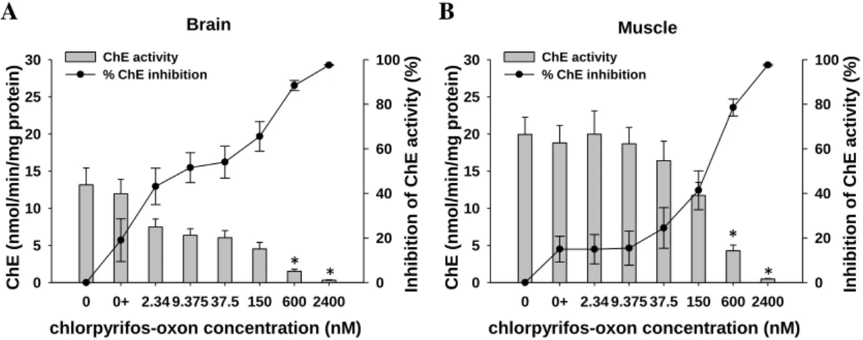
The results of ChE substrate preference in the brain and muscle tissues are shown in Figure 5.
Figure 5 – Cholinesterase substrate preferences in the brain (A) and muscle (B) of Prionace
glauca. Cholinesterases (ChE) activity is expressed as mean values ± standard error. ATCh =
Acetylthiocholine iodide. BTCh = S-Butyrylthiocholine iodide. PTCh = Propionylthiocholine iodide.
In brain tissue (Fig. 5A), the substrate with higher hydrolysis rate was ATCh (19.2 nmol/min/mg protein), followed by PTCh (10.8 nmol/min/mg protein) and BTCh (8.1 nmol/min/mg protein). The enzymatic catalytic efficiency indicated by the parameters of the Michaelis–Menten equation (Tab. 1) also demonstrates the preference for the substrate ATCh (higher Vmax/Km values). Furthermore, a decrease in ChE
A Brain Substrate concentration (mM) 0 5 10 15 20 C h E act iv it y ( n m o l/ m in /m g p ro tein) 0 10 20 30 40 ATCh PTCh BTCh Muscle Substrate concentration (mM) 0 5 10 15 20 C h E act iv it y ( n m o l/ m in /m g p ro tein) 0 10 20 30 40 ATCh PTCh BTCh B
39 activity caused by excess of the substrates ATCh and BTCh was also verified with concentrations higher than 2.56 mM and 5.12 mM, respectively.
Table 1 - Values of the Michaelis–Menten constant (Km), maximal velocity (Vmax) and the
catalytic efficiency (Vmax/Km) of Prionace glauca cholinesterases for the three tested substrates. Values of the Michaelis-Menten equation are expressed as the mean ± standard error.
Km (mM)
Vmax
(nmol/min/mg protein) Vmax/Km
Brain ATCh 0.045±0.0181 18.82±1.6499 422.90 PTCh 0.056±0.0257 9.09±0.7340 163.26 BTCh 0.027±0.0120 7.50±0.5637 279.76 Muscle ATCh 0.053±0.0140 31.62±2.0914 598.87 PTCh 0.034±0.0140 4.97±0.3371 147.36 BTCh - - -
In muscle tissue (Fig. 5B), there was a clear preference for the substrate ATCh, as seen by the higher hydrolysis rates and greater catalytic efficiency of ChE with this substrate (Tab. 1). Indeed, there were higher hydrolysis rates in the muscle than in the brain tissue with maximum ChE activities of 29.5 nmol/min/mg protein at 0.64 mM of ATCh when compared with the 19.2 nmol/min/mg protein maximum activity in brain at 2.56 mM of the same substrate. Almost no ChE activity was observed in muscle when using the substrate BTCh. In this tissue there was also an inhibition of hydrolysis by excess of ATCh at concentrations higher than 0.64 mM.
Regarding the results with the specific inhibitors, incubation with eserine, a generic inhibitor of ChE, significantly inhibited ChE activity in brain already at the lowest concentration tested of 0.781 µM (F6,49 = 115.1, p<0.001) whereas in muscle
significant inhibitions only occurred at concentrations higher than 12.5 µM (F6,49 =
179.5, p<0.001) (Fig. 6A). However, at concentrations higher than 50µM almost complete inhibitions were observed in both tissues (over 95% inhibition). Concerning the specific inhibitor for AChE (Fig. 6B), incubation with BW284C51 in brain only
40
significantly inhibited the enzyme activity at concentrations higher than 50 µM (F6,49 =
35.8, p<0.001) and inhibitions above 90% were observed only at 800µM, whereas in muscle a significant inhibition of 93% occurred already in the lowest concentration tested (F6,49 = 122.8, p<0.001). No effects on enzyme activity were observed with
iso-OMPA incubations, a specific inhibitor of BChE, either in brain (F6,49 = 9.69, p = 0.138)
or muscle (F6,49 = 0.47, p = 0.825) tissues (Fig. 6C). However, there was a
dose-response inhibition in the brain, albeit non-significant, reaching 40% inhibition in the highest iso-OMPA concentration tested.
Figure 6 – Effect of the inhibitors eserine (A), BW284C51 (B) and iso-OMPA (C) on Prionace
glauca cholinesterase (ChE) activities in brain and muscle tissues (expressed as mean values ±
standard error) using acetylthiocholine as substrate. Bars correspond to ChE activities and lines correspond to the percentage of ChE inhibition. An asterisk indicates a significant difference from the control at p ≤ 0.05 (ANOVA, Dunnett's test).
2.3.2. In vitro effects of chlorpyrifos-oxon
The effect of ethanol, the solvent used for chlorpyrifos stock solution, in the ChE activity was tested and compared with control and no statistical difference was observed either in brain (t(14) = 0.403, p = 0.693) or muscle (t(14) = 0.420, p = 0.681) tissues.
Eserine concentration (µM) 0 0.781 3.125 12.5 50 200 800 C h E ( n m o l/ m in /m g p ro tein) 0 10 20 30 40 In h ib it io n o f C h E act iv it y ( % ) 0 20 40 60 80 100 BW284C51 concentration (µM) 0 0.781 3.125 12.5 50 200 800 C h E ( n m o l/ m in /m g p ro tein) 0 10 20 30 40 In h ib it io n o f C h E act iv it y ( % ) 0 20 40 60 80 100 iso-OMPA concentration (mM) 0 0.0156 0.0625 0.25 1 4 16 C h E ( n m o l/ m in /m g p ro tein) 0 10 20 30 40 In h ib it io n o f C h E act iv it y ( % ) 0 20 40 60 80 100 Brain Muscle % inhibition in brain % inhibition in muscle A B C * * * * * * * * * * * * * * * * * * *
41 Regarding the effects of the in vitro exposure to chlorpyrifos-oxon, there was a dose-response pattern showing lower ChE activities with increasing pesticide concentrations, in both tissues tested, with almost complete inhibitions (over 97%) with the highest pesticide concentration (Fig. 7).
Figure 7 – Cholinesterase (ChE) activity values and percentage of activity inhibition (expressed
as mean values ± standard error) in the brain (A) and muscle (B) of Prionace glauca exposed in
vitro to chlorpyrifos-oxon. An asterisk indicates a significant difference from the solvent control
(0+) at p ≤ 0.05 (ANOVA, Dunnett's test).
Although significant inhibitions in relation to control were only detected at 600 nM of pesticide in both tissues (brain: F6,49 = 40.38, p<0.001; muscle: F6,49 = 37.77,
p<0.001), the lower concentrations of chlorpyrifos (until 150nM) caused statistically
higher ChE inhibitions in the brain than in the muscle (two-way ANOVA, p < 0.001). This higher sensitivity to chlorpyrifos in the brain tissue can also be seen by the estimated IC50 (±SE) values of 48.97 ± 3.79 nM (i.e. 16.39 µg/L) in brain and 204.97 ±
94.32 nM (i.e. 68.57 µg/L) in muscle.
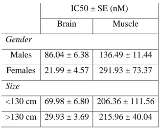
In order to address effects of gender and size on the susceptibility to chlorpyrifos-oxon, samples were divided in two groups of four according to gender (males and females) and size (larger or smaller than 130 cm). The IC50 values for these
separate groups, in brain and muscle tissues, were calculated and the results can be seen in Table 2. Brain chlorpyrifos-oxon concentration (nM) 0 0+ 2.34 9.375 37.5 150 600 2400 C hE (nm ol/ m in/ m g prot ein) 0 5 10 15 20 25 30 Inhibi tion of C hE act iv ity (% ) 0 20 40 60 80 100 ChE activity % ChE inhibition Muscle chlorpyrifos-oxon concentration (nM) 0 0+ 2.34 9.375 37.5 150 600 2400 C hE (nm ol/ m in/ m g prot ein) 0 5 10 15 20 25 30 Inhibi tion of C hE act iv ity (% ) 0 20 40 60 80 100 ChE activity % ChE inhibition B A * * * *
42
Table 2 – In vitro inhibition concentrations (IC50) of chlorpyrifos-oxon in Prionace glauca,
depending on gender and size of the organisms.
IC50 ± SE (nM) Brain Muscle Gender Males 86.04 ± 6.38 136.49 ± 11.44 Females 21.99 ± 4.57 291.93 ± 73.37 Size <130 cm 69.98 ± 6.80 206.36 ± 111.56 >130 cm 29.93 ± 3.69 215.96 ± 40.04
According to the IC50 values, the higher sensitivity of brain tissue when
compared to the muscle is visible independently of the organisms’ gender or size, and the differences between tissues are even more pronounced within females and in larger individuals (Tab. 2).
Regarding the effects of gender or size within each tissue, there were some differences in the IC50 values, e.g. lower IC50 for females or larger organisms in the
brain (not the same trend in muscle). These differences were however not statistically significant and therefore the response to chlorpyrifos-oxon in brain or muscle was not affected by either gender or size (two-way ANOVA, p > 0.05).
2.4. Discussion
2.4.1. Cholinesterase characterization
To use ChE as a biomarker of effect to pollutants in a particular species, it is vital to characterize this enzyme on different target tissues, as they may have several non-specific esterases that can mislead ecotoxicological studies (Gomes et al., 2014; Pestana et al., 2014).
Regarding the brain tissue, incubation with eserine sulfate, an organophosphorus compound well-known as a general inhibitor of ChE at low concentrations, resulted in an almost complete enzyme inhibition (Fig. 6A), meaning that the measured enzymatic activity is mostly due to ChE, and not to other nonspecific esterases (Eto, 1974; Pezzementi et al., 1991). The characterization of the brain’s ChE was performed by