Beneficial effects of Oltipraz, nuclear factor - erythroid -
2 - related factor 2 (Nrf2), on renal damage in unilateral
ureteral obstruction rat model
_______________________________________________
Emre Can Polat
1, Huseyin Besiroglu
2, Levent Ozcan
3, Alper Otunctemur
1, Ahmet Tugrul Eruyar
4, Adnan
Somay
5, Nurver Ozbay
5, Mustafa Cekmen
6, Ceyla Eraldemir
7, Emin Ozbek
81 Department of Urology, Okmeydani Training and Research Hospital, University of Health Sciences,
Istanbul, Turkey; 2 Department of Urology, Catalca Ilyas Cokay State Hospital, Istanbul, Turkey; 3 Department of Urology, Derince Training and Research Hospital, University of Health Sciences,
Kocaeli, Turkey; 4 Department of Pathology, Derince Training and Research Hospital, University of Health Sciences, Kocaeli, Turkey; 5 Department of Pathology, Fatih Sultan Mehmet Training and Research Hospital, University of Health Sciences, Istanbul, Turkey; 6 Department of Biochemistry, Istanbul Medeniyet University, Istanbul, Turkey; 7 Department of Biochemistry, Kocaeli University, Kocaeli, Turkey; 8 Department of Urology, Cerrahpasa Medical School, Istanbul University, Istanbul, Turkey
ABSTRACT
Introduction: We investigated whether Oltipraz (OPZ) attenuated renal fibrosis in a unilateral ureteral obstruction (UUO) rat model.
Materials and Methods: We randomly divided 32 rats into four groups, each consisting of eight animals as follows: Rats in group 1 underwent a sham operation and received no treatment. Rats in group 2 underwent a sham operation and received OPZ. Rats in group 3 underwent unilateral ureteral ligation and received no treatment. Group 4 rats were subjected to unilateral ureteral ligation plus OPZ administration. Transforming growth factor beta-1 (TGF-β1), E-cadherin, nitric oxide (NO) and hydroxyproline levels were measured. Histopathological and immunohistochemical examinations were car-ried out.
Results: TGF-β1, NO and E-cadherin levels in the UUO group were significantly higher than the sham group and these values were significantly different in treated groups compared to the UUO group. In rats treated with UUO + OPZ, despite the presence of mild tubular degeneration and less severe tubular necrosis, glomeruli maintained a better morphology when compared to the UUO group. Expressions of α-SMA in im-munohistochemistry showed that the staining positivity decreased in the tubules of the OPZ-treated group.
Conclusions: While the precise mechanism of action remains unknown, our results demonstrated that OPZ exerted a protective role in the UUO-mediated renal fibrosis rat model highlighting a promising therapeutic potency of Nrf2-activators for alleviating the detrimental effects of unilateral obstruction in kidneys.
ARTICLE INFO
Keywords:
Oltipraz [Supplementary Concept]; Renal Insufficiency; Ureter
Int Braz J Urol. 2018; 44: 1243-51
_____________________
Submitted for publication: March 28, 2018
_____________________
Accepted after revision: July 16, 2018
_____________________
INTRODUCTION
Ureteral obstruction occurs at any stage of life from fetal development to adulthood in any segment of the ureter between ureteral orifices and the renal pelvis. The histopathologic changes are characterized by tubular dilatation or atrophy, inflammatory cell infiltration, fibroblast activa-tion and proliferaactiva-tion, increases in matrix pro-teins, and progressive tubulointerstitial fibrosis. These histopathologic processes might eventually result in the loss of renal parenchyma leading to permanent renal function deterioration.
One of the most prevalent molecular me-chanisms of tubulointerstitial fibrosis due to ure-teral obstruction is epithelial - to - mesenchymal transition. Numerous studies have proposed that under pathologic conditions renal tubular epithe-lial cells may undergo a phenotypic transforma-tion into matrix - producing myofibroblasts by an epithelial - mesenchymal transition (EMT) process (1-3). Activated by several growth factors such as transforming growth factor β (TGFβ), myofibro-blasts function as the primary source for produ-cing extracellular matrix (ECM), including colla-gen and fibronectin. Furthermore, the expression of intercellular epithelial adhesion molecules such as E - cadherin decrease and mesenchymal cell markers such as α - smooth muscle actin (α - SMA), N - cadherin and vimentin increase. For this rea-son, EMT is recognized as a molecular component of renal fibrosis (4-6).
Reactive oxygen species (ROS) have a ma-jor role in the development of renal fibrosis indu-cing epithelial - mesenchymal transition (EMT) in the presence of UUO. Nitric oxide (NO), a vasodi-lator, has been implicated in late renal hemody-namic changes and observed upregulated in the kidney during the UUO process. Even though the excessive amounts of nitric oxide (NO) are oxidi-zed into ROS, they have proven to be useful in in-terrupting signaling and controlling inflammation (7, 8). In addition, renal hydroxyproline (Hyp), which is commonly used to measure the collagen content in biological tissue values, has been used to assess fibrosis (9).
Oltipraz (OPZ), 5 - (2 - pyrazynyl) - 4 -
dithiolethione that targets Nrf2 (nuclear factor - erythroid - 2 - related factor 2), an agent that plays a pivotal role in cellular defense against oxidative stress by promoting the transcription of various antioxidant genes (10). Several agents have been used to prevent UUO - induced renal injury in ani-mal models but there is no experiment for the role of oltipraz (OPZ) in the literature. A randomized, double-blind, placebo - controlled phase II trial demonstrated that reductions in inflammation, oxidative stress and fibrosis could be achieved using oltipraz (OPZ), nuclear factor - erythroid - 2 - related factor 2 (Nrf2) activator in patients with liver fibrosis or cirrhosis (11).
Based on these findings, we investigated the potential effect of OPZ in attenuating renal fibrosis induced by UUO in rats.
MATERIALS AND METHODS
Animals
Male Wistar albino rats, weighing 200 to 250 g and six to seven weeks old, were housed in clean plastic cages in a temperature - and humidi-ty - controlled facilihumidi-ty under constant 12 - hour li-ght / 12 - hour dark photoperiods with free access to food and water. The Institutional Animal Care and Use Committee approved the use of animals and the experimental protocol and animals were treated according to the Guide for the Care and Use of Laboratory Animals of Research Council.
Treatment and experimental protocols
St. Louis, MO) (p.o. 30 mg / kg body weight / day). Rats in group 3 underwent unilateral ureteral liga-tion and received no treatment. Group 4 rats were subjected to unilateral ureteral ligation and recei-ved OPZ. The OPZ dose was based on the previous studies (13).
At 14 days after UUO, all rats were sacri-ficed by high - dose ketamine. Kidneys were rea-ched with an abdominal midline incision. The left kidney was immediately excised and separated from the surrounding tissues, washed twice with cold saline, and stored at - 800 C to determine the markers of renal fibrosis, EMT and tubular injury.
A portion of the left renal tissue was sto-red in formol solution for the histopathologic and immunohistochemical examinations. Paraffinized tissue samples were examined for leukocyte infil-tration and renal fibrosis.
Measurement of transforming growth fac-tor beta - 1, E - cadherin, nitric oxide and hydro-xyproline levels.
The TGFβ - 1 ELISA kit (ref MB100B, R & D Systems) was used to measure TGFβ - 1 in re-nal tissue 50 microns of total proteins from each tissue were assayed. This assay detects activated TGFβ-1 with a sensitivity of 4.6 pg / mL. The co-efficient of variation intra-assay is < 4%, and the coefficient inter - assay is < 8%.
The concentration of soluble E - cadherin was measured with a commercially available san-dwich enzyme - linked immunosorbent assay kit based on monoclonal antibodies (Zymed Labora-tories Inc., CA). The coefficient of variation intra - assay is < 5%, and the coefficient inter - assay is 7%. The sensitivity is 2.0 ng / mL.
The nitrate concentrations in samples were assayed by enzymatically reducing nitrate. 50 microL of samples were incubated with the same volume of reductase buffer (0.1M) potassium ade-nine dinucleotide and four units of nitrate curve were obtained by incubating sodium nitrate (10 - 200 µM) with the buffer. The total amount of nitrite and the amount of nitrite in the samples was then determined using the Griess method (14). The samples were incubated with the same volume of Griess reagent (1% sulphanilamide and 0.1% naphthyl ethylenediamine dihydrochloride in 5%
phosphoric acid). The absorbance at 550 nm was determined using a multiwell plate reader. The re-sults were reported as the concentration of nitrate plus nitrite (microM NO3 + NO2) for samples of nitrite for supernatants.
Renal tissue fragments were homogenized in saline 0.9%, frozen and lyophilized. The assay was performed with 40 mg of the lyophilized tis-sue that was subjected to alkaline hydrolysis in 300 micros plus 75 microLNaOH 10 moL / L at 120 degrees C for 20 minutes. An aliquot of 50 microL of the hydrolyzed tissue was added to 450 microL of chloramine T oxidizing reagent (Chloramine T 0.056 moL / L, n - propanol 10% in acetate / citrate buffer pH 6.5) and allowed to react for 20 minutes. A hydroxyproline standard curve with the highest concentration of 400 microns was prepared in a similar fashion. The color was developed by the addition of 500 microL of the Ehrlich reagent (p - dimethylamine - benzaldehyde, 1 moL / L) diluted in n - propanol / perchloric acid, 2: 1 supernatant was transferred to 96 - well plates, and the absor-bance was read at 550 nm.
Histopathological and immunohistochemical examination
The histopathological and immunohisto-chemical evaluation were performed on left kid-ney tissues. Paraffin - embedded specimens were cut into 5 - mm thick sections and stained with hematoxylin and eosin, Masson’s trichrome and α - smooth muscle actin (α - SMA) were used for examination under the light microscope (BH - 2; Olympus, Tokyo, Japan).
To evaluate leukocyte infiltration, the wide-ning of interstitial spaces with focal leukocyte infil-tration was assessed in five randomly chosen sec-tions prepared from each kidney sample. For each section, the average number of leukocytes per 0.28 mm2 was calculated from these leukocyte - infiltrated
foci using a high - power microscopic field (x 400). To estimate the grade of interstitial fibro-sis, the interstitial area that was stained green with Masson’s trichrome was evaluated as a percentage of the total examined area in five randomly chosen sections prepared from each kidney sample using an image analyzer (Leica; Leica Micros Imaging Solu-tions, Cambridge, UK). For each section, interstitial space widening with focal leukocyte infiltration and interstitial fibrosis was assessed in high - power fields (x 400) to quantify the results. The Banff classifica-tion of kidney pathology was used to score the degree of mononuclear cell infiltration and interstitial fibro-sis. The score was graded from 0 to 3, depending on the severity of histological characteristics (17).
Statistical analyses
Continuous variables of all groups were demonstrated as mean values ± standard devia-tion (SD). Statistical analyses of the
histopatholo-gic evaluation of the groups were carried out by the Chi - square test, and biochemical data among four groups were analyzed by Kruskal - Wallis test. The p - value of < 0.05 was accepted as sta-tistically significant. In case a statistical signifi-cant was observed between three or four groups in Kruskal - Wallis test, Mann - Whitney U test was utilized for the detection of the significance between two groups.
RESULTS
Transforming growth factor beta - 1, E - cadherin, nitric oxide and hydroxyproline levels
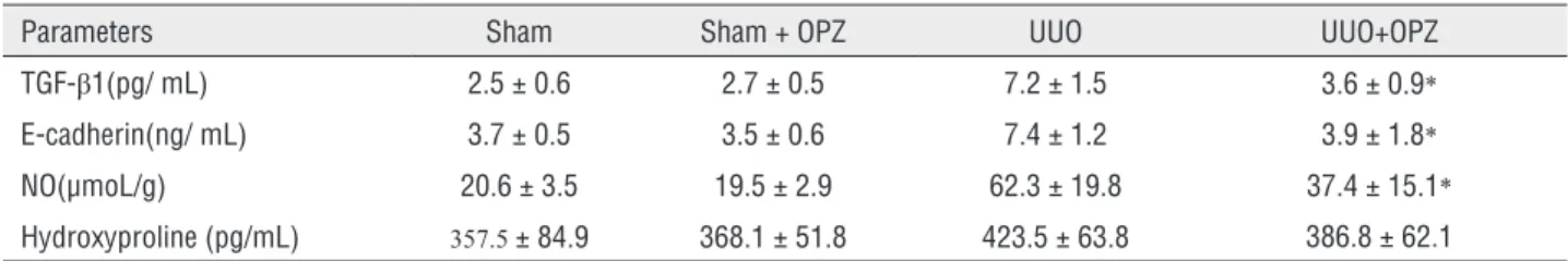
TGF - β1 level in sham, UUO and UUO + OPZ
groups were 2.5 ± 0.6; 7.2 ± 1.5 and 3.6 ± 0.9 res-pectively. NO level in sham, UUO and UUO + OPZ groups were 20.6 ± 3.5; 62.3 ± 19.8 and 37.4 ± 15.1 respectively and E - cadherin level in those groups were 3.7 ± 0.5; 7.4 ± 1.2 and 3.9 ± 1.8 respectively. TGF - β1, NO, and E - cadherin levels were signi-ficantly higher in the UUO group than the sham group. UUO + OPZ group had lower levels of these markers compared to the UUO group. Hydroxypro-line level was 423.5 ± 63.8 in UUO group, whereas the remaining three groups had lower levels; but this difference did not reach a statistical significan-ce. The details are shown in Table-1.
Histopathologic and immunohistochemical exa-mination results
Histopathologic examination of kidneys showed no pathologic findings in the sham and sham + OPZ groups (Figures 1A and 1B). Mild and severe tubular necrosis in the proximal
tubu-Table 1 - TGF-β1, E-cadherin, NO and Hydroxyproline levels in kidney.
Parameters Sham Sham + OPZ UUO UUO+OPZ
TGF-β1(pg/ mL) 2.5 ± 0.6 2.7 ± 0.5 7.2 ± 1.5 3.6 ± 0.9*
E-cadherin(ng/ mL) 3.7 ± 0.5 3.5 ± 0.6 7.4 ± 1.2 3.9 ± 1.8*
NO(µmoL/g) 20.6 ± 3.5 19.5 ± 2.9 62.3 ± 19.8 37.4 ± 15.1*
Hydroxyproline (pg/mL) 357.5 ± 84.9 368.1 ± 51.8 423.5 ± 63.8 386.8 ± 62.1
TGF = transforming growth factor β1; NO = nitric oxide.
Values are expressed as mean ± SD for eight rats in each group. * Significantly different from UUO group (p < 0.05).
les was found in rats with UUO compared to the sham group (Figure-1C). Despite the presence of mild tubular degeneration and less severe tubular necrosis in rats treated with UUO + OPZ, glomeruli maintained a better morphology compared to the UUO group (Figure-1D).
Histopathologic examination was normal in rats with the sham operation (group 1). Severe leukocyte infiltration was observed in the peri-glomerular and peritubular interstitium of the rat kidneys in group 3 with UUO (Figures 2A and B). Quantitative analysis of the focal leukocyte infiltra-tion area in the interstitium showed that leukocyte infiltration was significantly reduced in rats admi-nistered with UUO + OPZ (group 4) (Figure-2C).
UUO caused significant interstitial fibro-sis in rats that received no treatment (group 3).
The percentage of the area of interstitial fibrosis in the rats with UUO that received no treatment was significantly greater than that of rats with UUO that received OPZ (group 4) (Figures 3A-C). These changes are summarized in Table-2. Expressions of α - SMA in immunohistochemistry showed that the staining positivity decreased in the tubules of the OPZ - treated group (Figures 4A-C).
DISCUSSION
In this study, we analyzed the protective effect of OPZ against renal fibrosis in a rat UUO model, a well - established in vivo model of renal fibrosis. Our study confirmed the protective role of OPZ through a quantitative examination of renal tis-sue damage after the induction of UUO in rats. To
Figure 2 - A) Normal kidney morphology in a sham group. B) Leukocyte infiltration was observed in the peritubular interstitium of the UUO. C) Leukocyte infiltration was reduced in the OPZ-treated group (hematoxylin&eosin,*400).
A B C
Figure 1 - A) Normal tubulus and glomeruli in kidney cortex H&Ex100 (sham group). B) Normal tubulus and glomeruli in kidney cortex H&Ex100(sham+OPZ group). C) Severe tubular total necrosis, tubular degeneration and epithelial vacuolization in the proximal tubules H&Ex400(UUO group). D) Mild epithelial vacuolization in the proximal tubules and normal glomeruli H&Ex100 (UUO+OPZ treated group).
A B C
Figure 3 - A) Normal kidney morphology in a sham group. B) Severe fibrosis was observed in the peritubular interstitium of the UUO. C) Mild fibrosis was reduced in the OPZ-treated group (Masson&Trichrome, *400).
Table 2 - Scoring of tubular necrosis.
Tubular necrosis Interstitial fibrosis Mononuclear cell infiltration
n 0 1 2 3 0 1 2 3 0 1 2 3
Sham 8 8 0 0 0 8 0 0 0 8 0 0 0
Sham+OP 8 8 0 0 0 8 0 0 0 8 0 0 0
UUO 8 0 1 3 4 0 1 4 3 0 1 2 5
UUO+OPZ* 8 0 6 2 0 2 5 1 0 1 5 0 2
Score 0 = no degeneration; 1 = mild degeneration; 2 = moderate degeneration; 3 = severe degeneration
* Statistical significant difference from the UUO group and P < 0.05.
Figure 4 - A) α-SMA staining was not observed in the tubules of the sham group. B) Moderate -SMA positivity was observed
in more than 50% of the tubules in the UUO group. C) mild -SMA positivity was observed in 25-50% of the tubules in the OPZ-treated group ( -SMAx100).
the best of our knowledge, this report is the first to show that OPZ has a preventive effect on functional, histological kidney injury caused by UUO.
The molecular mechanisms of tubuloin-terstitial fibrosis owing to UUO include several components such as inflammatory cell infiltra-tion, fibroblasts and extracellular matrix produc-tion, and epithelial to mesenchymal transition. Among these, EMT has been of great interest to researchers during recent decades. EMT has been asserted as a functional and phenotypic change of epithelial cells that is reminiscent of mesenchymal cells reflecting a global process affecting adjacent cells (18). During the EMT process, expression of epithelial adhesion molecules such as E - cadherin is decreased whereas mesenchymal marker pro-teins such as α - smooth muscle actin (α - SMA) and N - cadherin are up - regulated. The EMT process has been viewed as a pathological predi-cator of renal fibrosis as myofibroblasts transfor-med from epithelial cells act as the main origin of ECM production (19). Moreover, the phenotype transformation of renal epithelial cells can cau-se dysfunction of the kidney, eventually resulting in glomerulosclerosis. Furthermore, accumulating lines of evidence support the concept that ROS affects EMT changes. Higher levels of ROS may aid the EMT process of epithelial cells and lead to fibrosis in the context of transcription factors and the multifunctional role of ROS in cellular signa-ling pathways (20, 21).
Nuclear factor - erythroid - 2 - related factor 2 (Nrf2) is a vital molecule of the endoge-nous antioxidant system that plays a central role in stimulating expression of various antioxidant - associated genes in cellular defense against oxi-dative stress. In the absence of oxioxi-dative stress, Nrf2 resides in the cytoplasm together with its repressor kelch - like ECH - associated protein 1 (KEAP1). Many agree that the upregulating in the production of antioxidant enzymes, which follo-ws the detaching of Nrf2 from KEAP1 and sub-sequent movement into the nucleus, is a result of ROS overproduction or results from responses to electrophilic reagent treatment (22). Debates con-tinue as to whether the EMT process is a real and direct contributor to renal fibrosis pathology in vivo (23, 24).
Based on the results showing the linka-ge between Nrf2 and TGFβ1 signaling, we hypo-thesized that Nrf2 could have a potential role in protecting the kidney in unilateral ureteral obs-truction. It is believed that oltipraz, a prototype dithiolethione, might support the joining of the Nrf2 to the antioxidant response element (25).
We measured renal cortical E - cadherin, TGF - β1, hydroxyproline and NO levels bioche-mically as evidence of EMT. Except for the hydro-xyproline level, all these markers were heightened in UUO rats and OPZ attenuated their levels. These results suggest that OPZ ameliorates the renal fi-brosis by inhibiting these markers, which are the indicators of renal fibrosis. The possible expla-nation for the level of hydroxyproline not being significant among the groups might be the fact that the duration of UUO may be insufficient for observing the exact fibrotic changes in the kidney. It is probable that hydroxyproline, being a com-ponent of collagen, would be detected as increa-sed in UUO kidney rats if they were examined one month or later after the obstruction date.
Additionally, pooled urine in UUO is ma-rkedly hypotonic (26). In theory, hypotonic cell growths could be caused by the hypotonic nature of pooled UUO urine. Increases in E - cadherin expressions and the induction of connective tis-sue growth factors (CTGF) were instigated through hypotonic media. It has been proposed that CTGF is an element of renal EMT via evidence of upre-gulation in UUO (27, 28).
We think that E - cadherin can be simul-taneously upregulated in response to hypotonic stretch, suggesting that profibrotic activation of tubular cells might not require a ‘classical’ EMT process.
for slight desquamation and atrophy of the tubu-lar epithelial cells. Also, expressions of α - SMA in immunohistochemistry showed that the staining positivity decreased in the tubules of the OPZ - treated group. Scientific evidence from the va-rious field of diseases including pulmonary hyper-tension, liver ischemia / reperfusion injury yielded promising results on the potential effect of OPZ (29, 30). In a cell culture study, Atilano - Roque A et al. demonstrated that Nrf2 activating agent, OPZ had a beneficial effect on the viability of hu-man kidney cells and expression of antioxidant and efflux transporter genes in proximal tubules which was exposed to cisplatin. Our results have similar findings compared to these studies. Given the similar pathophysiologic mechanisms, we may propose that OPZ is a promising agent for allevia-ting kidney injury in the presence of the ureteral obstruction.
We should address some potential limita-tions of this study. Firstly, we measured NO levels and reported that rats with UUO had increased levels of NO. However, our study lacks the measurement of NO isoforms including neuronal, endothelial, and inducible NOS. Secondly, although we exerted the conventional methods including the detection of the levels of molecules as mentioned earlier and histopathologic examination of renal tissue, we did not use quantitative RT - PCR, western blot, and immunofluorescence analysis to analyze further and demonstrate the expression of cell markers. Thirdly, although UUO is a well - accepted animal renal fibrosis model, it is criticized for the fact that the contralateral kidney may compensate for the deteriorated function of the obstructed kidney pre-cluding meaningful renal function measurements (31). Lastly, whether the sensitivity and specificity of the markers used for EMT in our study are higher than some other EMT markers such as vimentin, cytokeratin, and β catenin is not known. Further studies on these markers will probably yield more insights to this debate.
CONCLUSIONS
Taken all together, while the mechanism of actions remains unknown, our results show that
renal fibrosis. However, further well - designed animal and clinical studies are needed to confirm our results.
CONFLICT OF INTEREST
None declared.
REFERENCES
1. Burns WC, Kantharidis P, Thomas MC. The role of tubular epithelial-mesenchymal transition in progressive kidney disease. Cells Tissues Organs. 2007;185:222-31.
2. Healy E, Brady HR. Role of tubule epithelial cells in the pathogenesis of tubulointerstitial fibrosis induced by glomerular disease. Curr Opin Nephrol Hypertens. 1998;7:525-30.
3. Strutz F, Okada H, Lo CW, Danoff T, Carone RL, Tomaszewski JE, et al. Identification and characterization of a fibroblast marker: FSP1. J Cell Biol. 1995;130:393-405.
4. Iwano M, Plieth D, Danoff TM, Xue C, Okada H, Neilson EG. Evidence that fibroblasts derive from epithelium during tissue fibrosis. J Clin Invest. 2002;110:341-50.
5. Kalluri R, Neilson EG. Epithelial-mesenchymal transition and its implications for fibrosis. J Clin Invest. 2003;112:1776-84. 6. Wynn TA. Cellular and molecular mechanisms of fibrosis. J
Pathol. 2008;214:199-210.
7. Huang HS, Ma MC, Chen CF, Chen J. Changes in nitric oxide production in the rat kidney due to CaOx nephrolithiasis. Neurourol Urodyn. 2006;25:252-8.
8. Aviram M, Dornfeld L, Rosenblat M, Volkova N, Kaplan M, Coleman R, et al. Pomegranate juice consumption reduces oxidative stress, atherogenic modifications to LDL, and platelet aggregation: studies in humans and in atherosclerotic apolipoprotein E-deficient mice. Am J Clin Nutr. 2000;71:1062-76.
9. Wang Q, Usinger W, Nichols B, Gray J, Xu L, Seeley TW, et al. Cooperative interaction of CTGF and TGF-β in animal models of fibrotic disease. Fibrogenesis Tissue Repair. 2011;4:4. 10. Noorafshan A, Kardeh S, Ashkani-Esfahani S, Namazi MR,
Saleh E. The Effects of Oltipraz on Tissue Regeneration in the Process of Wound Healing: A Stereological Study. Bull Emerg Trauma. 2014;2:161-5.
11. Kim SG, Kim YM, Choi JY, Han JY, Jang JW, Cho SH, et al. Oltipraz therapy in patients with liver fibrosis or cirrhosis: a randomized, double-blind, placebo-controlled phase II trial. J Pharm Pharmacol. 2011;63:627-35.
13. Cho IJ, Sung DK, Kang KW, Kim SG. Oltipraz promotion of liver regeneration after partial hepatectomy: The role of PI3-kinase-dependent C/EBPbeta and cyclin E regulation. Arch Pharm Res. 2009;32:625-35.
14. Menaka KB, Ramesh A, Thomas B, Kumari NS. Estimation of nitric oxide as an inflammatory marker in periodontitis. J Indian Soc Periodontol. 2009;13:75-8.
15. Sun Y, Oberley LW, Li Y. A simple method for clinical assay of superoxide dismutase. Clin Chem. 1988;34:497-500. 16. Allen CT. Laboratory methods in histochemistry. In: Prophet
EB, Mills B, Arrington JB, Sobin LH (eds) American registry of pathology, 1st edn. Washington DC. 1992; pp. 53. 17. Kinugasa F, Noto T, Matsuoka H, Urano Y, Sudo Y, Takakura
S, et al. Prevention of renal interstitial fibrosis via histone deacetylase inhibition in rats with unilateral ureteral obstruction. Transpl Immunol. 2010;23:18-23.
18. Galichon P, Hertig A. Epithelial to mesenchymal transition as a biomarker in renal fibrosis: are we ready for the bedside? Fibrogenesis Tissue Repair. 2011;4:11.
19. Kalluri R, Weinberg RA. The basics of epithelial-mesenchymal transition. J Clin Invest. 2009;119:1420-8. Erratum in: J Clin Invest. 2010;120:1786.
20. Adler V, Yin Z, Tew KD, Ronai Z. Role of redox potential and reactive oxygen species in stress signaling. Oncogene. 1999;18:6104-11.
21. Kinnula VL, Myllärniemi M. Oxidant-antioxidant imbalance as a potential contributor to the progression of human pulmonary fibrosis. Antioxid Redox Signal. 2008;10:727-38. 22. Kaspar JW, Niture SK, Jaiswal AK. Nrf2:INrf2 (Keap1)
signaling in oxidative stress. Free Radic Biol Med. 2009;47:1304-9.
23. Fragiadaki M, Mason RM. Epithelial-mesenchymal transition in renal fibrosis - evidence for and against. Int J Exp Pathol. 2011;92:143-50.
24. Jiang YS, Jiang T, Huang B, Chen PS, Ouyang J. Epithelial-mesenchymal transition of renal tubules: divergent processes of repairing in acute or chronic injury? Med Hypotheses. 2013;81:73-5.
25. Choi SH, Kim YM, Lee JM, Kim SG. Antioxidant and mitochondrial protective effects of oxidized metabolites of oltipraz. Expert Opin Drug Metab Toxicol. 2010;6:213-24. 26. Quinlan MR, Perez-Barriocanal F, Wright E et al. Analysis of
urinary and plasma electrolytes in a rat model of unilateral ureteric obstruction (UUO). Open J Urol Nephrol 2008; 1: 16–21. 27. Yokoi H, Mukoyama M, Sugawara A, Mori K, Nagae T,
Makino H, et al. Role of connective tissue growth factor in fibronectin expression and tubulointerstitial fibrosis. Am J Physiol Renal Physiol. 2002;282:F933-42.
28. Wahab NA, Mason RM. A critical look at growth factors and epithelial-to-mesenchymal transition in the adult kidney. Interrelationships between growth factors that regulate EMT in the adult kidney. Nephron Exp Nephrol. 2006;104:e129-34.
29. Eba S, Hoshikawa Y, Moriguchi T, Mitsuishi Y, Satoh H, Ishida K, et al. The nuclear factor erythroid 2-related factor 2 activator oltipraz attenuates chronic hypoxia-induced cardiopulmonary alterations in mice. Am J Respir Cell Mol Biol. 2013;49:324-33.
30. Rao J, Qian X, Li G, Pan X, Zhang C, Zhang F, et al. ATF3-mediated NRF2/HO-1 signaling regulates TLR4 innate immune responses in mouse liver ischemia/reperfusion injury. Am J Transplant. 2015;15:76-87.
31. Atilano-Roque A, Wen X, Aleksunes LM, Joy MS. Nrf2 activators as potential modulators of injury in human kidney cells. Toxicol Rep. 2016;3:153-9.
_______________________ Correspondence address:
Emre Can Polat, MD, FEBU Department of Urology Okmeydani Training & Research Hospital University of Health Sciences,