Catarina Vizetto Guerreiro Duarte
Reactivity of human fetal and adult
immortalized hepatocytes to potentially
toxic bilirubin and bile acid species
LISBOA
DEPARTAMENTO DE CIÊNCIAS DA VIDA
Catarina Vizetto Guerreiro Duarte
Reactivity of human fetal and adult
immortalized hepatocytes to potentially toxic
bilirubin and bile acid species
Dissertação apresentada para a obtenção do Grau de Mestre
em Genética Molecular e Biomedicina, pela Universidade
Nova de Lisboa, Faculdade de Ciências e Tecnologia
Orientador:
Profa. Doutora Dora Maria Tuna de Oliveira Brites Brites (FF/UL)
Co-orientadora:
Doutora Adelaide Maria Afonso Fernandes (FF/UL)
LISBOA
NOTA DE ABERTURA
A presente dissertação destina-se à obtenção do grau de Mestre em Genética Molecular e Biomedicina, pela Faculdade de Ciências e Tecnologia, da Universidade Nova de Lisboa.
O trabalho prático foi realizado no grupo “Neuron Glia Biology in Health and Disease” do Centro de Patogénese Molecular (CPM)/iMed da Faculdade de Farmácia da Universidade de Lisboa (FFUL), tendo a orientação sido da responsabilidade da Professora Doutora Dora Maria Tuna de Oliveira Brites, Investigadora Coordenadora do referido grupo e da Doutora Adelaide Maria Afonso Fernandes.
O trabalho prático teve início dia 1 de Setembro de 2008, com a integração nas actividades desenvolvidas no Laboratório e após uma fase de aprendizagem das técnicas laboratoriais utilizadas.
O presente trabalho de investigação (não integral) foi apresentado, em comunicação oral, no XXIX Congresso Nacional de Gastrenterologia e Endoscopia Digestiva e publicado na forma de resumo no Jornal Português de Gastrenterologia.
1
Vizetto Duarte C, Fernandes A, Brites D. A icterícia como factor de risco da colestase crónica: manifestações citopatológicas numa linha humana saudável de hepatócitos.
XXIX Congresso Nacional de Gastrenterologia e Endoscopia Digestiva, Porto, 2009.
2
Vizetto Duarte C, Fernandes A, Brites D. A icterícia como factor de risco da colestase crónica: manifestações citopatológicas numa linha humana saudável de hepatócitos.
AGRADECIMENTOS
Pela primeira vez sentimo-nos desassossegados. As palavras ganham um novo sentido quando não se tornam previsíveis; ganham nova vida quando propõem olhares e despertam perplexidades, quando inquietam o pensamento. E tornam-se sempre insuficientes quando com elas queremos dizer o que nos vai para lá da alma. Como agora.
Esta dissertação resulta não apenas de extensas horas de estudo, reflexão e trabalho durante as diversas etapas que a constituem. É igualmente o culminar de um objectivo académico a que me propus e que não seria possível sem a ajuda de um vasto número de pessoas.
Estou especialmente agradecida à Professora Doutora Dora Brites por me ter acolhido no grupo “Neuron Glia Biology in Health and Disease” e me ter proporcionado a oportunidade e as condições necessárias para a elaboração deste trabalho. O seu elevado padrão de rigor e exigência, a sua perspicácia, o vasto conhecimento e as sugestões transmitidas durante a elaboração desta dissertação foram essenciais para o desenvolvimento desta tese. À sua hábil direcção, instrução e apoio na superação dos diversos obstáculos, o meu muito obrigado.
À Doutora Adelaide Fernandes pelos seus sábios conselhos, recomendações, correcções e contagioso entusiasmo. Obrigado por sempre me ter apoiado, para além das suas obrigações profissionais, e por me ter ajudado a adaptar a toda a realidade do laboratório e às novas técnicas com que me deparei. O seu apoio, críticas e sugestões foram imprescindíveis na construção deste trabalho.
Ao Professor Doutor Rui Silva e à Professora Doutora Alexandra Brito gostaria de manifestar a minha gratidão por todos os ensinamentos científicos ao longo do decorrer deste trabalho. Gostaria ainda de agradecer o vosso permanente apoio, ideias, sugestões e espírito crítico.
Palmela, e também àqueles que se encontravam na mesma altura em processo de Mestrado, Filipa Cardoso, Eduarda Coutinho e Ema Torrado, pela ajuda e intercâmbio de ideias para a elaboração do trabalho. A vossa constante ajuda contribuiu para um excelente desenvolvimento dos estudos experimentais deste trabalho. Aprendi muito com vocês!
Às colegas do Laboratório de Bilirrubina e Ácidos Biliares, Sónia e Andreia, obrigada pela boa disposição e sentido de entreajuda, que fazem do nosso local de trabalho um local onde existe prazer em estar.
Aos restantes grupos e colegas do Centro de Patogénese Molecular gostaria de agradecer a forma acolhedora como me receberam.
Aos meus amigos de longa data pois sempre me apoiaram e fizeram sorrir nos bons e maus momentos.
Não podia deixar de agradecer às pessoas mais importantes da minha vida. À minha família. À minha mana Fernanda, obrigada por partilhares a minha vida desde sempre, me ajudares a crescer e a me tornar no que sou e por me apoiares de forma incondicional. Sem ti, não era eu. Ao meu cunhado Fernando obrigado pelo apoio e por sempre me fazer ver a vida pelo lado optimista. Aos meus sobrinhos, os meus amores, Tomás e Diogo, um muito obrigada por darem um novo sentido à minha vida.
Aos meus avós, João e Florinda, por sempre me apoiarem e ajudarem em tudo. Adoro-vos!
CONTENTS
Abbreviations ... vii
Abstract ... ix
Resumo ... xi
1. Introduction ... 1
1.1. The liver ... 1
1.1.1. Liver development ... 1
1.1.2. Liver structure ... 3
1.1.3. Liver functions ... 5
1.2. Pathophysiology of cholestasis ... 7
1.2.1. Main clinical biomarkers of chronic cholestasis: from diagnosis to prognosis ... 8
1.2.1.1. Bile acids ... 9
1.2.1.2. Bilirubin ... 12
1.2.2. Acute vs. chronic cholestasis... 15
1.3. Mechanisms of hepatocyte injury during cholestasis ... 17
1.3.1. Major modes of cell death in the liver ... 17
1.3.2. Cell death and the development of inflammation ... 19
1.3.3. Inflammatory signaling pathways ... 20
1.4. Aims of the thesis ... 22
2. Materials and Methods ... 23
2.1. Chemicals ... 23
2.2. Equipment ... 24
2.3. Fetal and adult hepatocyte cell culture ... 24
2.4. Hepatocyte treatment with bilirubin and bile acid species ... 27
2.5. Cytotoxicity evaluation ... 28
2.5.1. LDH release ... 28
2.5.2. Apoptosis assessment ... 29
2.6. Western blot assay ... 30
2.7. Immunofluorescence detection of NF-κB ... 31
2.8. Statistical Analysis ... 31
3. Results ... 33
3.1. Decreased viability of hepatocytes is induced by bilirubin and bile acid species, mainly at 48 h of incubation ... 33
3.2. Apoptosis is enhanced in hepatocytes treated with bilirubin and bile acid species ... 35
3.2.1. Increase of caspase-3 activity ... 35
3.2.2. Increase of apoptotic features ... 37
3.3. MTS reduction is enhanced in hepatocytes treated with bilirubin and bile acid mlspecies ... 41
3.4. JNK1/2 signaling pathway is activated in hepatocytes treated with bilirubin and bile acid species while p38 signaling pathway is decreased. ... 42
3.5. NF-κB signaling pathway is activated in hepatocytes treated with bilirubin and bile acid species ... 46
4. Discussion ... 51
ABBREVIATIONS
AFP α-fetoprotein
AGQDC ácido glicoquenodesoxicólico [N-(3α,7α-dihydroxy-5β-cholan-24-oyl)glycine]
BC bilirrubina conjugada
BNC bilirrubina não conjugada
BSA bovine serum albumin
CA cholic acid (3α,7α,12α-trihydroxy-5β-cholan-24-oic acid)
CB conjugated bilirubin
CDCA chenodeoxycholic acid (3α,7α-dihydroxy-5β-cholan-24-oic acid)
DCA deoxycholic acid (3α,12α-dihydroxy-5β-cholan-24-oic acid)
DMEM Dulbecco’s modified Eagle’s medium
GCDCA glycochenodeoxycholic acid [N-(3α,7α-dihydroxy-5β-cholan-24-oyl)glycine]
HHL-5 human hepatocyte line 5
HSA human serum albumin
HSC hepatic stellate cell
IL interleukin
IκB inhibitory protein of NF-κB
JNK 1/2 c-Jun N-terminal kinases 1 and 2
LCA lithocholic acid (3α-hydroxy-5β-cholan-24-oic acid)
LDH lactate dehydrogenase / lactato desidrogenase
MAPKs mitogen-activated protein kinases
MEKKs members of the MAPK kinase family
MTS 3-(4,5-dimethylthiazol-2-yl)-5-(3-carboxymethoxyphenyl)-2-(4-sulfophenyl)-2H-tetrazolium, inner salt
NF-κB nuclear factor κB
PBS phosphate-buffered saline
SDS sodium dodecyl sulphate
TBS trisbuffered saline
T-TBS trisbuffered saline containing Tween-20
TCDCA taurochenodeoxycholic acid [2-[(3α,7α-dihydroxy-24-oxo-5β -cholan-24-oyl)amino]ethanesulfonic acid]
UCB unconjugated bilirubin
UDP
WRL-68
uridine diphosphate
ABSTRACT
apoptosis, in a more marked way than when incubated with GCDCA, CB or UCB alone. The results showed similar data to those previously obtained for caspase-3 activity. Next, we evaluated the ability of hepatocytes to reduce the MTS compound which can either identify loss of cell viability or cellular proliferation upon a stimulus. In general, all the treatments slightly elevated MTS reduction. Having verified that the exposure to GCDCA in combination with CB and UCB promoted cellular death, we decided to evaluate the activation of JNK1/2 and p38 MAPKs (by western blot assay) and NF-κB (by immunocytochemistry). These molecules are typical main effectors of the inflammatory response and key mediators of specific intracellular programs that coordinate the cellular response to a variety of extracellular stimuli, also involved in the modulation of cell fate. In both cell lines, JNK1/2 were activated mainly after CB and UCB treatment, and also after GCDCA+CB+UCB. In general, GCDCA alone did not exert any effect in JNK1/2 activation. Activated p38 was, on the contrary, consistently reduced by CB and UCB incubations, in the two cell lines. P-p38 was also reduced in GCDCA+CB+UCB treatment in WRL-68 cell line, but not in HHL-5 cell line, where it slightly increased after 24 h. Regarding the NF-κB translocation to the nucleus, it was evidenced that CB was the main activator of NF-κB in our study model for both HHL-5 and WRL-68 cell lines, and that the activation of this transcription factor increased when cells were co-treated with GCDCA+CB+UCB. In addition, a major and sustained effect was observed in HHL-5 cells when compared to WRL-68 cells. As NF-κB has been considered to have anti-apoptotic functions in hepatocytes, by inducing transcription of survival genes, these data contribute, at least in part, to explain the higher levels of UCB-induced cell death in our model comparing to CB. Higher NF-κB activation in the adult cell line may also indicate less predisposal to cell death by apoptosis. Altogether, it is demonstrated that the fetal cells generally respond rapidly and in a more marked manner to the various stimuli, suggesting a more immature phenotype. Collectively, our results point out that the poor prognosis related with the presence of bilirubin in a chronic cholestatic situation is due to the marked toxicity that these molecules exert together, literally destroying hepatocytes by processes of cellular death. In this regard, the prevention of cellular demise or the induction of survival processes by means of pharmacological intervention will be of great interest in the clinical approach of jaundice-associated cholestasis.
RESUMO
permanecendo elevada até às 24 h (P<0.01 vs. controlo, P<0.01 vs. AGQDC, P<0.05 vs. BC,
quando associado com a BC+BNC. A maior e mais persistente actividade do NF-κB nos hepatóctios adultos pode também sugerir uma menor susceptibilidade destas células à morte celular por apoptose. No conjunto, é demonstrado que os hepatócitos fetais apresentam uma resposta mais rápida e mais marcada aos vários estímulos, confirmando a sua maior susceptibilidade como células mais imaturas. Colectivamente, os resultados alcançados evidenciam que o mau prognóstico atribuído à presença da bilirrubina nas colestases hepáticas crónicas se deve à marcada toxicidade que estas moléculas exercem em conjunto, levando à destruição dos hepatócitos por processos de morte celular por apoptose e citólise. Desta forma, a prevenção da morte celular ou a indução de processos de sobrevivência mediante indução farmacológica será de grande interesse na abordagem clínica da icterícia colestática.
1. INTRODUCTION
1.1. The liver
The liver is one of the largest as well as one of the most important organs inside the body. Indeed, it is considered a vital organ and there is currently no effective approach to compensate for the absence of liver function leading to the need of liver transplantation.
1.1.1. Liver development
Organogenesis of the fetal liver begins in the third to fourth week of gestation with the development of an outpouching of endodermal epithelium from the ventral surface of the posterior foregut (Diehl-Jones and Askin, 2002). At this point, these endodermal cells are already capable of secreting proteins such as α-fetoprotein (AFP) and are specified to enter the liver lineage (determination). The morphology of the cells then changes to that of the hepatoblast (an early progenitor cell) and form the hepatic diverticulum (Zaret, 1996). The hepatic diverticulum, or liver bud, consists of rapidly growing hepatoblasts that penetrate mesodermal mesenchymal cells arising from the septum transversum of the diaphragm (Shafritz and Dabeva, 2002). The resulting interaction between these two types of cells forms cords of hepatocellular tissue, separated by sinusoids, which receive blood from the vitalline vessels in the yolk sac. These vessels are eventually incorporated into the growing liver and form the portal and hepatic venous systems (Kaufman, 1992). As development continues, the connection between the hepatic bud and the duodenum narrows, originating the common bile duct (Sadler, 2000).
POSTERIOR FOREGUT
DETERMINATION COMMITMENT DIFFERENTIATION
Fig.1. Schematic diagram of hepatocytes and bile duct cells differentiation. A bud of endodermal epithelium cells, already AFP+, is primarily formed from the ventral surface of the posterior foregut. Then, these endodermal cells are specified to enter the liver lineage (determination). Their morphology changes to that of the hepatoblast which are AFP+, ALB+ and CK-19+, identifying the commitment stage. As the cells begin to differentiate they follow one of two patterns, they may differentiate into fully mature hepatocytes (which are only ALB+) or differentiate into bile duct cells (which do not produce AFP nor ALB, but only CK-19). AFP, α -fetoprotein; ALB, albumin; CK-19, cytokeratin-19. Adapted from Shafritz and Dabeva (2002).
In the following weeks (weeks 5–6), hematopoietic stem cells arising from the mesoderm of the septum transversum can be found in the liver. This is the beginning of the shift in hematopoiesis, from the yolk sac to the liver. The liver continues to be the main site of hematopoiesis until approximately six months of gestation, after which the bone marrow becomes the primary site of blood cell formation.
In the second month of gestation, the structure responsible for bile secretion, the canaliculus, develops. At week ten of embryonic life, the liver constitutes approximately 10 percent of the total body weight and by the third month of gestation, cholesterol and glycogen synthesis can be detected in the liver tissue (Sadler, 2000).
1.1.2. Liver structure
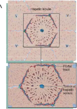
The adult liver is a voluminous organ (1200–1500 g), highly vascularized (Young et al., 2006), and it can be functionally divided into structures termed hepatic lobules. The hepatic lobule (Fig. 2A) is a polyhedral prism with its boundaries limited by six portal triads (Fig. 2B) prolonged by connective tissue. The portal triads contain the hepatic portal vein and the hepatic artery, the main blood vessels running into the liver, as well as a lymphatic vessel. The liver is therefore an unusual organ having both arterial and venous blood supplies. Besides the hepatic portal vein and the hepatic artery, the portal triad also contains a bile duct that transports bile away from the liver to be secreted and stored in the gallbladder. The centre of the lobule contains the terminal hepatic venule (centrolobular vein) that drains the blood from the liver. The portal triads are connected to the central veins by plates of hepatocytes separated by the sinusoids.
Fig. 2. The hepatic lobule is the smallest functional unit of the liver, a mass of liver parenchyma that is supplied by terminal branches of the portal vein and hepatic artery and drained by a terminal branch of the bile duct. (A) Schematic diagram showing the definition of a lobule, outlined by a hexagonal array of portal triads (T) arranged around a central hepatic venule (V). (B) Micrograph showing the overall structure of the human liver which is a solid organ composed of tightly packed plates of epithelial cells termed hepatocytes. In the human liver, a well-defined structural definition does not exists, although it can be seen the portal triads roughly defining a hexagon around the central hepatic vein. H & E staining (× 20). Adapted from Young et al. (2006).
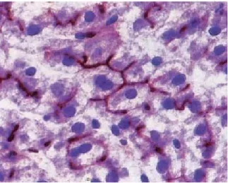
Hepatocytes, the chief parenchymal cells of the liver, are responsible for maintaining a wide range of specialized functions including storage, synthesis and detoxification/excretion of various molecules (Khan et al., 2007). These cells are large, multifaceted and polyhedral cells, arranged in plate-like cords separated by adjacent vascular sinusoids (Weibel et al., 1969). Within the hepatic cords, between adjacent hepatocytes, lies a network of bile canaliculi, allowing the passage of bile through intercellular channels, which drain into the nearest branch of the bile duct system (Fig. 3).
Fig. 3. Bile is secreted into a system of canaliculi which form a network within the hepatocyte plates. Bile then drain into the bile ductules of the portal tracts. The canaliculi are merely formed by the plasma membranes of adjacent hepatocytes. Enzyme histochemical method for ATPase of the bile canalicular membranes (stained in brown). Original magnification × 480. From Young et al. (2006).
This specialized architecture optimizes the liver’s parallel functions as an exocrine gland, an endocrine gland as well as a blood filter. Owing to the liver’s unique vascular organization, whereby blood percolates through the sinusoids from the place of inflow (the portal triad) to outflow (the terminal hepatic vein system), hepatocytes are exposed to a gradient of oxygen, nutrients, toxins and other biologically active molecules.
(Wake, 1995). Blood in the sinusoids is separated from hepatocytes by endothelial cells in the space of Dissé (Greenwel and Rojkind, 2001).
Fig.4. Micrograph demonstrating the main ultrastructural features of liver. Erythrocytes (E) can be seen within the liver sinusoids. Hepatocytes (H) are in contact to the sinusoids by a discontinuous layer of sinusoid lining endothelial cells (S). Space of Dissé (D) is located between the lining cells and the hepatocyte surface. Via the gaps in the sinusoid lining, the space of Dissé is continuous with the sinusoid lumen, thus bathing the hepatocyte surface with plasma. Numerous irregular microvilli extend from the hepatocyte surface into the space of Dissé, greatly increasing the surface area for metabolic exchanges. The hepatocyte cytoplasm is crowded with organelles, particularly mitochondrias and lysosomes, thus reflecting their range of biosynthetic and degradative activities. Lipid droplets (L) are present in variable numbers depending on nutritional status. Bile canaliculi (BC) are formed from the membranes of adjacent hepatocytes. From Young et al. (2006).
1.1.3. Liver functions
The liver is involved in synthesis of cholesterol, lipoproteins and phospholipids. It also oxidizes fatty acids to provide energy. Lipids and aminoacids are converted into glucose in the liver by gluconeogenesis. It synthesizes many proteins, including most of the plasma proteins such as albumin and blood clotting factors (for instance, fibrinogen and prothrombin). The liver is also the main site of detoxification of exogenous compounds such as drugs and toxins. The hepatocytes’ smooth endoplasmic reticulum possesses a large number of enzymes that breakdown and conjugate metabolites or toxic substances (e.g. alcohol, barbiturates, etc). This process, known as biotransformation, is able to convert lipophilic substances to more hydrophilic ones for subsequent elimination. Another major function of the liver is the production of bile, which is an alkaline secretion containing water, ions, phospholipids, bile acids and bile pigments (mainly bilirubin diglucuronide). Most attractive is the ability of the liver to regenerate, a unique property among solid organs in mammalian species. Following two-thirds partial hepatectomy, there is a compensatory growth by the remaining liver, resulting in restoration of the total parenchymal cell number and mass within 1-2 weeks (Michalopoulous and DeFrances, 1997). Liver transplantation is currently the only successful treatment for acute hepatic failure or end stage liver disease. At
the present time, however, a serious donor shortage is a major limitation to its use. Hepatocyte
transplantation may have the potential to solve this problem. Several studies using rat models
of primary hepatocyte transplantation revealed that transplantation leads to efficacious donor
chimerism that can rescue animals from lethal hepatic failure (Rajvanshi et al., 1996;
Gagandeep et al., 2000). Additionally, human cell lines have also been shown to improve the
survival rate in an acute liver failure model (Kobayashi et al., 2000). Hepatic stem cells from
fetal livers may also have the ability to repopulate the liver successfully and promote
long-term engraftment, given that they possess active proliferative capacity and the competence for
differentiate into hepatic and cholangiocytic lineages (Kakinuma et al., 2009a). It has been demonstrated that defined populations in mid-gestational fetal liver contain hepatic stem cells (Kakinuma et al., 2009b).
substances (drugs) and endogenous compounds (bile acids and bilirubin), and a cholestatic phase of liver development (physiological cholestasis) (Belknap et al., 1981; Suchy et al., 1981). In fact, evidence shows different patterns of perinatal hepatic enzymatic activity, which can affect the infant’s capacity for normal metabolic processes such as oxidation,
reduction, hydrolysis, and conjugation, therefore influencing its ability to metabolize,
detoxify, and excrete xenobiotics (Heubi et al., 1982; Emerick and Whitington, 2002). Some
of these differences may have relevance to understanding neonatal susceptibility to liver
disease.
1.2. Pathophysioly of cholestasis
The term “cholestasis” was generalized by Hans Popper in order to describe the retention of biliary constituents (Popper, 1981). Thus, cholestasis can also be defined as a decrease or cessation of canalicular bile flow that results in accumulation of bile components in hepatocytes and canaliculi (Elferink, 2003). This condition may result either from a functional defect in bile formation at the level of the hepatocyte (hepatocellular cholestasis) or from an impairment in bile secretion and flow at the level of bile ductules or ducts (extrahepatic cholestasis) (Trauner et al., 1999). Hepatocellular cholestasis may be caused by acute inflammation (hepatitis), cancer that has spread to the liver, inflammation or blockade of the bile ducts, genetic disorders, hormonal effects on bile flow during pregnancy (a condition called intrahepatic cholestasis of pregnancy) and/or drugs (Ling, 2007; Lee and Brady, 2009). On the other hand, causes of extrahepatic cholestasis are usually diseases of the bile ducts due to stones, abnormal narrowing of a bile duct (strictures) or tumors. Indeed, cholestasis is a common feature of many chronic human liver diseases leading to impaired bile formation and damage of target liver cells such as hepatocytes.
Cholestasis is also a frequent symptom of liver disease in newborns. The neonate develops cholestasis in response to a wide variety of insults, hepatic and systemic, indicating
a relative sensitivity of the mechanisms of bile formation and excretion compared with
children and adults.
The enterohepatic circulation in newborn animals of various species is characterized
by a decrease of bile acid secretion, bile flow, bile acid synthesis, bile acid pool size, uptake
of portal bile acids, and inefficient ileal uptake of bile acids (Balistreri et al., 1983). At birth,
basal bile acid secretion is decreased significantly compared with the mature animal and
humans, studies have also demonstrated that duodenal bile acids concentrations do not reach
the critical micellar concentration and are particularly low in premature infants, reflecting
immature bile acid secretion (Heubi et al., 1982; Suchy et al., 1987).
Strong evidence also shows that hepatocyte uptake of bile acids and of other anions
from the portal blood is decreased in the immature liver (Suchy et al., 1981; Suchy et al.,
1987). Also, bile acid synthesis is decreased in neonates compared with adults (Balistreri et
al., 1983). Altogether, decreased rate of bile secretion with decreased bile acid synthesis is
likely due to the immaturity of several steps of the bile acid synthetic pathway and enzymes
involved in bile acid conjugation (Subbiah and Hassan, 1982). Furthermore, major changes
occur during development in the volume densities of the cellular organelles that are involved
in bile acid metabolism. The volume density of the smooth endoplasmic reticulum is
markedly less in the neonate than in the adult rat liver and proliferates rapidly postnatally
(Rohr et al., 1971; Daimon et al., 1982).
The immaturity of the bile acid synthetic pathway of normal infants is also evidenced
by the presence of ‘‘atypical’’ bile acids in the meconium and stool of infants. These
‘‘atypical’’ bile acids are characterized by multiple hydroxylations and completely novel
species. Some of these species may be hepatotoxic (Back and Walter, 1980; Strandvik and
Wikstrom, 1982).The presence of potentially hepatotoxic bile acids could be a potentiating
factor that may cause amplification of any cholestatic process in the infant.
Moreover, the developing liver also has immature mechanisms for hepatoprotection as
animal studies reveal that detoxification mechanisms, such as sulfation, are not fully
developed at birth (Balistreri et al., 1984; Suchy et al., 1985), and glucuronidation is also
reduced in the developing liver (Klinger, 1982). The immaturity of these processes may
theoretically play a role in the pathophysiology of cholestasis during childhood.
1.2.1. Main clinical biomarkers of chronic cholestasis: from diagnosis to prognosis
1.2.1.1. Bile acids
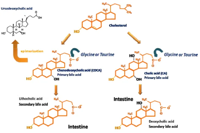
Bile acids, the major constituents of bile, are synthesized from cholesterol by a complex series of chemical reactions (for review see Björkhem, 1985; Russell and Setchell, 1992; Setchell and Russell, 1994). As represented in Figure 5, primary bile acids are synthesized from cholesterol by the addition of hydroxyl groups and the oxidation of its side chain to form a more water soluble end product. The two primary bile acids synthesized in humans and most animal species are cholic acid (CA) and chenodeoxycholic acid (CDCA). Prior to secretion and storage in the gallbladder bile, CA and CDCA are conjugated to the aminoacids glycine and taurine at a 3:1 ratio (Chiang, 2003), resulting in glycocholic acid (GCA) and taurocholic acid (TCA) or glycochenodeoxycholic acid (GCDCA) and taurochenodeoxycholic acid (TCDCA), respectively. Conjugation significantly alters the physiochemical properties of bile acids, markedly increasing the polarity of the molecule and, thereby, facilitating renal excretion (Hofmann and Roda, 1984). Furthermore, the greater hydrophilicity of the conjugated species minimizes the membrane-damaging potential of the more hydrophobic unconjugated species (Scholmerich et al., 1984).
Fig. 5. Schematical representation of the biochemical pathways involved in the conversion of cholesterol to bile acids. Cholesterol is transformed into the “primary” bile acids cholic and chenodeoxycholic acids by a complex series of chemical reactions. Prior to secretion and storage in gallbladder bile, cholic and chenodeoxycholic acids are conjugated at C-24 to the aminoacids glycine and taurine. The bile acids referred to as “secondary”, the lithocholic acid and the deoxycholic acid are formed from chenodeoxycholic acid and cholic acid respectively, in the intestine. Chenodeoxycholic acid can also undergo 7β-epimerization to originate the tertiary ursodeoxycholic acid, used in the treatment of hepatobiliary disorders. Although the described pathways are considered the most important for bile acid synthesis in humans, there are several alternative pathways (for review, see Setchell and Russell., 1994).
unknown once fetuses do not contain intestinal bacteria, it has been suggested that they are derived either from maternal bile via transplacental transport or from primary synthesis through an alternative pathway (Balistreri, 1991).
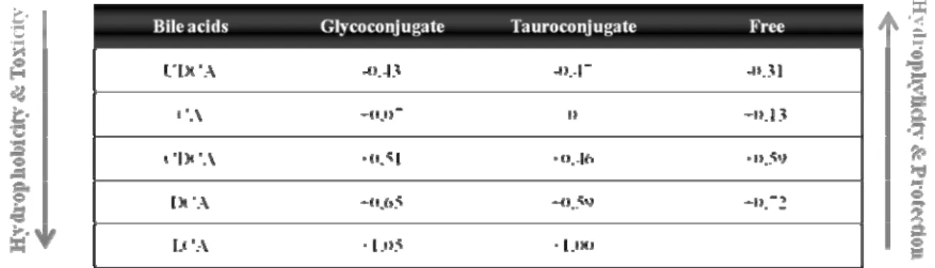
It is worth noting that not all bile acids are toxic and previous studies suggest that this may be related to slight changes in their chemical structure (Hofmann and Roda, 1984). Hydrophobicity is an important determinant of the toxicity and protection of bile acids, two biological properties of these compounds. Bile acids hydrophobicity depends on the number, position and orientation of the hydroxyl groups, as well as amidation at the C-24 position. Therefore, the magnitude of bile acids hydrophobicity and consequently their toxicity are UDCA < CA < CDCA < DCA < LCA as ilustrated in Table 1 (Carulli et al., 2000). Thus, UDCA is the more hydrophilic and the most universally used in the treatment of hepatobiliary disorders (Beuers and Paumgartner, 2002). Since conjugation with taurine and glycine increases their hydrophilicity, the conjugated species also show increased protective properties.
Table 1. Hydrophobic index of bile acids and their conjugated species.
Adapted from Heuman (1989).
hepatocytes as several studies in models of cholestasis have demonstrated mitochondrial dysfunction and caspase activation (Rodrigues et al., 2003; Maher, 2004).
1.2.1.2. Bilirubin
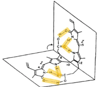
Bilirubin synthesis starts with the lysis of senescent or hemolyzed erythrocytes in the reticuloendothelial system. When erythrocytes are degraded, heme is released from hemoglobin and following its catabolism unconjugated bilirubin (UCB) is produced (Berk, 1994). This molecule presents a nearly symmetrical tetrapyrrolic structure, consisting of two rigid planar dipyrrole units (dipyrrinones) joined by a methylene bridge at carbon 10 and stabilized by intracellular hydrogen bonds (Fig. 6).
Fig. 6. Structure of unconjugated bilirubin (UCB). The molecule consists of two rigid, planar dipyrrole units joined by a methylene (-CH2) bridge at carbon 10, and is stabilized by hydrogen bonds (highlighted in yellow). Adapted from Ostrow et al. (1994).
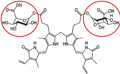
glutathione S-transferase) or protein Z during times of increased bilirubin load to the liver, to be carried into the endoplasmic reticulum for conjugation (Brito et al., 2006). Conjugation occurs inside the smooth endoplasmic reticulum, where each molecule of bilirubin combines with one or two molecules of glucuronic acid by the enzyme uridine diphosphate (UDP)-glucuronosyl transferase to produce bilirubin monoglucuronide and diglucuronide pigments (Fig. 7). This conjugation renders a higher solubility in aqueous medium to the UCB molecule.
Fig. 7. Structure of bilirubin diglucuronide. The pigment structure remains the same with the addition of two molecules of glucuronic acid (highlighted with red circles). Adapted from http://en.wikipedia.org/wiki/Bilirubin_diglucuronide.
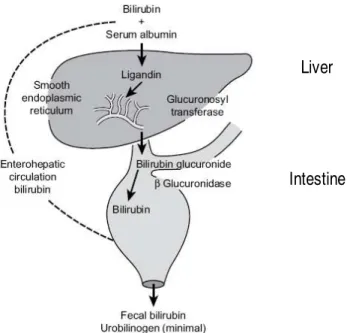
Liver
Intestine
Fig. 8. Bilirubin uptake, transport and secretion by the liver, followed by intestinal excretion. Adapted from Watson (2009).
In fetal life, bilirubin production begins as early as 12 weeks’ gestation. In children and adults, approximately two thirds of the monoglucuronides are conjugated to diglucuronides. However in neonates, monoglucuronide is the predominante conjugate specie. Owing to the immaturity of the fetal liver, the fetus has a limited ability to conjugate bilirubin and limited excretory function, and therefore physiologically relevant hepatobiliary elimination of bilirubin does not occur (Briz et al., 2006). The circulating fetal UCB readily crosses the placenta to the maternal circulation, where it is excreted by the maternal liver. However, the concentrations of UCB are higher in fetal than in maternal serum (Monte et al., 1995). Two facts contribute to this difference: a very active heme catabolism together with a very low UDP-glucuronosyl transferase activity in the fetal liver (Kawade and Onishi, 1981). Thus, during intrauterine life, the placenta is the major route for the excretion of fetal biliary pigments (for a review, see Marin et al., 2003).
intestinal absorption, thus increasing the enterohepatic circulation of UCB (Vitek et al., 2000). As a result, virtually all newborn infants will have mild to moderate UCB levels within the first days of life, a condition known as “physiologic jaundice”. Therefore, this neonatal jaundice reflects the transition from intrauterine to extrauterine bilirubin metabolism and is linked to normal development; it is considered benign, and is usually resolved by the end of the first week of life with no treatment requirement (Reiser, 2004). However, it is important to confirm that plasma bilirubin is reducing after 14 days (Beath, 2003). The duration of exposure to overstated hyperbilirubinemia is believed to represent increased risks for neurologic sequelae (Dennery et al., 2001; Hansen, 2002) and is one of the most common factors related with the readmission of term and near-term infants (Brown et al., 1999).
Yellow coloration of skin and eyes, dark urine and light-colored stools are also characteristic symptoms of cholestatic or obstructive jaundice. Jaundice results from excess bilirubin deposited in the skin and dark urine results from excess bilirubin that accumulates in systemic circulation and is excreted by the kidneys. Usually, during cholestasis, jaundice occurs as a consequence of insufficient bile flow (Elferink, 2003).
UCB and CB have also been demonstrated to promote hepatocyte and canalicular toxicity. Indeed, both species have been implicated in the inhibition of biliary phospholipid secretion (Labori et al., 2002; Labori et al., 2009), while UCB showed to induce canalicular membrane damage and consequently to promote intrahepatic cholestasis (Labori et al., 2009), which may aggravate the already established cholestatic condition. In accordance, elevation of serum bilirubin concentration is now considered a poor prognosis indicator in acute liver failure (Hadem et al., 2008) and primary biliary cirrhosis (Krzeski et al., 1999).
1.2.2. Acute vs. chronic cholestasis
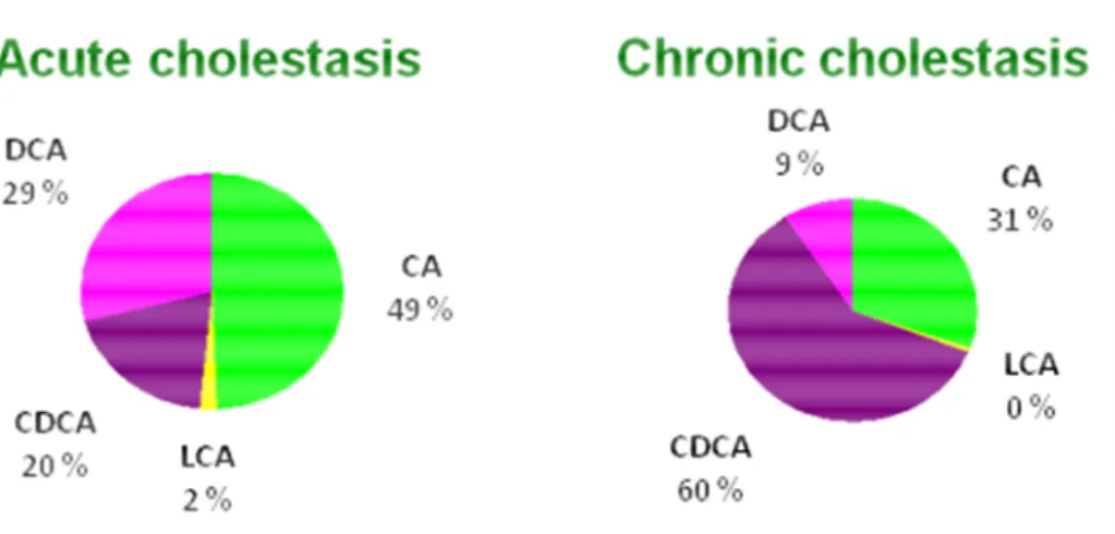
Fig. 9. Pie charts demonstrating the distribution of each bile acid during acute and chronic cholestasis (Brites, personal communication). CA, cholic acid; CDCA, chenodeoxycholic acid; DCA, deoxycholic acid; LCA, lithocholic acid.
Hepatocyte damage by toxic bile acids is assumed to represent a key event for progression of cholestatic liver diseases (Hofmann, 2002). In this regard, the GCDCA and the TCDCA, predominant dihydroxy bile acids present in chronic cholestatic patients due to conjugation of CDCA, have been held responsible for cholestasis associated liver injury (Schmucker et al., 1990). Exposure of hepatocytes to GCDCA, at concentrations representative of those found in cholestatic human liver injury, are thought to induce hepatocyte necrotic and apoptotic cell death (Patel et al., 1994; Gonzalez et al., 2000; Yerushalmi et al., 2001). Moreover, engulfment of the hepatocyte apoptotic bodies by hepatic stellate cell (HSC) and Kupffer cells enhances their expression of pro-fibrogenic genes and death ligands. Persistent activation of these cells promotes further hepatocyte death, which culminates in hepatic inflammation, with sustained HSC activation. If liver injury continues chronically, hepatic fibrosis develops as a result of the activation of HSC, which are the main cellular elements involved in extracellular matrix deposition (Friedman, 2000).
1.3. Mechanisms of hepatocyte injury during cholestasis
1.3.1. Major modes of cell death in the liver
The balance between cell division and cell death is a basic feature in the development and maintenance of liver homeostasis. Disturbances in this balance can cause liver diseases: too much cell death can cause liver injury; too little cell death is a prerequisite for the development of hepatocellular carcinoma. Thus, a tight control of the equilibrium between life and death in the liver is necessary.
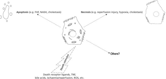
During cholestatic liver diseases, hepatocytes are exposed to increased levels of cytotoxic compounds like bile acids, bilirubin or even cytokines if inflammation is present. Although hepatocytes have an enormous capacity to defend themselves against these agents, excessive exposure will result in cell death (Schoemaker and Moshage, 2004). Cell death is typically discussed dichotomously as either apoptosis or necrosis (Fig. 10), although recently there have been described alternate types of cell death (for review see Fink and Cookson, 2005).
The word apoptosis was proposed by Kerr et al. (1972) to describe a controlled physiologic process of removing individual unnecessary components of an organism without destruction or damage to the organism. Apoptosis was initially confirmed as a specific form of cell death that served to eliminate excessive or unwanted cells during embryonic development and normal tissue growth (Williams, 1991), but had been clarified to be also induced in cellular injury with inflammatory disease (Haslett, 1992).
Samali et al. (1999) and others (Blagosklonny, 2000) have proposed that apoptosis should be defined as caspase (asparate-specific cysteinyl protease)-mediated cell death with the following morphological features: cytoplasmic and nuclear condensation, chromatin cleavage, formation of apoptotic bodies, maintenance of an intact plasma membrane, and exposure of surface molecules targeting cell corpses for phagocytosis. More specifically, the molecular definition of apoptosis can logically be based on the proteolytic activity of some caspases (caspase-2, -3, -6, -7, -8, -9, and -10) since they mediate the process of apoptotic cell death. Among them, caspase-3 has been identified as being a key mediator of apoptosis of mammalian cells (Samali et al., 1999). Caspase-3 is one of the effectors’ caspases from apoptosis that are activated by upstream initiator caspases and are responsible for the cleavage of the key cellular proteins, such as cytoskeleton proteins, that leads to the typical morphological changes observed in cells undergoing apoptosis.
Although apoptosis of hepatocytes can be triggered by several different stimuli, apoptotic signaling is mainly transduced by two major molecular pathways, an extrinsic pathway mediated by death receptors (i.e. TNF-α/TNFR1 signaling) on the cell surface and an intrinsic pathway, which is triggered at the mitochondrial level. Both pathways culminate in the activation of caspases and endonucleases, which ultimately degrade the cellular constituents. Deregulation of the apoptotic program is pathophysiologically involved in acute as well as in chronic liver diseases, including cholestasis (Miyoshi et al., 1999).
consequences for the cell (Majno and Joris, 1995). It results from metabolic disruption with energy depletion and loss of adenosine triphosphate (ATP), ion deregulation and enhanced degradative hydrolase activity.
The term necrosis has been generally used to portray non-apoptotic accidental death with features of oncosis. However, the distinction between the structural and biochemical processes occurring in a dying cell and the endpoint of death itself is a subject that has been disregarded in the literature and must be clearly distinguished. Pathologists use the word necrosis to designate the presence of dead tissue or cells, being considered the sum of changes that have occurred in cells after they died, regardless of the prelethal process (Levin et al., 1999). Necrosis, therefore, involves cell destruction with extravasation of intracellular components, an event observed after a cell has already died. By definition, this is manifested biochemically as the release of cytosolic enzymes including lactate dehydrogenase (McCarthy and Evan, 1998). The release of cellular contents triggers an inflammatory response in the surrounding tissue (Kerr et al., 1972).
Multiple types of death can be observed simultaneously in tissues or cell cultures exposed to the same stimulus (Fink and Cookson, 2005). An understanding of the processes leading to liver cell death will be important for development of effective interventions to prevent hepatocellular death and consequent liver failure (Malhi et al., 2006).
1.3.2. Cell death and the development of inflammation
associated with their activation and with production of transforming growth factor (TGF)-β, a potent profibrogenic cytokine, ultimately leading to fibrosis (Canbay et al., 2003).
1.3.3. Inflammatory signaling pathways
The hepatocyte, early injured in the course of cholestasis, is in part responsible for many of the subsequent inflammatory and fibrinogenic responses of nonparenchymal cells. Damaged hepatocytes may secrete molecules (cytokines, chemokines, growth factors, lipid peroxide products, etc.) that amplify the inflammatory response, stimulate fibrogenesis by HSC, or directly injure other nearby cells. Thus, understanding the events that initiate liver injury during cholestasis should focus to a large extent on the hepatocyte and the effects of toxic compounds such as bile acids and bilirubin on hepatocyte response and survival (Sokol
et al., 2006).
Cytokines are multifunctional pleiotropic proteins that play crucial roles in cell-to-cell communication and cellular activation. Functionally, cytokines have been classified as being either proinflammatory or anti-inflammatory depending on the final balance of their effects on the immune system (Mosmann et al., 1986). They are mediators that initiate multiple signaling pathways that, although independent, may interact with each other and influence the magnitude and duration of the inflammatory response. In some cases, different cytokines may present synergistic, redundant or even opposite actions. In healthy liver, constitutive production of cytokines is absent or very low. Irrespective of its etiology, inflammation-induced cholestasis is mediated by cytokines (Trauner et al., 1999), and hepatocytes are exposed to increased levels of cytokines such as tumor necrosis factor alpha (TNF-α) and various interleukins (IL) (Wullaert et al., 2007). In vivo studies have shown that bile acids are capable of inducing Kupffer cells to release proinflammatory cytokines and subsequently affect transcriptional alterations in the neighboring parenchymal cells (Miyake et al., 2000).
cytokines (Kyriakis and Avruch, 2001). MAPKs activation is catalyzed by members of the MAPK kinase family such as the MAP kinase kinase kinases (MEKKs) and it consists in the phosphorylation of tyrosine and threonine residues (Cobb and Goldsmith, 1995). Once activated, MAPKs phosphorylate target substrates including some transcription factors or other molecules involved in transcription factors activation such as the IκB/NF-κB signaling (Johnson and Lapadat, 2002).
MAPK cascade selectivity is conferred by specific interaction motifs located on physiological substrates, allowing distinct biological functions for each activated MAPK. Thus, the p38 activity is considered critical in normal immune and inflammatory responses (Ono and Han, 2000), whereas JNK1/2 phosporylation is associated with apoptosis (Davis, 2000). Of 3 known mammalian JNK genes, 2 are expressed in the liver: JNK1 and JNK2 (Czaja, 2003). Both can be activated by death receptor and endoplasmic reticulum stress pathways of apoptosis and may also be the pathway of caspase-independent reactive oxygen species–mediated cell death (Malhi and Gores, 2008).
The MAPK pathways have been shown to be strongly activated after partial hepatectomy and presumably play a key role in regulating hepatocytes proliferation during hepatic regeneration (for review see Fausto, 2000). Interestingly, activation of MAPK pathways also precedes the process of HSC proliferation and activation that is associated with tissue remodeling and leads to hepatic fibrosis (Svegliati-Baroni et al., 2003).
The general function of MAPK cascades is the regulation of gene expression. In this way, MAPKs regulate cell proliferation and cell survival, but also mediate cell death. However, the actual roles of each MAPK cascade are cell-type and context-dependent. Importantly, studies in non-proliferating cells or primary cultures are scarce and rather reveal the physiological role of MAPKs.
Besides MAPKs, other signaling pathways modulate cell death in hepatocytes, thereby influencing the balance between pro- and anti-apoptotic signals. One of them is the NF-κB signaling cascade (Schoemaker and Moshage, 2004).
the translocation of NF-κB to the nucleus. Phosphorylated IκB is consequently ubiquitinated and degraded in the proteasomes. In the nucleus, NF-κB binds to κB binding sites in promoters of target genes and induces transcription of these genes. Many NF-κB-regulated genes are survival or antiapoptotic genes that protect cells against harmful compounds released during inflammation (Schoemaker et al., 2003). NF-κB-inducible anti-apoptotic genes expressed in hepatocytes are prime candidates for novel therapies in liver diseases (Schoemaker et al., 2002).
A coordinate activation of these pathways, ordered in space and time, orchestrates the complex response to injury by inducing genes that regulate cell survival, proliferation, differentiation and tissue specific functions. On this basis, pharmacological or molecular modulation of intracellular kinases and NF-κB have been under consideration as an approach to therapy of neoplastic as well as non-neoplastic conditions (Sebolt-Leopold et al., 1999; Zhu et al., 1999; Sebolt-Leopold, 2000).
1.4. Aims of the thesis
Numerous studies have investigated the mechanisms and pathways of liver damage after exposure to bile acids mimicking a situation of cholestasis. However, the stimulation of these mechanisms associated with hiperbilirrubinemia has not been considered in most of the
in vitro studies. In addition, the study of hepatotoxic mechanisms seldom considers the fetal/neonatal conditions.
Thus, the main aims of this project are: (a) to evaluate the role of GCDCA alone in human hepatocyte injury; (b) to investigate the effects of the additional presence of UCB and CB on human hepatocyte response and cytotoxicity; (c) to assess the involved intracellular pathways leading to injury in our experimental models; and (d) to explore the reasons behind the different susceptibility of fetal and adult hepatic cells to jaundice, cholestasis or both.
2. MATERIALS AND METHODS
2.1. Chemicals
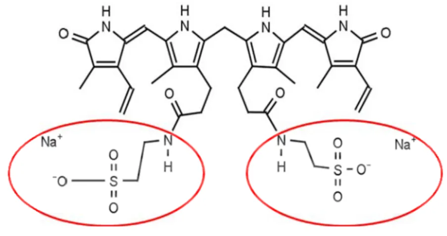
Dulbecco’s modified Eagle’s medium (DMEM), fetal bovine serum (FBS), non-essential aminoacids (100×) and L-glutamine were purchased from Biochrom AG (Berlin, Germany). Glycochenodeoxycholic acid (GCDCA) [N-(3α,7α-dihydroxy-5β-cholan-24-oyl) glycine] minimum 96% pure was from Calbiochem (Darmstadt, Germany). Antibiotic antimycotic solution (20×), human serum albumin (HSA) (fraction V, fatty acid free), bovine serum albumin (BSA), trypsin, Hoechst dye 33258, mouse antibody anti-β-actin and goat antibody anti-rabbit labeled with fluorescein isothiocyanate (FITC) were acquired from Sigma Chemical Co (St. Louis, MO, USA). Unconjugated bilirubin (UCB), also from Sigma Chemical Co, was purified according to the method of McDonagh and Assisi (1972). Bilirubin ditaurate [ditaurine amide of bilirubin (disodium salt)] showed in Figure 11, was purchased from Frontier Scientific (Logan, UT, USA) and used as conjugated bilirubin (CB) as described previously by Kajihara et al. (2000), Labori et al. (2002) and (2009), since it is the only conjugated species of bilirubin that is commercially available.
Fig. 11. Bilirubin ditaurate, resulting from the conjugation of one molecule of bilirubin with two molecules of taurine amide sodium salt (red circles). Adapted from http://www.frontiersci.com/detail.php?FSIcat=B850. .
Biosciences (Piscataway, NJ, USA). LumiGLO® was acquired from Cell Signalling (Beverly, MA, USA). Sodium dodecyl sulphate (SDS) was acquired from VWR-Prolabo. Acrylamide, bis-acrylamide and Tween-20 were from Merck (Darmstadt, Germany). Primary specific monoclonal antibodies were: rabbit anti-phospho-p38 MAPK (P-p38) from Cell Signaling; mouse anti-phospho-JNK1/2 (P-JNK1/2) and rabbit anti-p65 NF-κB subunit from Santa Cruz Biotechnology (Santa Cruz, CA, USA). Depex-Polystyrene dissolved in xylene (DPX) mountant for microscopy was obtained from BDH, Laboratory Supplies, Poole, UK. Caspase-3 substrate was purchased from Calbiochem (San Diego, CA, USA). Horseradish peroxidase-labelled goat anti-rabbit IgG and anti-mouse IgG were from Santa Cruz Biotechnology (Santa Cruz, CA, USA). Protein assay kit (for protein quantification) was from Bio-Rad Laboratories (Hercules, CA, USA). 75-cm2 flasks as well as 6-well and 12-well tissue culture plates were from Orange Scientific (Braine-l'Alleud, Belgium).
2.2 Equipment
Axioskop® microscope was obtained from Zeiss, Germany. The phase contrast microscope, model CK2-TR, was from Olympus Optical Co. Ltd. Western blot apparatus and spectrophotometer PR 2100 were purchased from Bio-Rad Laboratories (Hercules, CA, USA).
2.3. Fetal and adult hepatocyte cell culture
WRL-68, a human epithelial fetal liver cell line, was first deposited in the American Type Culture Collection, Rockville, MD (ATCC accession number CL48) by Apostolov, who also registered its patent (USA patent no. 3 935 066) in 1976. The patent states that WRL-68 cells: (i) form individually separated islands on discrete clumps when cultured in a growth medium; (ii) have a morphology closely resembling that of hepatocytes of the human liver; (iv) show increased production of glycogen in the presence of 1% glucose in the medium; (v) are capable of supporting viruses for the preparation of viral vaccines; and that (vi) their generation time is not more than 24 h.
Fig. 12. Typical polygonal arrangement of the epithelial cells WRL-68 observed under a phase contrast microscope. When WRL-68 cells are seeded at low density, polygonal-to-spindle shape and some rounded cells are detected. At higher magnification these cells exhibit prominent, round, or oval-shaped nuclei containing one or more nucleoli. The cytoplasm appears to be granular and dense. They do not grow as a monolayer and began to detach before they cover the entire available surface for the culture plate. Their morphologic characteristics and epithelial cell shape are compatible with those of liver parenchymal cells. Scale bar = 50 µm.
The synthesis of liver-specific serum proteins, particularly albumin and AFP, has been the benchmark for considering whether differentiated functions in hepatocytes cultures had been preserved. Thus, WRL-68 ability to produce albumin and AFP indicates that they retain functions of normal liver parenchymal cells. In addition, the expression of AFP has been described to be also a characteristic of fetal or cancerous liver cells. In this context, WRL-68 cells have been described as a fetal cell line (Gutiérrez-Ruiz et al., 1994). In resume, since WRL-68 cells maintain their fetal hepatic properties, they are considered an useful system for the study of hepatic functions and development in vitro (Gutiérrez-Ruiz et al., 1991; Gutiérrez-Ruiz et al., 1992). The WRL-68 cell line used in the present study was purchased from the European Collection of Cell Cultures (ECACC), catalogue no.89121403, lot no.04A010.
oncoproteins, that are known to immortalize human epithelial cells (Clayton et al., 2005). These cells retain primary hepatocyte healthy phenotype, suggesting the maintenance of a large degree of hepatic function without the presence of tumorigenic characteristics (Clayton
et al., 2005). In agreement, AFP, a marker associated with a fetal or tumorigenic phenotype, was either absent or expressed at low levels in the HHLs confirming its origin from a non-neoplastic tissue. HHLs also present effective contact inhibition (Clayton et al., 2005), a trait lost in tumour cells. Moreover, the observation that these cells can be maintained in a monolayer status for a considerable period of time (in this case, 7 days) could indicate that they would be of value in repopulating a damaged or depleted liver, without the generation of metastatic tumours (Clayton et al., 2005). We must take into account that immortalized cell lines de-differentiated somewhat in culture after many passages, resulting in expression of AFP and other tumour-related proteins (Woodworth et al., 1988). However, HHLs have not shown the presence of AFP even after 80 passages (Clayton et al., 2005). The HHL-5 cell line used in the present study was kindly provided by Dr. Arvind Patel from Institute of Virology, University of Glasgow, UK.
Fig. 13. The arrangement of the HHL-5 cell line was observed using a phase contrast microscope. HHL-5 morphology is compatible with those of liver cells, presenting a typical polygonal arrangement of epithelial cells but, contrary to WRL-68, this cell line grows as a monolayer. Scale bar = 50 µm.
WRL-68 and HHL-5, respectively) of non-essential aminoacids (NEA) and 1% antibiotic/antimycotic solution. The cells were trypsinized when cultures were sub-confluent (70-80%) using 0.5% trypsin and seeded at a density of 2.0 × 10⁵ cells/mL on 75-cm2 flasks. For the experimental studies, cells were seeded at a density of 1.0 × 10⁵ cells/mL either on 6-well tissue culture plates or on glass coverslips placed in 12-6-well tissue culture plates and maintained at 37ºC in a humidified atmosphere of 5% CO2 for 24 h prior to treatment. All the cells used in this work were between passages 5 and 15.
2.4. Hepatocyte treatment with bilirubin and bile acid species
Hepatocytes were stimulated with 100 µM GCDCA, 100 µM CB, 100 µM UCB, 100 µM GCDCA + 100 µM CB + 100 µM UCB or vehicle alone, in the presence of 100 µM HSA from 1 to 48 h, at 37ºC as described in Figure 14. A concentrated solution of 5 mM GCDCA was prepared in sterile phosphate-buffered saline (PBS) and appropriate dilution in the incubation medium was made. Concentrated solutions of 10 mM CB and 10 mM UCB were prepared in 0.1 M NaOH immediately before use and appropriate dilutions were made in the incubation medium restoring the pH to 7.4 by addition of equal amounts of 0.1 M HCl. All the experiments with CB and UCB were performed with light protection (vials wrapped in tin foil and dim light) to avoid photodegradation.
Fig. 14. Schematic representation of the experimental design. Cultured hepatocytes were incubated with 100 µM GCDCA, 100 µM CB, 100 µM UCB, 100 µM GCDCA + 100 µM CB + 100 µM UCB (all in the presence of 100 µM HSA) or vehicle alone (100 µM HSA) from 1 to 48 h, at 37ºC. After 1, 4, 8 and 24 h of treatment, MAPKs activation was evaluated by western blot assay. At 6, 12, 24 and 48 h after treatment we assessed caspase-3 activity using a specific substrate. At 12, 24 and 48 h after incubation we appraised cellular parameters as LDH release and MTS reduction. The estimation of apoptotic nuclei by Hoescht staining and evaluation of NF-κB translocation to the nucleus by immunocytochemistry was also made after 12, 24 and 48 h incubation.
2.5. Cytotoxicity evaluation
Standard evaluation of cytotoxicity was performed by measuring: (i) the lactate dehydrogenase (LDH) released by nonviable cells; (ii) the apoptotic cell death either by determining the activity of caspase-3, a known effector caspase (Samali et al., 1999) or the number of apoptotic nuclei; and (iii) the ability of viable cells to reduce the MTS compound.
2.5.1. LDH release
incubation medium was transferred into corresponding wells of a 96-well microplate. Then 100 µL of the reaction mixture [catalyst solution (Diaphorase/NAD+ mixture) plus dye solution (iodotetrazolium chloride and sodium lactate), in a proportion of 1:45] was added to each well and the plate was incubated for 10 min at 15-25°C. At the end of the incubation the reaction was stopped by adding 50 µL per well of 1 N HCl and the absorbance measured at 490 nm with a reference wavelength of 620 nm. All readings were corrected for the possible interference of UCB and CB absorption and the results expressed as percent of LDH release, obtained by treating nonincubated cells with 2% Triton X-100 in DMEM for 30 min.
2.5.2. Apoptosis assessment
The caspase-3 activity was assayed using the caspase-3 substrate, according to the manufacturer´s instructions. In brief, following incubation, the medium was discarded and adherent cells were harvested in chilled cell lysis buffer [50 mM HEPES (pH 7.4); 100 mM NaCl; 0.1% (w/v) CHAPS; 1 mM DTT; 0.1 mM EDTA] following a 30 min incubation on ice. Cell lysate was centrifuged at 10,000×g for 10 min and supernatant transferred to a new eppendorf. Then, 20 µL of sample supernatant was transferred into corresponding wells of a 96 well assay plate and 80 µL of the reaction mixture containing 0.2 mM of Ac-DEVD-pNA, a specific substrate of caspase-3, in protease buffer assay [50 mM HEPES (pH 7.4); 100 mM NaCl; 0.1% (w/v) CHAPS; 10 mM DTT; 0.1 mM EDTA; 10% (v/v) glycerol] was added, and incubated at 37°C. The amount of pNA released by enzyme reaction was measured at 405 nm every 30 min until 2 h. The absorbance results obtained for each sample were normalized to protein concentration measured in cell lysate supernatant using the protein assay kit according to the manufacturers’ instructions, and presented as fold change versus control.
described by Kerr and collaborators (Kerr et al., 1972). Results are expressed as the percentage of apoptotic cells.
2.5.3. MTS reduction
The ability of viable cells to reduce the MTS compound was evaluated as previously described (Riss and Moravec, 1992). In brief, after the treatment, incubation media were removed and cells were incubated for 1 h, at 37°C, with 500 µL of the reaction mixture containing 0.2 mg/mL MTS plus 45 µg/mL PMS in DMEM. At the end of incubation, 100 µL of media was transferred into corresponding wells of a 96 well assay plate and absorbance measured at 490 nm. Results were expressed as percentage of control.
2.6. Western blot assay
Phosphorylation of p38 and JNK1⁄2 was analyzed following 1, 4, 8 and 24 h treatment.
Total cell extracts were obtained by lysing cells in ice-cold cell lysis buffer [20 mM Tris-HCl (pH 7.5), 150 mM NaCl, 1 mM EDTA, 1 mM EGTA, 1% Triton X-100, 2.5 mM sodium pyrophosphate, 1 mM b-glycerophosphate, 1 mM Na3VO4, 1 µg/mL Leupeptin, 1 mM PMSF] for 5 min on ice followed by sonication. The lysate was centrifuged at 14 000 g for 10 min at 4ºC, and the supernatants were collected and stored at -80ºC. Protein concentrations were determined using a protein assay kit, according to the manufacturer’s specifications. Equal amounts of protein (50 µg) were subjected to SDS–polyacrylamide gel electrophoresis and transferred to a nitrocellulose membrane. Membranes were washed with Tris-buffered saline containing Tween 20 (T-TBS; 10 mM Tris-HCl, 150 mM NaCl, 0.1% Tween 20) and blocked for 1 h at room temperature (22–25ºC) in blocking buffer [T-TBS plus 5% (w/v) non-fat dried milk]. Membranes were incubated with primary antibody overnight at 4ºC [rabbit anti-P-p38 MAPK (1:1000), mouse anti-P-JNK1/2 (1:500), or mouse anti-β-actin (1:10 000) in 5% (w/v) bovine serum albumin]. After repeated washes in T-TBS, the membranes were incubated with horseradish peroxidase-labelled secondary antibody [anti-rabbit (1:5000) and anti-mouse (1:5000) in 5% (w/v) non-fat milk], for 1 h at room temperature. Protein bands were detected by LumiGLO® and visualized by autoradiography with Hyperfilm ECL. The
relative intensities of protein bands were analyzed using the Quantity One® 1-D densitometric
2.7. Immunocytochemistry
Nuclear translocation of NF-κB was assessed following 12, 24 and 48 h treatment by NF-κB immunostaining performed as usual in our laboratory (Fernandes et al., 2006). In brief, fixed cells on coverslips were permeabilized using blocking buffer [1% (w/v) BSA and 0.4% (v/v) Triton x-100 in PBS] for 1 h at room temperature and primary antibody (polyclonal rabbit anti-p65 NF-κB subunit (1:200) in blocking buffer] incubated overnight at 4°C. Cells were then incubated with FITC-labeled goat anti-rabbit antibody (1:160) as the secondary antibody for 1 h at room temperature, washed with PBS, and mounted as previously described (2.4.3). To identify the total number of cells, hepatocyte nuclei were stained with Hoechst dye 33258 as above mentioned. Fluorescence was visualized using a Leica DC 100 camera adapted to an Axioskop® microscope. Pairs of U.V. and green-fluorescence images of 10 random microscopic fields (original magnification: 400×) were acquired per sample. NF-κB-positive nuclei, was identified by localization of the NF-κB p65 subunit staining exclusively at the nucleus, and total cells were counted (~400 cells per sample) to determine the percentage of NF-κB-positive nuclei.
2.8. Statistical Analysis
3. RESULTS
3.1. Decreased viability of hepatocytes is induced by bilirubin and bile acid species,
mainly at 48 h of incubation
As previously reported, GCDCA induce cytolysis of rat hepatocytes, releasing the lactate dehydrogenase enzyme from the cytoplasm to the surrounding media (Spivey et al., 1993; Benz et al., 1998; Yerushalmi et al., 2001). To investigate the ability of GCDCA, CB, UCB and GCDCA+CB+UCB to induce membrane disruption in our culture of fetal (WRL-68) and adult (HHL-5) hepatocyte cell lines , cells were treated for periods of 12, 24 and 48 h, and the levels of LDH activity in the incubation media were determined. As demonstrated in Figure 15, the release of LDH by HHL-5 cells remained unchanged after 12 h and 24 h incubations, while it significantly increased after 48 h incubation but only for GCDCA+CB+UCB treatment (P<0.01). Interestingly, this value was markedly different when compared to GCDCA, CB or UCB incubation alone (P<0.05). Relatively to WRL-68 cells (Fig. 16), the release of LDH also increased significantly after 48 h incubation with GCDCA+CB+UCB when compared to control (P<0.05). Once again, these values were significant when compared to GCDCA (P<0.05) and UCB (P<0.01) incubations.
Fig. 16. Incubation of WRL-68 cells with glycochenodeoxycholic acid (GCDCA) + conjugated bilirubin (CB) + unconjugated bilirubin (UCB) induces cytolysis at 48 h. Hepatocytes were treated with 100 µM GCDCA, 100 µM CB, 100 µM UCB, 100 µM GCDCA+CB+UCB, in the presence of 100 µM human serum albumin, or vehicle alone (Control) for the indicated time periods. The incubation medium was collected for determination of released lactate dehydrogenase (LDH). Data are means ± SEM from four independent experiments. **P<0.01 vs. control, *P<0.05 vs. control, §P<0.05 vs. GCDCA, ††P<0.01 vs. CB.
Comparing the two cell lines at 48 h (controls, Fig 15 and 16), HHL-5 showed an increased susceptibility to cytolysis. In addition, all treatments induced a significantly higher LDH leakage to the media in HHL-5 cell line. By evaluating the relative answer of the two cell lines (normalizing to the controls) at 48 h upon bilirubin and bile acid species treatment (Table 2), WRL-68 showed less respond to GCDCA and CB, while slightly more vulnerable than HHL-5 to UCB and GCDCA+CB+UCB. These findings indicate that HHL-5 may be more prone to cytolysis in an obstructive jaundice and that WRL-68 may be, to some extent, more sensitive to a condition of unconjugated hyperbilirubinemia.
Table 2 – Relative answer of the two cell lines (WRL-68/HHL-5) in terms of released lactate dehydrogenase (LDH) upon 48 h treatment with bilirubin and bile acid species, alone or in association.
WRL-68/HHL-5
GCDCA 0.78
CB 0.59
UCB 1.2
GCDCA+CB+UCB 1.1
3.2. Apoptosis is enhanced in hepatocytes treated with bilirubin and bile acid species
3.2.1. Increase of caspase-3 activity
Based on previous studies indicating that GCDCA induces mixed features of cell death in primary cultures of rat hepatocytes (Benz et al., 1998; Yerushalmi et al., 2001), we next examined the occurrence of apoptosis by assessing caspase-3 activity. We investigated if GCDCA, CB, UCB or GCDCA+CB+UCB induced caspase-3 activity in our study model at periods of 6, 12, 24 and 48 h. Regarding HHL-5 cells (Fig. 17), although no increase was evident for individual treatments with GCDCA, CB or UCB, caspase-3 activity increased ~1.5-fold after GCDCA+CB+UCB exposure at 6 h (P<0.01 vs. control, P<0.01 vs. GCDCA,
P<0.05 vs. UCB). Interestingly, this increment was magnified to ~3.5-fold at 12 h, and remained significantly elevated until 48 h. For this later time-point, it was already observed some caspase-3 activation following GCDCA, CB or UCB incubation, although without statistical significance.
Fig. 17. Incubation of HHL-5 cells with glycochenodeoxycholic acid (GCDCA) + conjugated bilirubin (CB) + unconjugated bilirubin (UCB) increases caspase-3 activity. Hepatocytes were treated with 100 µM GCDCA, 100 µM CB, 100 µM UCB, 100 µM GCDCA+CB+UCB, plus 100 µM human serum albumin or vehicle alone (Control) for the indicated time periods. Total cell lysates were used to detect caspase-3 activity. Data are means ± SEM from five independent experiments. **P<0.01 vs. control, *P<0.05 vs. control, §§P<0.01 vs. GCDCA, §
Concerning WRL-68 cells (Fig. 18), caspase-3 activity increased 4-fold after GCDCA+CB+UCB at 6 h incubation (P<0.05) and this activation was sustained until 12 h incubation (P<0.01). In these cells, GCDCA also enhanced caspase-3 activity after 6 and 12 h after treatment, though not in a statistical significant manner. With CB and UCB incubations, caspase-3 activity also increased significantly after 12 h (P<0.05 and P<0.01, respectively). At 24 h, caspase-3 activity remained significantly high only with GCDCA-CB+UCB treatment. After 48 h of incubation, caspase-3 activity decreased in each condition.
Fig. 18. Incubation of WRL-68 cells with glycochenodeoxycholic acid (GCDCA) + conjugated bilirubin (CB) + unconjugated bilirubin (UCB) increases caspase-3 activity. GCDCA+CB+UCB increases caspase-3 activity in the WRL-68 cell line, from 6 to 24 h. Hepatocytes were treated with 100 µM GCDCA, 100 µM CB, 100 µM UCB, 100 µM GCDCA+CB+UCB, plus 100 µM human serum albumin or vehicle alone (Control) for the indicated time periods. Total cell lysates were used to detect caspase-3 activity. Data are means ± SEM from five independent experiments. **P<0.01 vs. control, *P<0.05 vs. control.
Altogether, in both cell lines, co-incubation of GCDCA with CB and UCB after 6, 12 and 24 h showed a significant increase in caspase-3 activity. However, WRL-68 cell line showed globally higher answer to treatments in terms of caspase-3 activity. As shown in Table 3, WRL-68 cells demonstrated an earlier increase in caspase-3 activity namely by both bilirubin species followed by that of GCDCA+CB+UCB treatment with a peak at 6 h, and lasting at least 12 h.